. 1998 Jun 1;12(11):1705-13.
doi: 10.1101/gad.12.11.1705.

Notochord repression of endodermal Sonic hedgehog permits pancreas development

Affiliations

Notochord repression of endodermal Sonic hedgehog permits pancreas development

M Hebrok et al. Genes Dev. .

Abstract

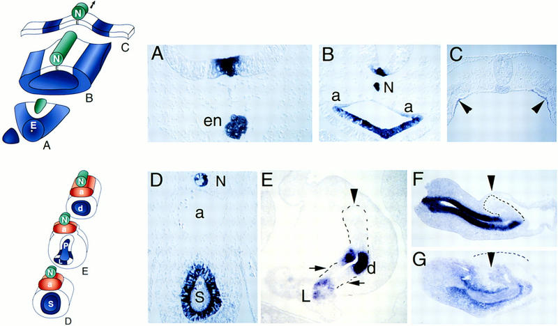
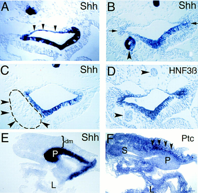
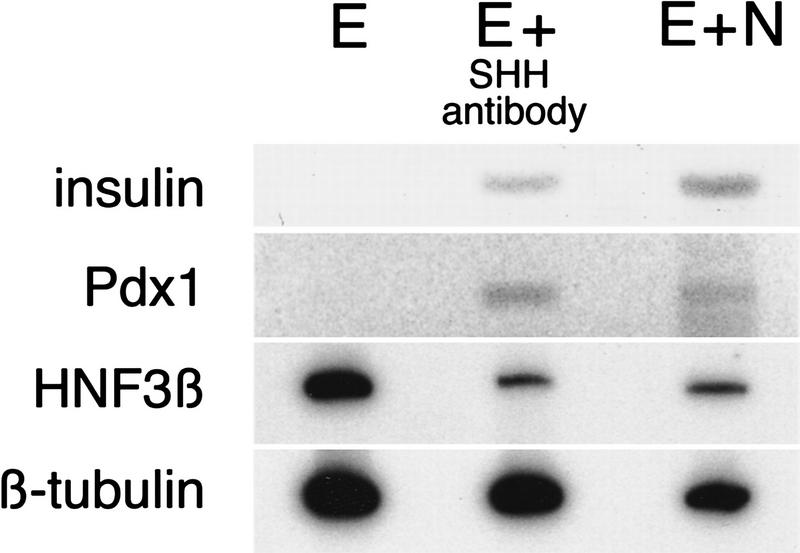
Notochord signals to the endoderm are required for development of the chick dorsal pancreas. Sonic hedgehog (SHH) is normally absent from pancreatic endoderm, and we provide evidence that notochord, in contrast to its effects on adjacent neuroectoderm where SHH expression is induced, represses SHH expression in adjacent nascent pancreatic endoderm. We identify activin-betaB and FGF2 as notochord factors that can repress endodermal SHH and thereby permit expression of pancreas genes including Pdx1 and insulin. Endoderm treatment with antibodies that block hedgehog activity also results in pancreatic gene expression. Prevention of SHH expression in prepancreatic dorsal endoderm by intercellular signals, like activin and FGF, may be critical for permitting early steps of chick pancreatic development.


Figures

Figure 1
Figure 1
Patterns of Shh and Ptc expression in endoderm and pancreas. Upper schematic shows transverse sections through gut endoderm (E) and adjacent tissues including notochord (N) at stage 11 (A–C). Endoderm without Shh expression is white, endoderm with Shh expression is blue. Lower schematic shows transverse sections through notochord (N), aorta (a), stomach (S), pancreas (P), and duodenal (d) endoderm and adjacent mesenchyme at stage 18–19 (D–G). (A) In situ hybridization with Shh probe of transverse section through prechordal endoderm at the extreme rostral end of the digestive tract. Shh expression in the floor plate of the neural tube is detected dorsal to endodermal (en) cells also expressing Shh. Note absence of notochord at this rostrocaudal level. (B) Shh expression in foregut endoderm rostral to the anterior intestinal portal. Shh expression is evident in floor plate cells and notochord (N), dorsal to endoderm expression. Note absence of dorsal endodermal Shh expression and the transition from columnar shape of ventral endodermal cells to squamous shape of dorsal midline endoderm. Paired dorsal aortas are marked “a”. (C) Midgut/hindgut Shh expression. Arrowheads point to two stripes of endodermal Shh expression. Endoderm medial and lateral to these Shh-expressing cells does not express detectable Shh. Floor plate and regressing Hensen’s node cells at this caudal level do not express detectable Shh at this stage. (D) Shh expression in stomach (S) endoderm and notochord (N) after aortas (a) fuse. Expression of Shh is evident in dorsal and ventral columnar epithelial cells. (E) Endodermal Shh expression in the pancreatic anlage. Endoderm in the nascent dorsal (arrowhead) and two ventral pancreatic buds (arrows) does not express Shh in contrast to strong expression in duodenal (d) endoderm between the buds and ventral common bile duct endoderm in the liver (L). (F) Sagittal section of dissected foregut revealing Shh expression anterior to the dorsal pancreas bud (arrowhead and dotted outline) in stomach and duodenal endoderm. Space adjacent to the dorsal pancreas bud is the lumen of the right omphalomesenteric vein. (G) Sagittal section revealing foregut expression of Ptc in mesenchyme adjacent to stomach and duodenal endoderm and in dorsal pancreas endoderm (arrowhead), but little detectable Ptc in dorsal pancreas mesenchyme (outlined by dashed line). (A–G) Dorsal is at the top. (F,G) Anterior is at left.
Figure 2
Figure 2
Notochord signals repress Shh expression in adjacent endoderm. Transverse sections through foregut adjacent to the anterior intestinal portal at stage 16 (A–D). (A) Ectopic Shh expression in dorsal foregut endoderm after notochord deletion (n = 10) at stage 10–11. Arrowheads outline blue dorsal endoderm. Contrast to Fig. 1B. (B) Ectopic notochord graft decreases adjacent endodermal Shh expression; compare to contralateral control side (arrows). Ectopic notochord also expresses Shh (arrowhead). The endoderm adjacent to notochord grafts is reproducibly thinner than corresponding control endoderm on the contralateral side (n = 18) The apparent lumen in the transplanted notochord is an artifact of histologic preparation. (C) Endodermal Shh expression after insertion of the ninth somite (dashed line and arrowheads), from a stage 10 donor, adjacent to endoderm. Shh expression in endoderm is unaffected compared to the contralateral side (n = 4). (D) HNF3β expression in foregut endoderm after notochord grafting. Endogenous or grafted notochord (arrowheads) does not express HNF3β at this stage. Ventral endodermal HNF3β expression is unaffected by ectopic notochord. (E,F) Sagittal sections through dissected pancreatic anlage and adjacent anterior foregut at stage 19. (E) Ectopic endodermal Shh expression in the dorsal pancreas bud (P) after notochord deletion at stage 11. Some liver (L) is seen in this section but liver and stomach endoderm are out of the section plane. Mesenchyme (dm) overlying the dorsal pancreas bud is indicated. (F) shows stomach (S), liver (L), and ectopic Ptc expression in mesenchyme (arrowheads) adjacent to the dorsal pancreas bud (P) after notochord deletion. Dorsal is toward the top; anterior is toward the left in E and F.
Figure 3
Figure 3
Separation of pancreatic endoderm and notochord promotes Shh expression and prevents Pdx1 and insulin expression. (A) Schematic transverse section through stage 12 pancreatic anlage summarizing tissue-restricted expression patterns of Gnot1 (notochord), caudal (endoderm), GATA5 (endothelial and splanchnic mesoderm), GMHox (splanchnic mesoderm), and Pax1 (ventral somitic mesoderm), adapted from Patten and Carlson (1974). (B) Gene expression detected by RT–PCR in dorsal pancreatic endoderm isolated at stage 12. Total RNA was harvested and analyzed by RT–PCR for caudal, GNot1, GATA5, GMHox, Pax1, HNF3β, and β-tubulin RNAs in whole torso (WT), which includes ectoderm, mesoderm, and endoderm germ layers, freshly dissected midline endoderm (E0) or notochord (N). β-Tubulin was used as a loading control. No signal was detected in control samples of RNA from whole embryo trunk samples untreated with reverse transcriptase (−RT). (C) Schematic of in vitro pancreatic endoderm growth and RT–PCR analysis revealing pancreas marker gene induction by notochord. Endoderm and notochord were removed from stage-12 embryos as previously described (Kim et al. 1997a) and grown in contact in a collagen matrix. (D) RT–PCR analysis of RNA from freshly dissected endoderm (EO) shows no insulin, Pdx1, or Shh expression. Endoderm isolated from stage-12 embryos and grown for 3 days in vitro (E) without notochord expresses Shh but not insulin or Pdx1. Recombination and growth of endoderm with notochord (E+N) results in insulin and Pdx1 expression. High levels of Shh expression by notochord in the E+N sample are also detected.
Figure 4
Figure 4
Activin-βB and FGF2 mimic notochord activity in inducing pancreatic genes. (A) Notochord (N) isolated at stage 12 expresses FGF2, activin-βB, and Gnot1 assayed by RT–PCR. Absent GMHox or GATA5 signals indicate lack of adherent lateral mesoderm. Absence of adherent midline endoderm is demonstrated by lack of detectable caudal expression. Isolated lateral mesoderm (LM) expresses FGF2, GMHox, and GATA5, but not activin-βB, caudal or Gnot1. β-Tubulin was used as a loading control. (B) Endoderm isolated at stage 12 was grown in the absence (E) or presence of increasing amounts of activin-βB (E+activin) or recombinant FGF2 (E+FGF2), then analyzed by RT–PCR for expression of insulin, Pdx1, or β-tubulin. (C) Isolated stage 12 endoderm grown in the presence of 5 U/ml human activin-βB and recombinant Shh at increasing doses,or grown in the presence of FGF2 at increasing doses. Endoderm grown in activin and Shh was assayed for expression of insulin, Pdx1, and β-tubulin; explants grown in FGF2 were tested for expression of patched (ptc), Sonic hedgehog (shh) and β-tubulin. PCR products of β-tubulin transcripts were used as loading controls.
Figure 5
Figure 5
Ectopic activin down-regulates endodermal Shh. Black arrowheads indicate wild-type endogenous expression of endodermal Shh. White arrowheads indicate where expression is not present in embryos adjacent to ectopic activin-coated beads; yellow arrowheads and black circles indicate beads. (A–E) In situ hybridization with Shh probe at stage 17–18, (A–C) at the level of midgut and (D,E) in the stomach anlage. Control bead (B) soaked in phosphate buffered saline or activin-soaked beads (C,E) were implanted adjacent to midline foregut endoderm in stage 10 embryos. Shh expression in unmanipulated endoderm (A,D) is the same as in the embryos with control bead implants. In E, the beads became detached during processing for in situ hybridization.
Figure 6
Figure 6
Antibody-mediated induction of pancreatic gene expression by endoderm. Explanted stage 12 endoderm was grown alone (E), in contact with nickel–agarose beads preincubated in an affinity-purified antiserum specific to the 19-kD amino-terminal Shh peptide (E+SHH antibody) or in contact with stage 12 notochord (E+N). Samples were collected after 3 days and analyzed by RT–PCR for expression of insulin, Pdx1, HNF3β, and β-tubulin.

References

    1. Ahlgren U, Jonsson J, Edlund H. The morphogenesis of the pancreatic mesenchyme is uncoupled from that of the pancreatic epithelium in IPF1/PDX1-deficient mice. Development. 1996;122:1409–1416. - PubMed
    1. Ahlgren U, Pfaff SL, Jessell TM, Edlund T, Edlund H. Independent requirement for ISL1 in formation of pancreatic mesenchyme and islet cells. Nature. 1997;385:257–260. - PubMed
    1. Apelqvist Å, Ahlgren U, Edlund H. Sonic hedgehog directs specialised mesoderm differentiation in the intestine and pancreas. Curr Biol. 1997;7:801–804. - PubMed
    1. Basler K, Edlund T, Jessell TM, Yamada T. Control of cell pattern in the neural tube: Regulation of cell differentiation by dorsalin-1, a novel TGFβ family member. Cell. 1993;73:687–702. - PubMed
    1. Beaupain D, Dieterlen-Lièvre F. Etude immunocytologique de la differenciation du pancreas endocrine chez l’embryon de poulet. II. Glucagon. Gen Comp Endocrinol. 1974;23:421–423. - PubMed

Publication types

LinkOut - more resources