. 1998 Jun;180(12):3245-9.
doi: 10.1128/JB.180.12.3245-3249.1998.

Appropriate expression of filamentous phage f1 DNA replication genes II and X requires RNase E-dependent processing and separate mRNAs

Affiliations

Appropriate expression of filamentous phage f1 DNA replication genes II and X requires RNase E-dependent processing and separate mRNAs

R J Kokoska et al. J Bacteriol. 1998 Jun.

Abstract

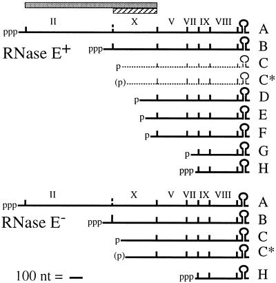
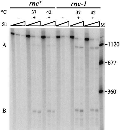
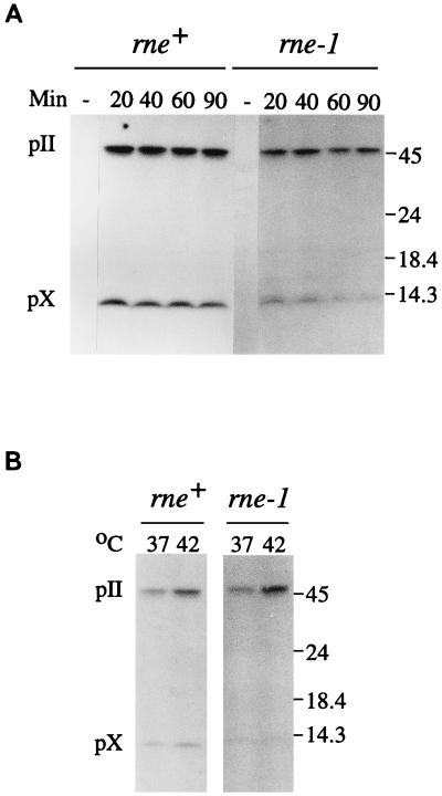
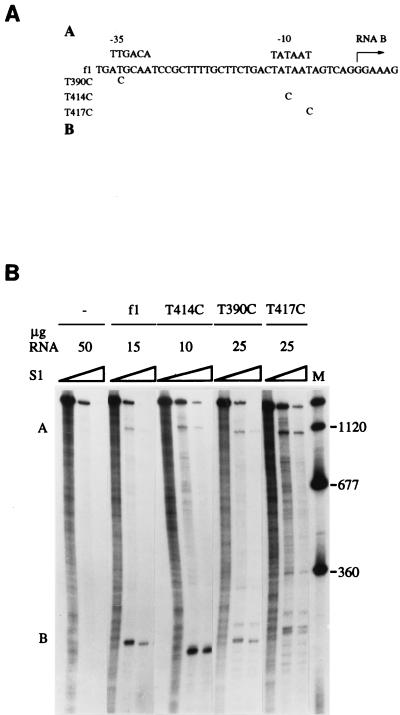
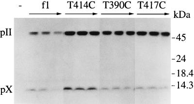
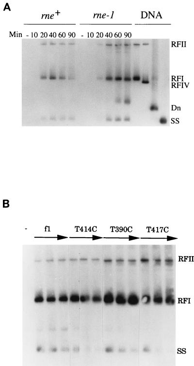
The products of in-frame overlapping genes II and X carried by the filamentous phage f1 genome are proteins with required but opposing functions in phage DNA replication. Their normal relative levels are important for continuous production of phage DNA without killing infected Escherichia coli hosts. Here we identify several factors responsible for determining the relative levels of pII and pX and that, if perturbed, alter the normal distribution of the phage DNA species in infected hosts. Translation of the two proteins is essentially relegated to separate mRNAs. The mRNAs encoding genes II and X are also differentially sensitive to cleavage dependent on rne, the gene encoding the only E. coli endo-RNase known to have a global role in mRNA stability. Whereas pII levels are limited at the level of mRNA stability, normal pX levels require transcription in sufficient amounts from the promoter for the smaller mRNA encoding only pX.


Figures

FIG. 1
FIG. 1
Bacteriophage f1 mRNA for RNase E+ and RNase E E. coli hosts. The extents of gene II (shaded bar) and gene X (hatched bar) are shown. The mRNA diagram for the RNase E+ hosts is based on the work of many authors (reviewed in reference 18), and that for the RNase E hosts is as described in reference . The f1 mRNAs, scaled as indicated, range in size from 370 to 2,000 nt. Coding regions are demarcated by vertical lines. Primary transcripts from strong constitutive promoters and posttranscriptional cleavage products are indicated by known or probable (in parentheses) phosphorylation status. RNAs C and C* are short-lived species observed at very low levels in wild-type hosts (1, 2) but higher levels in RNase E hosts (10). The f1 mRNAs have a common 3′ end generated by rho-independent termination.
FIG. 2
FIG. 2
Primary transcripts A and B in phage-infected rne+ and rne-1 hosts. DS211 (F′Tn10/rne+ λ) and DS212 (F′Tn10/rne-1 λ) were grown at 34°C to 2 × 10⁸ cells/ml in Luria-Bertani broth (27) containing 12 μg of tetracycline per ml and infected (multiplicity of infection of 50) with wild-type f1 (+) or left uninfected (−). They were shifted to the indicated temperatures at 15 min after infection. Samples (5 ml) were removed immediately prior to infection or 60 min after the temperature shift. The 5′-32P-labeled probe and size standards (nucleotides) (lane M) were generated in a similar manner by primer extension and purification on alkaline gels. Each lane contained the products of analysis of 2 μg (DS211) or 1 μg (DS212) of RNA and 0.1 pmol of probe. The S1 nuclease concentrations for each set of lanes were none or 0.1 and 0.35 U/μg of RNA. Samples were electrophoresed on 6% polyacrylamide sequencing gels containing a gradient of 0.5× to 2.5× TBE (89 mM Tris-borate, 2.5 mM EDTA [pH 8.3]).
FIG. 3
FIG. 3
Immunoblot analysis of pII and pX. The rne+ and rne-1 hosts were infected with wild-type f1 (multiplicity of infection of 300) or left uninfected (−) and shifted to 37°C (A) or 37 and 42°C (B). Samples removed immediately prior to infection or at the indicated times in minutes after the temperature shift were chilled in 0.5 ml of a combination of 10 mM (each) Tris-HCl (pH 7.5), EDTA, NaCl, and NaN3. Following centrifugation, cells were resuspended in sample buffer, incubated at 100°C for 5 min, and analyzed on sodium dodecyl sulfate-polyacrylamide gels containing gradients of 10 to 20% acrylamide and 0.26 to 1.0% bisacrylamide. Electrophoretic transfer was to 0.2-μm-pore-size BA83 nitrocellulose membranes (24, 30). Each lane contained an amount of extract equivalent to 108 cells. pII and pX were detected with anti-pX immunoglobulin G (100 μg) and 125I-labeled protein A (0.5 μCi). Antiserum against pX was made from protein overexpressed in strain K561 (7) containing a plasmid with the pII coding region under control of the tacI promoter. To a culture (50 ml) grown at 37°C to 2 × 10⁸ cells/ml in Luria-Bertani broth containing 0.1 mg of ampicillin per ml, isopropyl-β-d-thiogalactopyranoside (IPTG) was added to 2 mM. Ampicillin was replenished at the time of induction and 1 h later. Bacteria were harvested by centrifugation after 2 h and resuspended in 5 ml of sample buffer (30), and samples were fractionated on sodium dodecyl sulfate-polyacrylamide gels (13). pX was eluted electrophoretically. Antiserum was prepared by Pocono Rabbit Farm and Laboratory, Inc., and immunoglobulin G fractions were isolated by chromatography on protein A-Sepharose. The positions of pII, pX, and molecular mass standards (in kilodaltons) are indicated to the right.
FIG. 4
FIG. 4
Primary transcripts A and B in a wild-type host infected with phage bearing mutations in the promoter for RNA B. (A) Location of the point mutations in the f1 DNA sequence (8). Consensus sequences for the −35 and −10 regions of sigma 70 promoters and the probable start point for transcription of RNA B (+1) are shown. Oligonucleotide mutagenesis (12) used uracil-containing f1 (+) strand DNA generated in CJ236 (dut ung mutant). Following extension of the annealed primers and closure of new strands, the double-stranded circular molecules were introduced into JM109 (29) and incubated overnight at 37°C in Luria-Bertani top agar in a lawn of JM109. The desired mutations were identified by dideoxy sequencing (25). (B) S1 nuclease protection analysis of RNAs A and B following infection of S26r1eλ (Hfr Cavalli phoA4(Am) serU132 λ) with no phage (−), wild-type phage (f1), or the indicated mutants. Assays, performed with samples isolated 50 min after infection, contained the indicated quantities of RNA and 5′-end-labeled probe (0.05 pmol). The final S1 nuclease concentrations in each set of lanes were none or 0.33 and 1.0 U/μg of RNA. The panel is a composite that presents one RNA concentration of several tested for each phage strain. Sizes are shown to the right in nucleotides.
FIG. 5
FIG. 5
Immunoblot analysis of pII and pX produced by the phage promoter mutants. Cell extracts from S26r1eλ left uninfected (−) or infected with the indicated phage strains were prepared immediately prior to infection or 30, 60, and 90 min after infection.
FIG. 6
FIG. 6
Southern blot hybridization of phage DNA species. (A and B) DS211 (rne+) or DS212 (rne-1) (A) and S26r1eλ (B) left uninfected (−) or infected with the indicated phage strains (multiplicity of infection of 80). Subcultures of DS211 and DS212 grown at 30°C were shifted to 37°C just before infection. Samples (0.5 ml) were mixed with an equal volume of an ice-cold mixture of 10 mM Tris-HCl (pH 8.1), 10 mM EDTA, 10 mM NaCl, and 10 mM NaN3 and washed twice with the same buffer lacking NaN3 to remove free phage. Resuspended cells were lysed (14) at 55°C. EDTA (pH 9.3) was added to 25 mM, proteinase K was added to 150 μg/ml, and incubation was continued at 37°C for 60 min. Chromosomal DNA was sheared by passage through a 22-gauge needle. Purified DNA preparations (24) were resuspended in 20 μl of 10 mM Tris-HCl (pH 8.0)–1 mM EDTA. Standards included RFI (duplex supercoiled), RFII (nicked duplex), and ssDNA (SS) (24). RFIV DNA (relaxed) was generated by treatment of RFI DNA with Drosophila melanogaster topoisomerase II (9). Denatured DNA (Dn) was generated from RFI DNA by incubation in 1.5 N NaOH at room temperature for 15 min, followed by addition of sodium acetate (pH 4.9) to a concentration of 0.6 M. DNA samples (3 μl) were electrophoresed overnight at 40 V in 1% agarose gels (20 by 20 by 0.3 cm) in a mixture of 80 mM Tris-phosphate (pH 8.2) and 8 mM EDTA containing 0.5 μg of ethidium bromide per ml with continuous buffer recirculation. Transfer of DNA was to GeneScreen Plus after alkaline denaturation (24). The DNA probe (∼107 cpm) was generated by nick translation (24) of 0.1 μg of f1 RFI DNA in the presence of [α-32P]dCTP (3,000 Ci/mmol, DuPont NEN). DNA samples from uninfected cultures (−) and from time points in minutes after infection are indicated. In panel B, the three lanes shown for each phage strain represent samples isolated at 30, 60, and 90 min after infection.

Similar articles

Cited by

References

    1. Blumer K J, Steege D A. mRNA processing in Escherichia coli: an activity encoded by the host processes bacteriophage f1 mRNAs. Nucleic Acids Res. 1984;12:1847–1861. - PMC - PubMed
    1. Cashman J S, Webster R E, Steege D A. Transcription of bacteriophage f1: the major in vivo RNAs. J Biol Chem. 1980;255:2554–2562. - PubMed
    1. Cohen S N, McDowall K J. RNase E: still a wonderfully mysterious enzyme. Mol Microbiol. 1997;23:1099–1106. - PubMed
    1. Fulford W, Model P. Specificity of translational regulation by two DNA binding proteins. J Mol Biol. 1984;173:211–226. - PubMed
    1. Fulford W, Model P. Regulation of bacteriophage f1 DNA replication. I. New functions for genes II and X. J Mol Biol. 1988;203:49–62. - PubMed

Publication types

LinkOut - more resources