. 1998 Jul;72(7):5343-50.
doi: 10.1128/JVI.72.7.5343-5350.1998.

The latent herpes simplex virus type 1 genome copy number in individual neurons is virus strain specific and correlates with reactivation

Affiliations

The latent herpes simplex virus type 1 genome copy number in individual neurons is virus strain specific and correlates with reactivation

N M Sawtell et al. J Virol. 1998 Jul.

Abstract

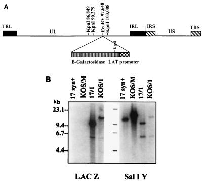
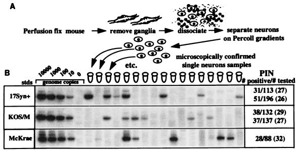
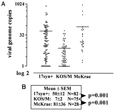
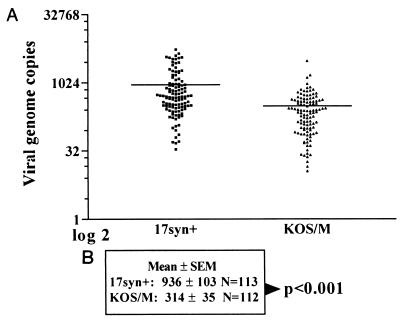
The viral genetic elements that determine the in vivo reactivation efficiencies of fully replication competent wild-type herpes simplex virus (HSV) strains have not been identified. Among the common laboratory strains, KOS reactivates in vivo at a lower efficiency than either strain 17syn+ or strain McKrae. An important first step in understanding the molecular basis for this observation is to distinguish between viral genetic factors that regulate the establishment of latency from those that directly regulate reactivation. Reported here are experiments performed to determine whether the reduced reactivation of KOS was associated with a reduced ability to establish or maintain latent infections. For comparative purposes, latent infections were quantified by (i) quantitative PCR on DNA extracted from whole ganglia, (ii) the number of latency-associated transcript (LAT) promoter-positive neurons, using KOS and 17syn+ LAT promoter-beta-galactosidase reporter mutants, and (iii) contextual analysis of DNA. Mice latently infected with 17syn+-based strains contained more HSV type 1 (HSV-1) DNA in their ganglia than those infected with KOS strains, but this difference was not statistically significant. The number of latently infected neurons also did not differ significantly between ganglia latently infected with either the low- or high-reactivator strains. In addition to the number of latent sites, the number of viral genome copies within the individual latently infected neurons has recently been demonstrated to be variable. Interestingly, neurons latently infected with KOS contained significantly fewer viral genome copies than those infected with either 17syn+ or McKrae. Thus, the HSV-1 genome copy number profile is viral strain specific and positively correlates with the ability to reactivate in vivo. This is the first demonstration that the number of HSV genome copies within individual latently infected neurons is regulated by viral genetic factors. These findings suggest that the latent genome copy number may be an important parameter for subsequent induced reactivation in vivo.


Figures

FIG. 1
FIG. 1
(A) Schematic of the HSV-1 genome. The terminal repeat long, unique long, internal repeat long, internal repeat short, unique short, and terminal repeat short portions of the genome are labeled TRL, UL, IRL, IRS, US, and TRS, respectively. Relevant KpnI restriction endonuclease sites and the location of the LAT promoter–β-Gal insert are indicated. (B) Viral DNAs were cleaved with KpnI, electrophoresed, blotted, and probed as described in Materials and Methods. Left, probe is a 624-bp HpaI fragment from the lacZ gene present in plasmid pCH110; right, probe is HSV-1 SalI fragment Y (bp 94853 to 98422 on the viral genome [19]).
FIG. 2
FIG. 2
(A) NIH 3T3 cells were infected at a low multiplicity (0.001 PFU/cell). At 24-h intervals, triplicate cultures were assayed for virus content. (B to D) Mice were infected with a total of 3 × 10⁵ PFU of the virus isolates on the corneas and snouts as described in Materials and Methods. At daily intervals, three mice from each group were sacrificed, and the eyes, snouts, and TG were homogenized and assayed for infectious virus.
FIG. 3
FIG. 3
Dual localization of HSV antigen and LAT promoter activity in TG during acute infection with 17/1 and KOS/1 (photomicrograph of sectioned TG on day 4). Both 17/1 (A)- and KOS/1 (C)-infected ganglia contain many HSV antigen-positive neurons and support cells. The fields shown were selected because several β-Gal-containing neurons were present. The difference in the number of antigen-positive cells in the 17/1 and KOS/1 sections shown is not representative. The vast majority (>90%) of β-Gal-expressing neurons were devoid of detectable HSV antigen in both 17/1 (B)- and KOS/1 (D)-infected ganglia. However, with both, there were a few neurons in which HSV antigen could be detected (inset). In agreement with previous findings, β-Gal staining was restricted to neurons (19, 31).
FIG. 4
FIG. 4
Analysis of latent infections at the single-cell level. Latently infected mice were processed for determination of PIN by using CXA-D. (A) Flow diagram of CXA-D. (B) Representative southern blots of the PCR products probed with a [γ-32P]ATP-labeled oligonucleotide internal to the amplification primers. The first four lanes are standards (stds) containing known amounts of HSV-1 DNA, representing the indicated genome equivalents. The fifth lane is a buffer blank performed on cell-free immobilized DNase-treated supernatant from the purified neuron preparations. As expected, the majority of the neurons tested did not contain any viral genomes. The number of neurons positive/the number tested and the PIN are shown on the right.
FIG. 5
FIG. 5
Analysis of HSV-1 latent genome copy number profiles. The data generated as in Fig. 3 were analyzed on a Molecular Dynamics PhosphorImager using ImageQuant software. The total counts per minute in each band was determined and compared to the values obtained for the standards included with each set of PCRs. (A) Scattergram of the results. Each point represents the number of HSV-1 genomes detected in a single neuron plotted on a semi-log2 graph to decompress the points. Horizontal bars within the columns mark the mean values. (B) Means ± standard errors. 17syn+ and McKrae groups were not different (P = 0.287, unpaired t test). The KOS/M samples were statistically different from both 17syn+ and McKrae (P = 0.001, unpaired t test).
FIG. 6
FIG. 6
Analysis of HSV-1 genome copy number profiles in samples of 10 latently infected neurons. The PIN generated in Fig. 3 was used to dilute the purified neurons such that each PCR contained approximately 10 latently infected cells (∼40 total neurons). As described in Materials and Methods, the number of PCR cycles was adjusted to maintain linearity in the standard sample between 100 and 10,000 copies. (A) Scattergram of results; (B) means ± standard errors.

Similar articles

Cited by

References

    1. Bloom D C, Hill J M, Devi-Rao G, Wagner E K, Feldman L T, Stevens J G. A 348-base-pair region in the latency-associated transcript facilitates herpes simplex virus type 1 reactivation. J Virol. 1996;70:2449–2459. - PMC - PubMed
    1. Cai W, Astor T L, Liptak L M, Cho C, Coen D M, Schaffer P A. The herpes simplex virus type 1 regulatory protein ICP0 enhances virus replication during acute infection and reactivation from latency. J Virol. 1993;67:7501–7512. - PMC - PubMed
    1. Davies A, Lumsden A. Relation of target encounter and neuronal death to nerve growth factor responsiveness in the developing mouse trigeminal ganglion. J Comp Neurol. 1984;223:124–137. - PubMed
    1. Dobson A T, Margolis T P, Gomes W A, Feldman L T. In vivo deletion analysis of the herpes simplex virus type 1 latency-associated transcript promoter. J Virol. 1995;69:2264–2270. - PMC - PubMed
    1. Glorioso J C, DeLuca N A, Fink D J. Development and application of herpes simplex virus vectors for human gene therapy. Annu Rev Microbiol. 1995;49:675–710. - PubMed

Publication types