. 1998 Jul;72(7):5351-9.
doi: 10.1128/JVI.72.7.5351-5359.1998.

In vivo immune evasion mediated by the herpes simplex virus type 1 immunoglobulin G Fc receptor

Affiliations

In vivo immune evasion mediated by the herpes simplex virus type 1 immunoglobulin G Fc receptor

T Nagashunmugam et al. J Virol. 1998 Jul.

Abstract

Herpes simplex virus (HSV) glycoproteins gE and gI form an immunoglobulin G (IgG) Fc receptor (FcgammaR) that binds the Fc domain of human anti-HSV IgG and inhibits Fc-mediated immune functions in vitro. gE or gI deletion mutant viruses are avirulent, probably because gE and gI are also involved in cell-to-cell spread. In an effort to modify FcgammaR activity without affecting other gE functions, we constructed a mutant virus, NS-gE339, that has four amino acids inserted into gE within the domain homologous to mammalian IgG FcgammaRs. NS-gE339 expresses gE and gI, is FcgammaR-, and does not participate in antibody bipolar bridging since it does not block activities mediated by the Fc domain of anti-HSV IgG. In vivo studies were performed with mice because the HSV-1 FcgammaR does not bind murine IgG; therefore, the absence of an FcgammaR should not affect virulence in mice. NS-gE339 causes disease at the skin inoculation site comparably to wild-type and rescued viruses, indicating that the FcgammaR- mutant virus is pathogenic in animals. Mice were passively immunized with human anti-HSV IgG and then infected with mutant or wild-type virus. We postulated that the HSV-1 FcgammaR should protect wild-type virus from antibody attack. Human anti-HSV IgG greatly reduced viral titers and disease severity in NS-gE339-infected animals while having little effect on wild-type or rescued virus. We conclude that the HSV-1 FcgammaR enables the virus to evade antibody attack in vivo, which likely explains why antibodies are relatively ineffective against HSV infection.


Figures

FIG. 1
FIG. 1
(A) Southern blot of wild-type, gE mutant, and rescued viruses. NS, NS-gEnull, rNS-gEnull, NS-gE339, rNS-gE339, and NS-gE406 were digested with NruI alone (lanes 1 to 6) or with NruI and XhoI to detect XhoI linkers in NS-gE339 and NS-gE406 (lanes 7 to 12). The blot was probed with a 1.1-kb HpaI-BglII gE fragment. The position of the 2.4-kb gE band is shown on the right, and positions (in kilobases) of DNA size markers are shown on the left. (B) Model of HSV-1 gE, a 550-amino-acid glycoprotein. Sig, predicted signal sequence, amino acids 1 to 23; # # #, the domain on gE, amino acids 235 to 264, that interacts with gI to form a hetero-oligomer complex (3); ∗∗∗, the region of gE, amino acids 235 to 380, which comprises the IgG Fc binding domain (3, 14); •••, the gE domain of homology with mammalian FcγRs. gE amino acids 322 to 359 have 46% identity and 66% similarity with domain 2 of human FcγRII (14). TM refers to the predicted transmembrane domain of gE, amino acids 420 to 444. Arrows at amino acids 339 and 406 indicate the positions of four amino acids (ARAA) inserted within and outside, respectively, the IgG Fc binding domain. HpaI and BglII sites were used to delete gE amino acids 124 to 508. The ICP6::lacZ cassette was cloned into this site to generate the gE null virus. The shaded balloons indicate potential N-linked glycosylation sites at gE amino acids 124 and 243. C’s mark the cysteine positions in the extracellular domain of gE at amino acids 63, 88, 271, 280, 289, 297, 314, 323, and 359.
FIG. 2
FIG. 2
Double-label flow cytometry for gE expression and monomeric IgG binding. Cells were infected with wild-type, gE mutant, and rescued viruses and analyzed for gE expression by using MAb anti-gE 1BA10 and FITC F(ab′)2 anti-mouse IgG (x axis) or for FcγR activity by using biotin-labeled monomeric nonimmune IgG and strepavidin-phycoerythrin (y axis). Fluorescence in the upper right quadrant indicates both gE expression and IgG binding, fluorescence in the lower left quadrant indicates neither gE expression nor IgG binding, while fluorescence in the lower right quadrant indicates gE expression but no IgG binding. (A) NS; (B) NS-gEnull; (C) NS-gE339; (D) NS-gE406; (E) rNS-gEnull; (F) rNS-gE339.
FIG. 3
FIG. 3
(A) Model showing the HSV-1 FcγR blocking complement-enhanced antibody neutralization. On the left, an antibody molecule (red) binds to its target antigen (shown in green as HSV-1 glycoprotein gD) by its Fab domain. The absence of an FcγR enables C1q (brown) to bind to the antibody Fc domain, leading to activation of complement and complement-enhanced antibody neutralization. On the right is shown an example of antibody bipolar bridging in which an antibody molecule (red) binds to its target antigen (green) by the Fab domain while the Fc domain of the same antibody molecule binds to the HSV-1 FcR (blue), which blocks the interaction of C1q (brown) with the IgG Fc domain. (B) Complement-enhanced antibody neutralization of FcγR+ viruses NS, rNS-gE339, and rNS-gEnull and FcγR viruses NS-gE339 and NS-gEnull. Each virus was incubated with pooled human IgG at 100 μg/ml, which resulted in 50% neutralization in the absence of complement. Then 10% nonimmune human serum or heat-inactivated serum was added, and complement-enhanced neutralization was calculated by determining the additional neutralization mediated by including complement in the reaction. Results are the mean (± SEM) of three experiments.
FIG. 3
FIG. 3
(A) Model showing the HSV-1 FcγR blocking complement-enhanced antibody neutralization. On the left, an antibody molecule (red) binds to its target antigen (shown in green as HSV-1 glycoprotein gD) by its Fab domain. The absence of an FcγR enables C1q (brown) to bind to the antibody Fc domain, leading to activation of complement and complement-enhanced antibody neutralization. On the right is shown an example of antibody bipolar bridging in which an antibody molecule (red) binds to its target antigen (green) by the Fab domain while the Fc domain of the same antibody molecule binds to the HSV-1 FcR (blue), which blocks the interaction of C1q (brown) with the IgG Fc domain. (B) Complement-enhanced antibody neutralization of FcγR+ viruses NS, rNS-gE339, and rNS-gEnull and FcγR viruses NS-gE339 and NS-gEnull. Each virus was incubated with pooled human IgG at 100 μg/ml, which resulted in 50% neutralization in the absence of complement. Then 10% nonimmune human serum or heat-inactivated serum was added, and complement-enhanced neutralization was calculated by determining the additional neutralization mediated by including complement in the reaction. Results are the mean (± SEM) of three experiments.
FIG. 4
FIG. 4
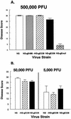
(A) Disease scores at the inoculation site in the mouse flank after infection by wild-type, NS, FcγR+ mutant, NS-gE406, FcγR mutants, NS-gE339 and NS-gEnull, and rescued virus, rNS-gE339. An inoculum of 5 × 10⁵ PFU was scratched onto the denuded flank of 10 BALB/c mice per group. The average (± SEM) cumulative disease scores from days 3 to 8 are shown at the inoculation site. (B) An inoculum of 5 × 10⁴ or 5 × 10³ PFU was scratched onto the denuded flank of each of five mice per group, and the mean (± SEM) cumulative disease scores from days 3 to 8 were calculated.
FIG. 5
FIG. 5
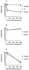
(A) Mice were passively immunized with 200 or 2,000 μg of human anti-HSV IgG, or saline (0 μg IgG) as a control, and then infected 16 h later with FcγR+ virus NS or rNS-gE339 or FcγR virus NS-gE339. Ten mice were evaluated at each data point. Disease scores in saline controls were set as 100%, and as shown in Fig. 4A, these scores were similar for all three viruses. Percent disease scores at 200 or 2,000 μg of human anti-HSV IgG were calculated as [mean (± SEM) disease score in animals receiving human anti-HSV IgG/mean disease score in saline-treated animals] × 100. (B) Mice were passively immunized with 200 or 2,000 μg of nonimmune human IgG, or saline (0 μg of IgG) as a control. Five mice were included at each data point. Percent disease scores were calculated as [mean (± SEM) disease score in animals receiving nonimmune IgG/mean disease score in saline-treated animals] × 100. (C) Mice were passively immunized with 200 or 2,000 μg of murine anti-HSV IgG, or saline (0 μg of IgG), as a control. Five mice were included at each data point. Percent disease scores were calculated as [mean (± SEM) disease score in animals receiving murine anti-HSV IgG/mean disease score in saline-treated animals] × 100.
FIG. 6
FIG. 6
(A) Viral titers in skin excised from the inoculation site 1, 2, 3, or 5 days after infection with FcγR+ virus NS or FcγR virus NS-gE339. Animals were passively immunized with 500 μg of human anti-HSV IgG or saline as a control and infected 16 h later. Data represent the mean (± SEM) of four (days 1 and 2), five (day 3), and eight (day 5) mice per group. (B) Viral titers in skin excised from the inoculation site 3 days postinfection with rescued FcγR virus rNS-gE339 or FcγR virus NS-gE339. Animals were passively immunized with 500 μg of murine anti-HSV IgG or saline as a control and infected 16 h later. Results are the mean (± SEM) of four mice except rNS-gE339 saline controls, which represent three mice.

Similar articles

Cited by

References

    1. Adler R, Glorioso J C, Cossman J, Levin M. Possible role of Fc receptors on cells infected and transformed by herpesvirus: escape from immune cytolysis. Infect Immun. 1978;21:442–447. - PMC - PubMed
    1. Balan P, Davis-Poynter N, Bell S, Atkinson H, Browne H, Minson T. An analysis of the in vitro and in vivo phenotypes of mutants of herpes simplex virus type 1 lacking glycoproteins gG, gE, gI or putative gJ. J Gen Virol. 1994;75:1245–1258. - PubMed
    1. Basu S, Dubin G, Basu M, Nguyen V, Friedman H M. Characterization of regions of herpes simplex virus type 1 glycoprotein E involved in binding the Fc domain of monomeric IgG and in forming a complex with glycoprotein I. J Immunol. 1995;154:260–267. - PubMed
    1. Basu S, Dubin G, Nagashunmugam T, Basu M, Goldstein L T, Wang L, Weeks B, Friedman H M. Mapping regions of herpes simplex virus type 1 glycoprotein I required for formation of the viral Fc receptor for monomeric IgG. J Immunol. 1997;158:209–215. - PubMed
    1. Baucke R B, Spear P G. Membrane proteins specified by herpes simplex viruses. V. Identification of an Fc-binding glycoprotein. J Virol. 1979;32:779–789. - PMC - PubMed

Publication types

Substances