Kaposi's sarcoma-associated herpesvirus viral interferon regulatory factor
- PMID: 9620998
- PMCID: PMC110176
- DOI: 10.1128/JVI.72.7.5433-5440.1998
Kaposi's sarcoma-associated herpesvirus viral interferon regulatory factor
Abstract
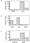
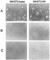
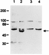
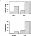
Interferons (IFNs) are a family of multifunctional cytokines with antiviral activities. The K9 open reading frame of Kaposi's sarcoma-associated herpesvirus (KSHV) exhibits significant homology with cellular IFN regulatory factors (IRFs). We have investigated the functional consequence of K9 expression in IFN-mediated signal transduction. Expression of K9 dramatically repressed transcriptional activation induced by IFN-alpha, -beta, and -gamma. Further, it induced transformation of NIH 3T3 cells, resulting in morphologic changes, focus formation, and growth in reduced-serum conditions. The expression of antisense K9 in KSHV-infected BCBL-1 cells consistently increased IFN-mediated transcriptional activation but drastically decreased the expression of certain KSHV genes. Thus, the K9 gene of KSHV encodes the first virus-encoded IRF (v-IRF) which functions as a repressor for cellular IFN-mediated signal transduction. In addition, v-IRF likely plays an important role in regulating KSHV gene expression. These results suggest that KSHV employs an unique mechanism to antagonize IFN-mediated antiviral activity by harboring a functional v-IRF.
Figures


Similar articles
-
Functional analysis of human herpesvirus 8-encoded viral interferon regulatory factor 1 and its association with cellular interferon regulatory factors and p300.J Virol. 1999 Sep;73(9):7334-42. doi: 10.1128/JVI.73.9.7334-7342.1999. J Virol. 1999. PMID: 10438822 Free PMC article.
-
Human herpesvirus 8 encodes an interferon regulatory factor (IRF) homolog that represses IRF-1-mediated transcription.J Virol. 1998 Jan;72(1):701-7. doi: 10.1128/JVI.72.1.701-707.1998. J Virol. 1998. PMID: 9420276 Free PMC article.
-
Transcriptional regulation of the Kaposi's sarcoma-associated herpesvirus viral interferon regulatory factor gene.J Virol. 2000 Sep;74(18):8623-34. doi: 10.1128/jvi.74.18.8623-8634.2000. J Virol. 2000. PMID: 10954564 Free PMC article.
-
Kaposi sarcoma herpesvirus-encoded interferon regulator factors.Curr Top Microbiol Immunol. 2007;312:185-209. doi: 10.1007/978-3-540-34344-8_7. Curr Top Microbiol Immunol. 2007. PMID: 17089798 Review.
-
[Cellular responses by cytokines--gene regulation in the IFN system].Rinsho Ketsueki. 1991 Apr;32(4):301-6. Rinsho Ketsueki. 1991. PMID: 1712401 Review. Japanese.
Cited by
-
Innate antiviral response: role in HIV-1 infection.Viruses. 2011 Jul;3(7):1179-203. doi: 10.3390/v3071179. Epub 2011 Jul 14. Viruses. 2011. PMID: 21994776 Free PMC article. Review.
-
Comparative analysis of the viral interferon regulatory factors of KSHV for their requisite for virus production and inhibition of the type I interferon pathway.Virology. 2020 Feb;541:160-173. doi: 10.1016/j.virol.2019.12.011. Epub 2019 Dec 30. Virology. 2020. PMID: 32056714 Free PMC article.
-
Sperm associated antigen 9 promotes oncogenic KSHV-encoded interferon regulatory factor-induced cellular transformation and angiogenesis by activating the JNK/VEGFA pathway.PLoS Pathog. 2020 Aug 10;16(8):e1008730. doi: 10.1371/journal.ppat.1008730. eCollection 2020 Aug. PLoS Pathog. 2020. PMID: 32776977 Free PMC article.
-
Interferon, Mx, and viral countermeasures.Cytokine Growth Factor Rev. 2007 Oct-Dec;18(5-6):425-33. doi: 10.1016/j.cytogfr.2007.06.001. Epub 2007 Aug 1. Cytokine Growth Factor Rev. 2007. PMID: 17683972 Free PMC article. Review.
-
Overlapping CRE and E box motifs in the enhancer sequences of the bovine leukemia virus 5' long terminal repeat are critical for basal and acetylation-dependent transcriptional activity of the viral promoter: implications for viral latency.J Virol. 2004 Dec;78(24):13848-64. doi: 10.1128/JVI.78.24.13848-13864.2004. J Virol. 2004. PMID: 15564493 Free PMC article.
References
-
- Alexander L, Lee H, Rosenzweig M, Jung J U, Desrosiers R C. EGFP-containing vector system that facilitates stable and transient expression assays. BioTechniques. 1997;23:64–66. - PubMed
-
- Arvanitakis L, Geras-Raaka E, Varma A, Gershengorn M C, Cesarman E. Human herpesvirus KSHV encodes a constitutively active G-protein-coupled receptor linked to cell proliferation. Nature. 1997;385:347–350. - PubMed
-
- Bovolenta C, Driggers P H, Marks M S, Medin J A, Politis A D, Vogel S N, Levy D E, Sakaguchi K, Appella E, Coligan J E, Ozato K. Molecular interactions between interferon consensus sequence binding protein and members of the interferon regulatory factor family. Proc Natl Acad Sci USA. 1994;91:5046–5050. - PMC - PubMed
-
- Cesarman E, Chang Y, Moore P S, Said J W, Knowles D M. Kaposi’s sarcoma-associated Herpesvirus-like DNA sequences in AIDS-related body-cavity-based lymphomas. N Engl J Med. 1995;332:1186–1191. - PubMed
-
- Chang Y, Cesarman E, Pessin M S, Lee F, Culpepper J, Knowles D M, Moore P S. Identification of herpesvirus-like DNA sequences in AIDS-associated Kaposi’s sarcoma. Science. 1994;266:1865–1869. - PubMed
Publication types
MeSH terms
Substances
Grants and funding
LinkOut - more resources
Full Text Sources
Other Literature Sources

