. 1998 Jul;72(7):5493-501.
doi: 10.1128/JVI.72.7.5493-5501.1998.

Influenza virus nucleoprotein interacts with influenza virus polymerase proteins

Affiliations

Influenza virus nucleoprotein interacts with influenza virus polymerase proteins

S K Biswas et al. J Virol. 1998 Jul.

Abstract

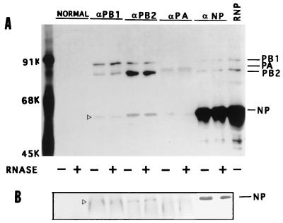
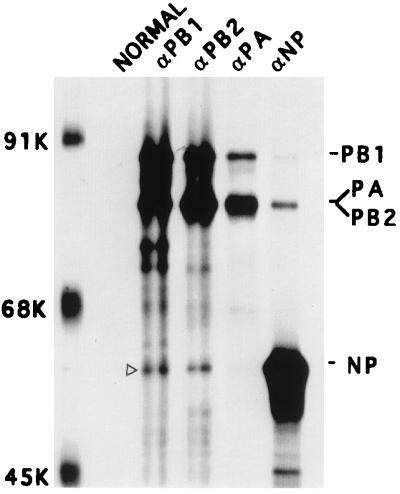
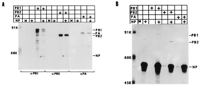
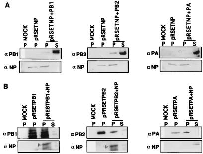
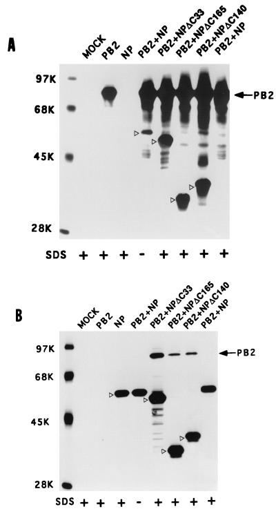
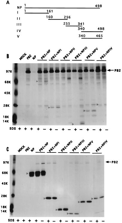
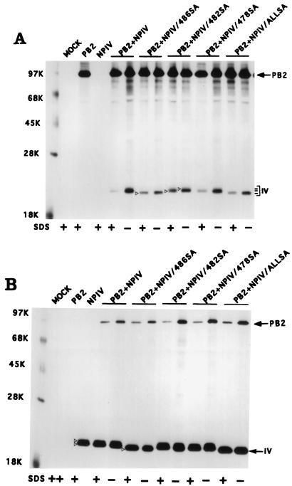
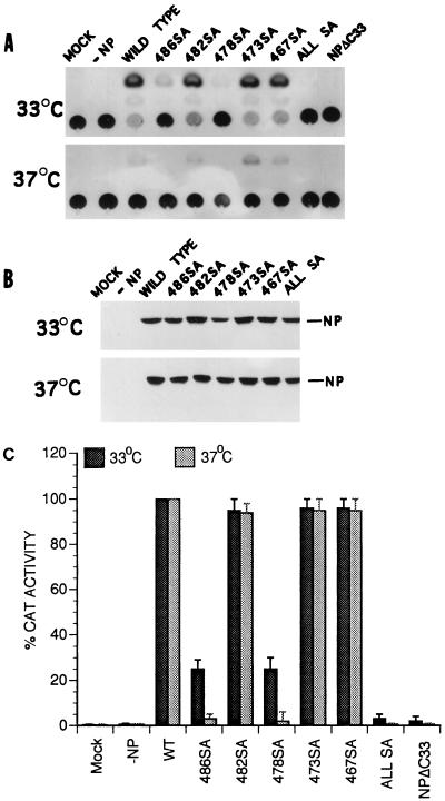
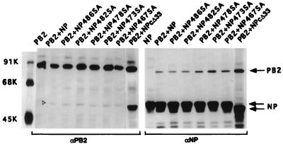
Influenza virus nucleoprotein (NP) is a critical factor in the viral infectious cycle in switching influenza virus RNA synthesis from transcription mode to replication mode. In this study, we investigated the interaction of NP with the viral polymerase protein complex. Using coimmunoprecipitation with monospecific or monoclonal antibodies, we observed that NP interacted with the RNP-free polymerase protein complex in influenza virus-infected cells. In addition, coexpression of the components of the polymerase protein complex (PB1, PB2, or PA) with NP either together or pairwise revealed that NP interacts with PB1 and PB2 but not PA. Interaction of NP with PB1 and PB2 was confirmed by both coimmunoprecipitation and histidine tagging of the NP-PB1 and NP-PB2 complexes. Further, it was observed that NP-PB2 interaction was rather labile and sensitive to dissociation in 0.1% sodium dodecyl sulfate and that the stability of NP-PB2 interaction was regulated by the sequences present at the COOH terminus of NP. Analysis of NP deletion mutants revealed that at least three regions of NP interacted independently with PB2. A detailed analysis of the COOH terminus of NP by mutation of serine-to-alanine (SA) residues either individually or together demonstrated that SA mutations in this region did not affect the binding of NP to PB2. However, some SA mutations at the COOH terminus drastically affected the functional activity of NP in an in vivo transcription-replication assay, whereas others exhibited a temperature-sensitive phenotype and still others had no effect on the transcription and replication of the viral RNA. These results suggest that a direct interaction of NP with polymerase proteins may be involved in regulating the switch of viral RNA synthesis from transcription to replication.


Figures

FIG. 1
FIG. 1
Presence of the NP-polymerase protein complex in influenza virus-infected cells. MDCK cells were infected with influenza virus (A/WSN/33) at an MOI of 5 and labeled with Express 35S (NEN) at 6 hpt for 1 h. Cells were lysed and fractionated into cytoplasmic and nuclear fractions, and vRNPs were removed from both fractions by ultracentrifugation (1, 6). Aliquots of RNP-free supernatants from the cytoplasmic fraction were treated with (+) or without (−) RNase. Each fraction was divided into five parts and immunoprecipitated with either normal serum or anti-PB1 anti-PB2, anti-PA, or anti-NP antibodies in the absence of SDS as noted in Materials and Methods. The RNP pellet was dissolved and immunoprecipitated with a mixture of anti-PB1, anti-PB2, and anti-NP antibodies. The immunoprecipitated complexes were separated by SDS-PAGE (8% gel) and autoradiographed (A). The gel in panel A was transferred to a membrane, and the relevant portion of the membrane was probed with anti-WSN antibodies and detected by chemiluminescence (B). The position of NP is shown with an open arrowhead. Similar results were obtained from the RNP-free nuclear supernatant (data not shown).
FIG. 2
FIG. 2
Interaction of NP with the polymerase protein complex in coexpressing cells. COS1 cells were infected with VTF7.3 at an MOI of 5 and transfected with a mixture of pGEM PB1, pGEM PB2 (3 μg of each DNA), pGEM PA (2 μg of DNA), and pGEM NP (1 μg of DNA). At 14 hpt, cells were labeled with Express 35S for 1 h, lysed, and divided into five parts. Each part was immunoprecipitated with either normal serum or anti-PB1, anti-PB2, anti-PA, or anti-NP antibodies. The immunoprecipitated complex was analyzed by SDS-PAGE (8% gel). The open arrowhead shows the position of NP.
FIG. 3
FIG. 3
Interaction of NP with PB1 and PB2 in coexpressing cells. COS1 cells were infected with VTF7.3 at an MOI of 5 and transfected alone with pGEM NP (1 μg), pGEM PB1 (3 μg), pGEM PB2 (3 μg), or pGEM PA (2 μg) or cotransfected pairwise as indicated (+). At 14 hpt, cells were labeled with Express 35S for 1 h and lysed. The lysate was divided into two parts; one part was immunoprecipitated with anti-NP (B), and the other was immunoprecipitated with either anti-PB1, anti-PB2, or anti-PA antibodies (A). The immunoprecipitated complex was separated by SDS-PAGE (8% gel) and autoradiographed. M, mock transfected with pGEM 3; +, DNA used for transfection. The open arrowhead shows the position of NP.
FIG. 4
FIG. 4
Copurification of the polymerase-NP protein complex, using either His-tagged NP or His-tagged PB1, PB2, or PA. COS1 cells in 60-mm-diameter dishes were infected with VTF7.3 at an MOI of 5 and transfected with pRSET NP (2 μg) alone or with pGEM PB1 (4 μg), pGEM PB2 (4 μg), or pGEM PA (2 μg) (A). In another set, VTF7.3-infected COS1 cells were transfected with pRSET PB1, (4 μg), pRSET PB2 (4 μg), or pRSET PA (2 μg) alone or with pGEM NP (2 μg). At 14 hpt cells were lysed as described in Materials and Methods. The lysate was incubated with TALON beads (Clontech) for 2 h with shaking in 4°C. The beads were then washed as described in Materials and Methods. TALON bead-bound (P) and unbound (S) proteins were analyzed by SDS-PAGE (8% gel) and Western blotted, and respective portions were probed with either anti-PB1, anti-PB2, anti-PA, or anti-WSN antibodies and developed in chemiluminescence solution. Open arrowheads show the positions of NP.
FIG. 5
FIG. 5
COOH-terminal deletion NP mutants bind strongly to PB2. COS1 cells in a 60-mm-diameter dish were infected with VTF7.3 at an MOI of 5 and transfected with pGEM PB2 and pGEM NP or NP mutants (2 μg of each). At 14 hpt, cells were labeled with Express 35S for 1 h, lysed by sonication, and clarified. The lysate was divided into two parts. One part was immunoprecipitated with anti-PB2 antibodies (A), and the other part was immunoprecipitated with anti-NP antibodies (B). The immunoprecipitated complex was analyzed by SDS-PAGE (8% gel). Positions of WT and mutant NP are shown with open arrowheads. +, immunoprecipitation and washing in the presence of 0.1% SDS.
FIG. 6
FIG. 6
Interaction of different NP fragments with PB2 protein. (A) Schematic diagram of different parts of NP expressed in pET vector. Numbers on the lines indicate amino acid residues of NP. These constructions were made by using appropriate restriction sites as stated in Materials and Methods. (B and C) Interactions of various parts of NP with PB2. COS1 cells were infected with VTF7.3 at an MOI of 5 and transfected with pET NP (or pET NP mutants) along with pGEM PB2 as described in Materials and Methods. At 14 hpt, cells were labeled with Express 35S, lysed, divided into two parts, and immunoprecipitated with either anti-PB2 (B) or T7 tag antibodies for NP mutants (C). The immunoprecipitated samples were analyzed by SDS-PAGE (10% [top half] and 15% [bottom half] polyacrylamide). Open arrowheads show the positions of WT and mutant NP.
FIG. 7
FIG. 7
Effects of SA mutations on the binding of NP IV to PB2. COS1 cells were infected with VTF7.3 at an MOI of 5 and then transfected with either pET NP IV or pET NP IV mutants along with pGEM PB2 DNA. Cells were labeled with Express 35S at 14 hpt for 1 h, lysed in the absence of SDS, and divided into four parts. Two parts were adjusted to 0.1% SDS (+). One part from each preparation was immunoprecipitated with anti-PB2 (A) or with anti-T7 tag antibodies for NP (B). The immunoprecipitated complex was analyzed by SDS-PAGE (10% [top half] and 15% [bottom half] polyacrylamide). Open arrowheads show the positions of NP IV mutants. Three lines on the right indicate the positions of different NP IV mutant proteins (open arrowhead) in the gel. ALLSA, all SA mutations (467SA, 473SA, 478SA, 482SA, and 486SA) combined.
FIG. 8
FIG. 8
In vivo polymerase activity of NP mutants. COS1 cells in 60-mm-diameter dishes were infected with VTF7.3 at an MOI of 5 and transfected with DNA containing pGEM PB1 (3 μg), pGEM PB2 (3 μg), pGEM PA2 (0.5 μg), Ribo-CAT (3 μg), and pGEM NP (or NP mutant) (5 μg) in duplicate plates. One set of plates was kept at 33°C, and other set was kept at 37°C. At 24 hpt, cells were lysed and assayed for CAT activity (A). Parts of the same lysates were analyzed by SDS-PAGE (8% gel), Western blotted on a membrane, and probed with anti-WSN antibodies. The membrane was developed by chemiluminescence reagent (B). The CAT activities of WT and mutant NP proteins in panel A were quantified and compared, using the activity of the WT NP at 33 or 37°C as 100% (C).
FIG. 9
FIG. 9
Interaction of SA mutants of NP with PB2. COS1 cells were infected with VTF7.3 at an MOI of 5 and cotransfected with either pGEM NP or mutant NP (1 μg) along with pGEM PB2 (3 μg). At 14 hpt, cells were labeled with Express 35S for 1 h, lysed, and divided into two parts; one part was immunoprecipitated with anti-NP antibodies, and the other part was immunoprecipitated with anti-PB2 antibodies in the absence of SDS. The immunoprecipitated complex was analyzed by SDS-PAGE (8% gel).

References

    1. Akkina R K, Chambers T M, Londo D R, Nayak D P. Intracellular localization of the viral polymerase proteins in cells infected with influenza virus and cells expressing PB1 protein from cloned cDNA. J Virol. 1987;61:2217–2224. - PMC - PubMed
    1. Albo C, Valencia A, Portela A. Identification of an RNA binding region within the N-terminal third of the influenza A virus nucleoprotein. J Virol. 1995;69:3799–3806. - PMC - PubMed
    1. Arrese M, Portela A. Serine 3 is critical for phosphorylation at the N-terminal end of the nucleoprotein of influenza virus A/Victoria/3/75. J Virol. 1996;70:3385–3391. - PMC - PubMed
    1. Avalos R T, Yu Z, Nayak D P. Association of influenza virus NP and M1 proteins with cellular cytoskeletal elements in influenza virus-infected cells. J Virol. 1997;71:2947–2958. - PMC - PubMed
    1. Barcena J, Ochoa M, De La Luna S, Melero J A, Nieto A, Ortin J, Portela A. Monoclonal antibodies against influenza virus PB2 and NP polypeptides interfere with the initiation step of viral mRNA synthesis in vitro. J Virol. 1994;68:6900–6909. - PMC - PubMed

Publication types

MeSH terms

LinkOut - more resources