. 1998 Jul;72(7):5535-44.
doi: 10.1128/JVI.72.7.5535-5544.1998.

Intracellular signaling by the chemokine receptor US28 during human cytomegalovirus infection

Affiliations

Intracellular signaling by the chemokine receptor US28 during human cytomegalovirus infection

M A Billstrom et al. J Virol. 1998 Jul.

Abstract

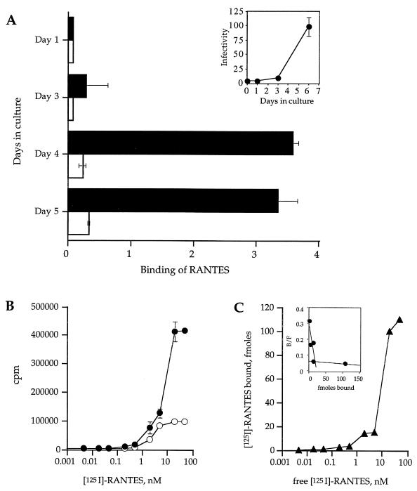
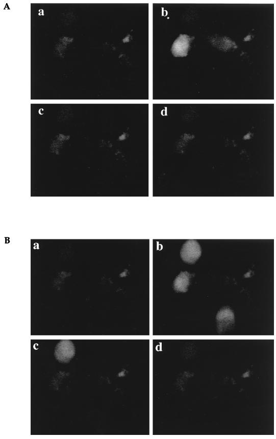
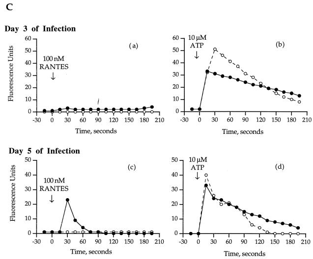
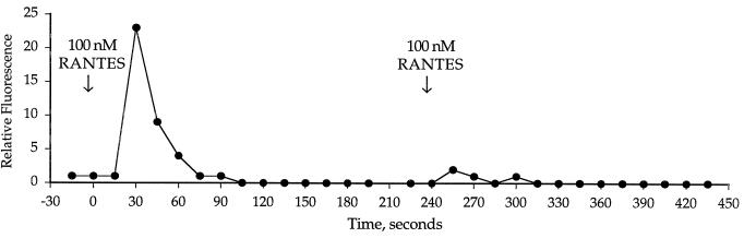
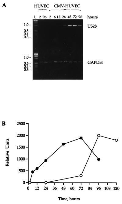
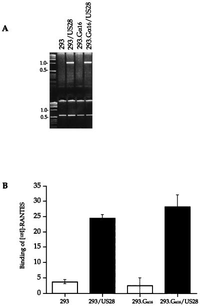
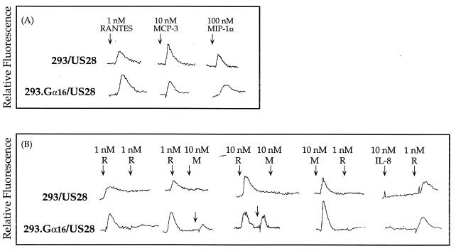
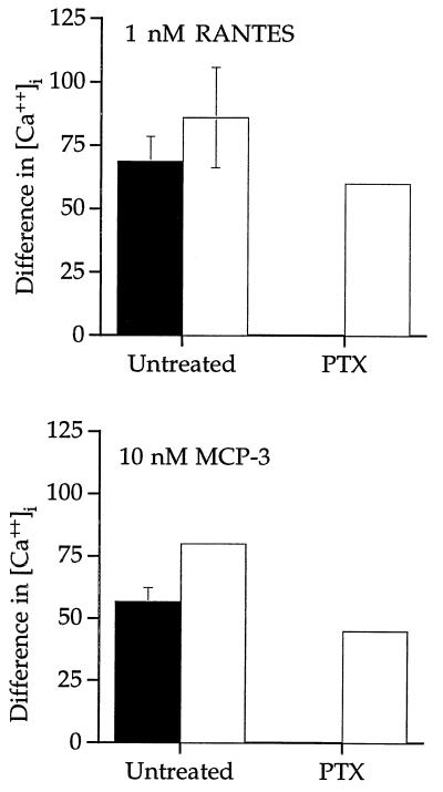
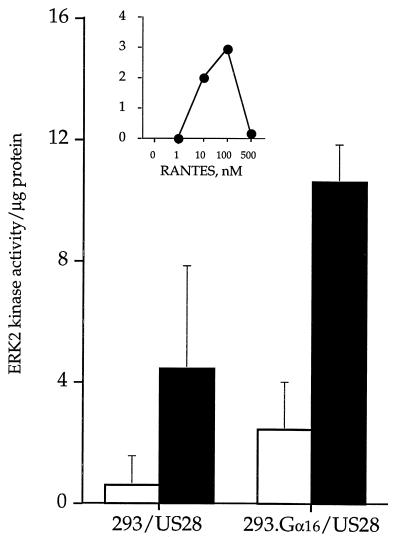
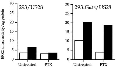
In patients with impaired cell-mediated immune responses (e.g., lung transplant recipients and AIDS patients), cytomegalovirus (CMV) infection causes severe disease such as pneumonitis. However, although immunocompetency in the host can protect from CMV disease, the virus persists by evading the host immune defenses. A model of CMV infection of the endothelium has been developed in which inflammatory stimuli, such as the CC chemokine RANTES, bind to the endothelial cell surface, stimulating calcium flux during late times of CMV infection. At 96 h postinfection, CMV-infected cells express mRNA of the CMV-encoded CC chemokine receptor US28 but do not express mRNA of other CC chemokine receptors that bind RANTES (CCR1, CCR4, CCR5). Cloning and stable expression of the receptor CMV US28 in human kidney epithelial cells (293 cells) with and without the heterotrimeric G protein alpha16 indicated that CMV US28 couples to both Galphai and Galpha16 proteins to activate calcium flux in response to the chemokines RANTES and MCP-3. Furthermore, cells that coexpress US28 and Galpha16 responded to RANTES stimulation with activation of extracellular signal-regulated kinase, which could be attributed, in part, to specific Galpha16 coupling. Thus, through expression of the CC chemokine receptor US28, CMV may utilize resident G proteins of the infected cell to manipulate cellular responses stimulated by chemokines.


Figures

FIG. 1
FIG. 1
Binding of 125I-RANTES to CMV-infected and uninfected HUVECs. (A) The inset shows the growth curve of CMV strain 4010 in primary endothelial cells (HUVECs). Subconfluent monolayers of HUVECs were infected with CMV strain 4010 stock at a multiplicity of infection of 0.1 and then harvested on days 1, 3, and 6 after infection. CMV-infected cells were subjected to titer determination on fresh HUVEC monolayers and assayed for infectivity. Infectivity is represented as 103 PFU per milliliter. Panel A shows binding of 10 pM 125I-RANTES in 100 pM unlabeled RANTES to CMV-infected (▪) or uninfected (□) monolayers. Binding is expressed as total bound labeled RANTES minus nonspecific binding of labeled RANTES relative to total free labeled RANTES and normalized to 20,000 cells. (B) Total binding of 125I-RANTES (•) and nonspecific binding (in the presence of 10 μM MCP-3) (○). (C) Specific binding was calculated by subtracting the nonspecific binding from the total binding and relating the resultant value to femtomoles (inset) by calculating the concentration from the specific activity of the 125I-RANTES.
FIG. 2
FIG. 2
Calcium flux in CMV-infected HUVECs. (A and B) Monolayers of CMV-infected HUVECs on day 5 of culture were loaded with Fluo-3, and a single field was analyzed by confocal microscopy for calcium flux after stimulation with 100 nM RANTES (A) or 10 μM ATP (B). Images were captured after stimulation at time zero (a), 30 s (b), 60 s (c), and 90 s (d). (C) Time course of the calcium flux in CMV-infected and mock-infected HUVECs. The relative fluorescence from the images on day 3 (a and b) and day 5 (c and d) of infection was estimated with National Institutes of Health Image software. •, CMV-infected HUVECs; ○, uninfected HUVECs.
FIG. 2
FIG. 2
Calcium flux in CMV-infected HUVECs. (A and B) Monolayers of CMV-infected HUVECs on day 5 of culture were loaded with Fluo-3, and a single field was analyzed by confocal microscopy for calcium flux after stimulation with 100 nM RANTES (A) or 10 μM ATP (B). Images were captured after stimulation at time zero (a), 30 s (b), 60 s (c), and 90 s (d). (C) Time course of the calcium flux in CMV-infected and mock-infected HUVECs. The relative fluorescence from the images on day 3 (a and b) and day 5 (c and d) of infection was estimated with National Institutes of Health Image software. •, CMV-infected HUVECs; ○, uninfected HUVECs.
FIG. 3
FIG. 3
Desensitization of the intracellular calcium flux in CMV-infected HUVECs in response to a second stimulation of RANTES.
FIG. 4
FIG. 4
Expression of US28 mRNA during CMV infection. (A) RNA was isolated from mock-infected or CMV-infected HUVECs at specific times (2, 6, 12, 24, 48, 72, and 96 h) after infection. RNA was transcribed to cDNA with Moloney murine leukemia virus reverse transcriptase, and specific fragments of US28 and GAPDH were identified by PCR. Samples were run on a 3.0% agarose gel and stained with Vistra Green. (B) Appearance of US28 mRNA and RANTES binding during CMV infection. The intensities of the bands in panel A were quantified by fluorescence imaging on a Storm phosphorimager. The arbitrary units of mRNA concentration are plotted against the time course of RANTES binding during infection.
FIG. 5
FIG. 5
Expression of CMV US28 in 293 cells. (A) Identification of mRNA transcripts of US28 in 293 and 293.Gα16 cells expressing US28. cDNA of mRNA collected from cell lysates was amplified with specific primers to HCMV US28 and GAPDH. (B) Binding of 10 pM 125I-RANTES to 293 and 293.Gα16 cells without US28 (□) or expressing US28 (▪). RANTES binding for each cell type was calculated relative to the total cpm and normalized to 5 × 10⁵ cells.
FIG. 6
FIG. 6
Tracings of intracellular Ca2+ flux in cells expressing US28 after stimulation with chemokines. Each tracing is representative of three separate runs. (A) Single stimulation of 293 and 293.Gα16 cells expressing US28 with RANTES, MCP-3, and MIP-1α. (B) Sequential stimulation of 293 and 293.Gα16 cells expressing US28 with either 1 or 10 nM RANTES (R) or 10 nM MCP-3 (M).
FIG. 7
FIG. 7
HCMV-US28 signaling of intracellular Ca2+ flux in the presence of 100 μg of PTX per ml. ▪, 293 cells expressing US28; □, 293.Gα16 cells expressing US28. The difference in intracellular calcium concentration is represented for each stimulation. Data are the mean of three experiments.
FIG. 8
FIG. 8
Activation of MAP kinase pathways by RANTES stimulation. Activation of ERK2 MAP kinase in 293 cells, and 293 cells expressing US28, 293.Gα16 cells, and 293.Gα16 cells expressing US28. MAP (ERK2) kinase was immunoprecipitated from unstimulated cells (□) and cells stimulated with 150 nM RANTES (▪). The data are the mean of three experiments. The inset represents a RANTES dose-response curve of ERK2 activation in 293.Gα16 cells expressing US28.
FIG. 9
FIG. 9
CMV US28 signaling of ERK2 MAP kinase activity in the presence of PTX. □, unstimulated cells; ▪, cells stimulated with 150 nM RANTES. The data represent a single experiment representative of three experiments.

References

    1. AbuBaker S, Boldogh I, Albrecht T. HCMV, stimulation of [3H] release from [3H]-arachidonic acid prelabelled cells. Arch Virol. 1990;113:255–266. - PubMed
    1. Albrecht T, Boldogh I, Fons M. Receptor-initiated activation of cells and their oncogenes by herpes-family viruses. J Invest Dermatol. 1992;98:29S–35S. - PubMed
    1. Albrecht T, Boldogh I, Fons M, AbuBakar S, Deng C. Cell activation signals and the pathogenesis of HCMV. Intervirology. 1990;31:68–75. - PubMed
    1. Albrecht T, Boldogh I, Fons M, Lee C, AbuBakar S, Russell J, Au W. Cell-activation responses to CMV infection. Relationship to phasing of CMV replication and to the induction of cellular damage. Subcell Biochem. 1989;15:157–202. - PubMed
    1. Avdi N, Winston B W, Russel M, Young S K, Johnson G L, Worthen G S. Activation of MEKK by FMLP in human neutrophils: mapping pathways for MAP kinase activation. J Biol Chem. 1996;271:33598–33606. - PubMed

Publication types

LinkOut - more resources