. 1998 Jul;72(7):5610-8.
doi: 10.1128/JVI.72.7.5610-5618.1998.

Persistent activation of RelA by respiratory syncytial virus involves protein kinase C, underphosphorylated IkappaBbeta, and sequestration of protein phosphatase 2A by the viral phosphoprotein

Affiliations

Persistent activation of RelA by respiratory syncytial virus involves protein kinase C, underphosphorylated IkappaBbeta, and sequestration of protein phosphatase 2A by the viral phosphoprotein

V Bitko et al. J Virol. 1998 Jul.

Abstract

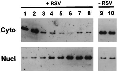
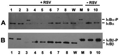
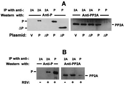
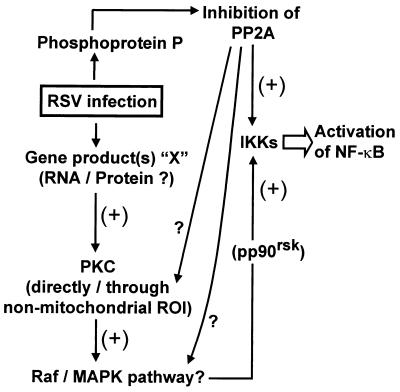
Respiratory syncytial virus (RSV) activated the RelA (p65) subunit of nuclear factor kappa B (NF-kappaB) over many hours postinfection. The initial activation coincided with phosphorylation and degradation of IkappaBalpha, the cytoplasmic inhibitor of RelA. During persistent activation of NF-kappaB at later times in infection, syntheses of inhibitors IkappaBalpha as well as IkappaBbeta were restored. However, the resynthesized IkappaBbeta was in an underphosphorylated state, which apparently prevented inhibition of NF-kappaB. Use of specific inhibitors suggested that the pathway leading to the persistent-but not the initial-activation of NF-kappaB involved signaling through protein kinase C (PKC) and reactive oxygen intermediates of nonmitochondrial origin, whereas phospholipase C or D played little or no role. Thus, RSV infection led to the activation of NF-kappaB by a biphasic mechanism: a transient or early activation involving phosphorylation of the inhibitor IkappaB polypeptides, and a persistent or long-term activation requiring PKC and the generation of hypophosphorylated IkappaBbeta. At least a part of the activation was through a novel mechanism in which the viral phosphoprotein P associated with but was not dephosphorylated by protein phosphatase 2A and thus sequestered and inhibited the latter. We postulate that this led to a net increase in the phosphorylation state of signaling proteins that are responsible for RelA activation.


Figures

FIG. 1
FIG. 1
Kinetics of RelA activation by RSV. Infection of A549 cells by RSV, preparation of nuclear (Nucl) and cytoplasmic (Cyto) extracts, and determination of RelA levels by immunoblotting were performed as described in Materials and Methods. At various times p.i., cells were harvested for lysis: lane 1, 0 h; lane 2, 2 h; lane 3, 4 h; lane 4, 6 h; lane 5, 8 h; lane 6, 10 h; lane 7, 20 h; lane 8, 30 h; lane 9, 10 h; lane 10, 30 h. ECL immunoblot analyses of cytosolic and nuclear fractions (as indicated) were carried out with anti-p65 antibody.
FIG. 2
FIG. 2
Time course of phosphorylation and degradation of IκBα (A) and IκBβ (B). ECL immunoblot analyses of total extracts of RSV-infected (and control, mock-infected) A549 cells were carried out with anti-IκBα and -IκBβ antibodies as described in Materials and Methods. The time points of harvest were as in Fig. 1. Cell extracts corresponding to lane 3 of panel A or lane 2 of panel B were incubated with 1 μg of wild-type (lane W) or mutant (lane M) PPλ in the presence of 4 mM MnCl2 at room temperature for 2 min before being subjected to SDS-PAGE; 20 μg protein was analyzed in each lane, except lane M in panel B, for which 40 μg was used.
FIG. 3
FIG. 3
Effect of signal transduction inhibitors on RSV-mediated activation of NF-κB. The inhibitors were added to RSV-infected A549 cells at the following final concentrations: D609, 20 μM; staurosporine (Stau.), 20 ng/ml; pseudosubstrate peptide (Pept.), 100 μM; PDTC, 40 μM; rotenone (Roten.), 40 μM; MG132, 80 μM; and U73122, 30 μM. Numbers above the lanes indicate the hours p.i. at which the cells were harvested for preparing extracts as described in Materials and Methods. (A) EMSA for NF-κB-binding activity in nuclear extracts; (B to D) immunoblots of nuclear RelA (B), IκBα (C), and IκBβ (D). The phosphorylated and nonphosphorylated forms of IκB polypeptides are indicated. To study the effect of inhibitors on NF-κB activity ex vivo, A549 cells were transfected with plasmid pBIIxluc, using Lipofectamine (Gibco-BRL), and then infected with RSV (or mock infected as a control) essentially as described elsewhere (13). The inhibitors were added at 2 h after addition of the virus. At 20 h thereafter, cells were processed for luciferase assay (13). Luciferase activities, expressed as percentages of the untreated activity (E), represent averages of three measurements.
FIG. 4
FIG. 4
Activation of RelA by recombinant RSV P protein. A549 cells at 75% confluency were transfected with pcDNA3-P alone (A, B, and D) or together with pBIIxluc (C) in multiple dishes and processed as indicated, using procedures described in Materials and Methods. The various pcDNA-3 plasmids are vector (V), full-length P clone (P), ΔC39 P clone (ΔP), and P clones with premature termination codons (T1 and T2). RelA and P protein levels were measured by immunoblotting of nuclear and cytoplasmic fractions, respectively (A and D). NF-κB activities in nuclear extracts were assayed by EMSA (B). Ex vivo activities of NF-κB were determined by reporter luciferase assay (C); average values from three experiments are shown with standard deviation bars. Where indicated, sodium salicylate (15 mM in panels A and B; 2 and 8 mM in panel C) was added to the transfected cells 4 h after addition of the DNA.
FIG. 5
FIG. 5
(A) Resistance of RSV P protein to PP2A. Bacterially expressed RSV P protein was phosphorylated by CK2 in the presence of [γ-32P]ATP (9, 39); 2 μg of the labeled P protein was incubated with 20 ng of purified enzyme (Enz.) PPλ or PP2A in 20-μl reaction mixtures under standard conditions (4). At the indicated time points, 4 μl of the reaction mixtures was removed and added to 20 μl of Laemmli sample buffer (34). All samples were heated in a boiling water bath for 5 min and analyzed by SDS-PAGE (39) followed by autoradiography as shown. (B and C) Inhibition of PP2A by phosphorylated P protein. Standard phosphatase reactions (20 μl) were carried out with 2 μg of 32P-labeled casein as the substrate and 4 μg of phosphate-free (P) or phosphorylated (P-PO4) RSV P protein. At indicated times, 4 μl of the reaction mixture was removed. Reactions were analyzed by SDS-PAGE (34) and autoradiography (B). The multiple bands of 32P-casein in each lane were scanned by densitometry, and their total intensities were plotted as percentages of the starting intensities (at 0 min) (C). Results are shown for assays with no P added (□), unphosphorylated P (○), and phosphorylated P (•).
FIG. 6
FIG. 6
Association of recombinant (A) and viral (B) P proteins with PP2A. A549 cells were transfected with plasmid pcDNA3-P (lane P), pcDNA3 clone of a ΔC39 deletion mutant of P (lane ΔP), or pcDNA3 vector without P gene (lane V) (A) or infected with RSV (Long) at an MOI of 3.0 (B). At 48 h posttransfection (or p.i.), the cells were processed for immunoprecipitation (IP) using antibodies against either PP2A or RSV P protein as indicated. The samples were analyzed by SDS-PAGE in which the immunoprecipitate from about 106 cells was applied in each lane. The proteins were transferred to a membrane and probed with the same antibodies in Western blot analysis as shown.
FIG. 7
FIG. 7
Postulated model for NF-κB activation by RSV. Unidentified RSV gene product(s) “X” intracellularly activates protein kinases such as PKC that may in turn activate other downstream kinases. Raf, members of the MAPK cascade, and ribosomal protein S6 kinase (pp90rsk) are some postulated examples (see text for details). Inhibition of PP2A by P may lead to a higher steady-state level of the phosphorylated product of any of these kinases; however, it is also possible that the kinases themselves are regulated by PP2A.

References

    1. Amtmann E. The antiviral, antitumoural xanthate D609 is a competitive inhibitor of phosphatidylcholine-specific phospholipase C. Drugs Exp Clin Res. 1996;22:287–294. - PubMed
    1. Ananaba G A, Anderson L J. Antibody enhancement of respiratory syncytial virus stimulation of leukotriene production by a macrophage-like cell line. J Virol. 1991;65:5052–5060. - PMC - PubMed
    1. Anderson L J, Heilman C A. Protective and disease-enhancing immune responses to respiratory syncytial virus. J Infect Dis. 1995;171:1–7. - PubMed
    1. Ansai T, Dupuy L C, Barik S. Interactions between a minimal protein serine/threonine phosphatase and its phosphopeptide substrate sequence. J Biol Chem. 1996;271:24401–24407. - PubMed
    1. Arnold R, Humbert B, Werchau H, Gallati H, Konig W. Interleukin-8, interleukin-6, and soluble tumour necrosis factor receptor type I release from a human pulmonary epithelial cell line (A549) exposed to respiratory syncytial virus. Immunology. 1994;82:126–133. - PMC - PubMed

Publication types

LinkOut - more resources