. 1998 Jul;72(7):5638-47.
doi: 10.1128/JVI.72.7.5638-5647.1998.

A small yeast RNA blocks hepatitis C virus internal ribosome entry site (HCV IRES)-mediated translation and inhibits replication of a chimeric poliovirus under translational control of the HCV IRES element

Affiliations

A small yeast RNA blocks hepatitis C virus internal ribosome entry site (HCV IRES)-mediated translation and inhibits replication of a chimeric poliovirus under translational control of the HCV IRES element

S Das et al. J Virol. 1998 Jul.

Erratum in

  • J Virol 1998 Nov;72(11):9419

Abstract

Hepatitis C virus (HCV) infection frequently leads to chronic hepatitis and cirrhosis of the liver and has been linked to development of hepatocellular carcinoma. We previously identified a small yeast RNA (IRNA) capable of specifically inhibiting poliovirus (PV) internal ribosome entry site (IRES)-mediated translation. Here we report that IRNA specifically inhibits HCV IRES-mediated translation both in vivo and in vitro. A number of human hepatoma (Huh-7) cell lines expressing IRNA were prepared and characterized. Constitutive expression of IRNA was not detrimental to cell growth. HCV IRES-mediated cap-independent translation was markedly inhibited in cells constitutively expressing IRNA compared to control hepatoma cells. However, cap-dependent translation was not significantly affected in these cell lines. Additionally, Huh-7 cells constitutively expressing IRNA became refractory to infection by a PV-HCV chimera in which the PV IRES is replaced by the HCV IRES. In contrast, replication of a PV-encephalomyocarditis virus (EMCV) chimera containing the EMCV IRES element was not affected significantly in the IRNA-producing cell line. Finally, the binding of the La autoantigen to the HCV IRES element was specifically and efficiently competed by IRNA. These results provide a basis for development of novel drugs effective against HCV infection.


Figures
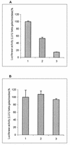
FIG. 1
FIG. 1
Effect of IRNA on HCV IRES-mediated translation in Huh-7 cells. Monolayer cells (106) were transfected with three different plasmid DNAs: pCDIR.Ribo.ΔT7 expressing IRNA, pCD HCV-luc reporter plasmid in which luciferase translation is programmed by the HCV IRES, and a β-Gal reporter gene to measure transfection efficiency. After 24 h of transfection, extracts were made and luciferase and β-Gal activities were measured. Luciferase activity (light units) is expressed as percentage of the control after normalizing for β-Gal activity and protein content for each transformation. (A) Vertical bars 1, 2, and 3 show the dose-response effect of pCDIR.Ribo.ΔT7 on HCV IRES-mediated translation of luciferase gene at 0, 1.25, and 1.88 μg of pCDIR.Ribo.ΔT7, respectively. Total DNA concentration was made up to 2.5 μg by adding 2.5, 1.25, and 0.62 μg of pCDNA3 (bars 1, 2, and 3, respectively). (B) A similar experiment was performed in which plasmid pCD HCV-luc was replaced by pCDNA3-luc conferring cap-dependent translation of luciferase. All transfection reactions contained 1 μg each of β-Gal and luciferase reporter plasmids.
FIG. 2
FIG. 2
IRNA selectively inhibits HCV IRES-mediated translation in vitro. The bicistronic construct containing the HCV IRES flanked by CAT and luciferase genes was transcribed by T7 RNA polymerase, and the bicistronic RNA was translated in vitro in 25 μl of HeLa cell lysate in the absence (A, lane 1) or presence of increasing concentrations of IRNA (lanes 2 to 5; 0.5, 1, 2, and 4 μg, respectively). (B) The band intensities of luciferase (LUC) and CAT were quantitated by densitometry, and the ratios of luciferase to CAT were calculated. The percentage of luciferase/CAT translation was plotted against the concentration of IRNA.
FIG. 3
FIG. 3
Schematic diagram of the constructs used for in vivo expression of IRNA. The diagram (not to scale) shows the cloning of IRNA encoding sequences into eukaryotic expression vector pCDNA3 (Invitrogen). The polylinker site following the cytomegalovirus (CMV) promoter sequence is illustrated above the parental plasmid. Additional construction of different IRNA-encoding plasmids are shown, and their names are given at the left. BGH, bovine growth hormone; pA, poly(A) site; SV40, simian virus 40; ori, origin.
FIG. 4
FIG. 4
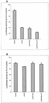
HCV IRES-mediated translation is inhibited in Huh-7 cells constitutively expressing IRNA. Plasmid pCD HCV-luc was cotransfected with a β-Gal reporter gene into either control Huh-7 cells or IRNA-expressing cell lines (pCDIR, pCDIR.Ribo, and pCDIR.Ribo.ΔT7), and luciferase activity was plotted as percentage of control (A). Similarly, plasmid pCDNA3-luc conferring cap-dependent translation of the luciferase gene was transfected into either the control Huh-7 or hepatoma-IRNA cell line (B). β-Gal activity was measured to normalize transfection efficiencies in both experiments.
FIG. 5
FIG. 5
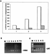
HCV-luciferase reporter dose response and quantitation of IRNA and luciferase mRNA in the cell line. (A) Increasing concentrations (1, 2, and 3 μg) of the pCD HCV-luc reporter plasmid were transfected into either Huh-7 hepatoma cells (dotted bars) or the IRNA-expressing hepatoma cell line (pCDIR.Ribo.ΔT7) (white bars). A control β-Gal plasmid was also cotransfected to normalize transfection efficiencies. Luciferase activity (103 light units [LU]) was plotted against increasing concentrations of the test plasmids. (B) Detection of IRNA in hepatoma cell line by RT-PCR. IRNA expression level was detected by RT-PCR using IRNA-specific oligonucleotide primers. Lane 1, no-RNA control; lanes 2 to 4, 1, 2.5, and 5 ng, respectively, of in vitro-transcribed, purified IRNA; lanes 5 to 7, 2, 1.5, and 1 μg, respectively, of total RNA from an IRNA-expressing hepatoma cell, pCDIR.Ribo.ΔT7. Lane 8 contained 2 μg of total RNA from the control Huh-7 cells. Lane M represents marker DNA. (C) Detection of luciferase (LUC) mRNA by RT-PCR. Total RNA isolated from control (Huh-7) and pCDIR.Ribo.ΔT7 cells transfected with the luciferase reporter plasmid was used to detect luciferase mRNA levels by using luciferase-specific oligonucleotide primers. Lane 1, no-RNA control; lanes 2 and 3, 1 and 10 ng of luciferase mRNA standard; lanes 4 and 5, 1 μg of total RNA isolated from Huh-7 cells and cells expressing IRNA, respectively, after transfection with pCDHCV-luc.
FIG. 6
FIG. 6
Effect of IRNA expression on cellular transcription and translation. (A and B) Detection of GAPDH (A) and β-actin (B) mRNAs by RT-PCR. Lane 1, no-RNA control; lanes 2 and 3, 3 μg of the total RNA isolated from Huh7 control cells and cell lines expressing IRNA (pCDIR.Ribo.ΔT7), respectively. (C) In vivo labeling of proteins. Monolayer Huh7 hepatoma cells (lane 1) and hepatoma pCDIR.Ribo.ΔT7 cells (lane 2) were labeled with [35S]methionine for 1 h, and in vivo-labeled proteins were analyzed on an SDS–14% polyacrylamide gel. Lane M shows the migration of 14C-labeled protein markers (Gibco/BRL), with approximate molecular masses as indicated to the left in kilodaltons.
FIG. 7
FIG. 7
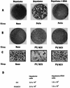
Hepatoma cells constitutively expressing IRNA prevent PV and HCV-PV chimera infection. Huh-7 control cells or the IRNA expressing hepatoma cell line (pCDIR.Ribo.ΔT7) (∼106 cells) were infected with 500 PFU of either PV (Polio) (A) or HCV-PV chimera (B and C). After 72 h, either cells were stained for the observation of cytopathic effects (C) or cell extracts were made to further infect HeLa monolayer cells for the plaque assay (A and B). Cells were stained by crystal violet. (D) Average virus titers obtained from three independent plaque assay experiments.
FIG. 8
FIG. 8
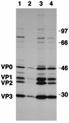
Hepatoma cells expressing IRNA inhibit translation of the PV-HCV chimera. Approximately 3.5 × 10⁵ monolayer hepatoma cells (Huh-7) (lanes 1 and 3) or IRNA-expressing hepatoma cells (lanes 2 and 4) were infected with approximately 3.75 × 10³ PFU of either PV-HCV (lanes 1 and 2) or PV-EMCV (lanes 3 and 4) chimera. After 24 h of infection, cells were labeled with [35S]methionine. In vivo-labeled proteins were immunoprecipitated with anti-PV capsid antibody and analyzed on an SDS–14% polyacrylamide gel. The positions of the PV capsid proteins are indicated on the left. Numbers at the right refer to the approximate molecular masses (in kilodaltons) of the 14C-labeled protein markers (Amersham).
FIG. 9
FIG. 9
IRNA binds proteins that interact with HCV 5′ UTR. (A) 32P-labeled IRNA (lanes 1 to 3) and HCV 5′ UTR RNA (lanes 4 and 5) were UV cross-linked to cellular polypeptides, using 30 μg of HeLa S10 fraction (lanes 2 and 4) and 0.3 μg of purified La protein (lanes 3 and 5). Numbers to the left correspond to the molecular masses (in kilodaltons) of the polypeptides indicated. Lane 1 contains the IRNA probe but no S10 extract. (B) Competition UV-cross-linking studies were performed with 32P-labeled HCV 5′ UTR RNA and 15 μg of HeLa S10 in the absence (lane 2) and presence of various unlabeled competitor RNAs (lanes 3 to 7). Lanes 3 and 4, 250- and 500-fold molar excess of unlabeled HCV 5′ UTR; lanes 5 and 6, 250- and 500-fold molar excess of unlabeled IRNA; lane 7, 500-fold molar excess of a nonspecific RNA (polylinker region of pSPluc; Promega). Lane 1 contains the probe but no S10 extract; lane M shows the migration (in kilodaltons) of marker proteins. The numbers to the right correspond to the molecular masses (in kilodaltons) of proteins which cross-link to the labeled HCV 5′ UTR probe. (C) 32P-labeled IRNA (lanes 1 to 4) and HCV 5′ UTR RNA (lanes 5 to 8) were UV cross-linked to cellular polypeptides, using 30 μg of S10 extract of either HeLa cells (lanes 2 and 6), Huh7 cells (lanes 3 and 7), or Huh7 cell line pCDIR.Ribo.ΔT7 (lanes 4 and 8). Lanes 1 and 5 contain the probe but no S10 extract. Numbers to the right correspond to the molecular masses (in kilodaltons) of the polypeptides indicated. Lane M shows the migration (in kilodaltons) of marker proteins. (D) Competition UV-cross-linking studies were performed with 32P-labeled HCV 5′ UTR RNA and purified La protein (150 ng) in the absence (lane 1) and presence of 100-fold (lanes 2, 4, and 6) and 200-fold (lanes 3, 5, and 7) molar excesses of unlabeled IRNA (lanes 2 and 3), nonspecific RNA (lanes 4 and 5), and HCV 5′ UTR RNA (lane 6 and 7). Lane M shows the migration (in kilodaltons) of marker proteins. The migration of La protein cross-linked with HCV 5′ UTR is indicated.

Similar articles

Cited by

References

    1. Ali N, Siddiqui A. Interaction of polypyrimidine tract binding protein with 5′ noncoding region of the hepatitis C virus RNA genome and its functional requirement in internal initiation of translation. J Virol. 1996;69:6367–6375. - PMC - PubMed
    1. Ali N, Siddiqui A. The La antigen binds 5′ noncoding region of the hepatitis C virus RNA in the context of the initiator AUG codon and stimulates internal ribosome entry site mediated translation. Proc Natl Acad Sci USA. 1997;94:2249–2254. - PMC - PubMed
    1. Blyn L B, Swiderek K M, Richards O, Stahl D C, Semler B L, Ehrenfeld E. Poly r(C) binding protein 2, binds to stem-loop IV of the poliovirus RNA 5′noncoding region: identification by automated ligand chromatography-tandem mass spectrometry. Proc Natl Acad Sci USA. 1996;93:11115–11120. - PMC - PubMed
    1. Blyn L B, Towner J S, Semler B L, Ehrenfeld E. Requirement of poly(rC) binding protein 2 for translation of poliovirus RNA. J Virol. 1997;71:6243–6246. - PMC - PubMed
    1. Brown E A, Zhang H, Ping L, Lemon S. Secondary structure of the 5′ nontranslated regions of hepatitis C virus and pestivirus genomic RNAs. Nucleic Acids Res. 1992;19:5041–5045. - PMC - PubMed

Publication types

LinkOut - more resources