. 1998 Jul;72(7):5735-44.
doi: 10.1128/JVI.72.7.5735-5744.1998.

Distinct roles of two binding sites for the bovine papillomavirus (BPV) E2 transactivator on BPV DNA replication

Affiliations

Distinct roles of two binding sites for the bovine papillomavirus (BPV) E2 transactivator on BPV DNA replication

T G Gillette et al. J Virol. 1998 Jul.

Abstract

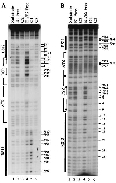
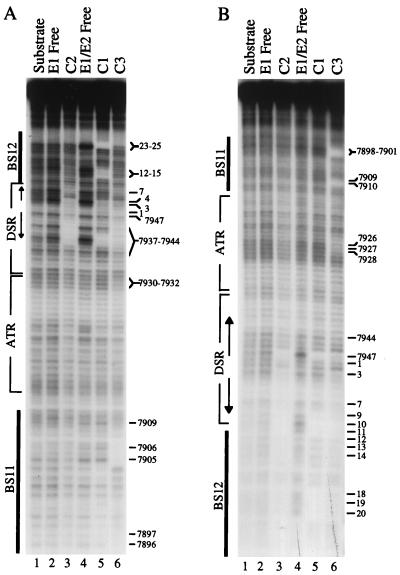
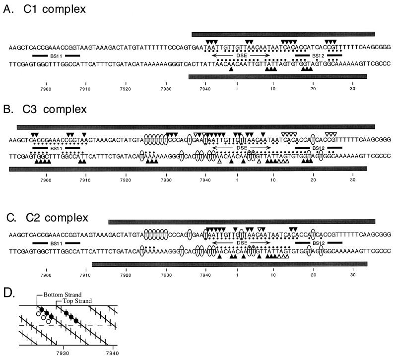
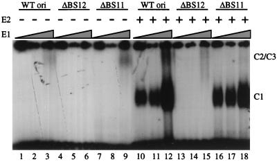
The modulation of DNA replication by transcription factors was examined by using bovine papillomavirus type 1 (BPV). BPV replication in vivo requires two viral proteins: E1, an origin-binding protein, and E2, a transcriptional transactivator. In the origin, E1 interacts with a central region flanked by two binding sites for E2 (BS11 and BS12), of which only BS12 has been reported to be essential for replication in vivo. Using chemical interference and electrophoretic mobility shift assays, we found that the binding of E2 to each site stimulates the formation of distinct E1-origin complexes. A high-mobility C1 complex is formed by using critical E2 contacts to BS12 and E1 contacts to the dyad symmetry element. In contrast, interaction of E2 with the BS11 element on the other origin flank promotes the formation of the lower-mobility C3 complex. C3 is a novel species that resembles C2, a previously identified complex that is replication active and formed by E1 alone. The binding of E1 greatly differs in the C1 and C3 complexes, with E1 in the C1 complex limited to the origin dyad symmetry region and E1 in the C3 complex encompassing the region from the proximal edge of BS11 through the distal edge of BS12. We found that the presence of both E2-binding sites is necessary for wild-type replication activity in vivo, as well as for maximal production of the C3 complex. These results show that in the normal viral context, BS11 and BS12 play separate but synergetic roles in the initiation of viral DNA replication that are dependent on their location within the origin. Our data suggest a model in which the binding of E2 to each site sequentially stimulates the formation of distinct E1-origin complexes, leading to the replication-competent complex.


Figures

FIG. 1
FIG. 1
Complexes formed on the wild-type BPV origin by E1 and E2. E1 alone (100 ng) (lane 1) or E1 (100 ng) plus E2 (60 ng) (lane 2) were incubated for 15 min at 37°C with a 32P-labeled DNA fragment containing the wild-type origin. The complexes were cross-linked by treatment with glutaraldehyde, separated by electrophoresis through a native 5% polyacrylamide gel, and visualized by autoradiography. The locations of the C1, C2, and C3 complexes are indicated.
FIG. 2
FIG. 2
Interference of the formation of E1- and E1-E2-origin complexes by partial origin depurination. Duplex DNA fragments, 5′-32P labeled on either the top (A) or bottom (B) strand, were subjected to partial depurination by treatment with formic acid. The DNA fragments were then incubated with E1 alone (300 ng) (lanes 2 and 3) or E1 (75 ng) plus E2 (60 ng) (lanes 4 to 6). Each reaction mixture was then subjected to native gel electrophoresis to isolate the C2 complex (lane 3), or the C1 (lane 5) and C3 (lane 6) complexes from the corresponding free (unbound) DNA (lanes 2 and 4, respectively). After separation, the free DNA pools, the DNA molecules within each complex, and the initial origin substrate DNA (lane 1) were isolated and chemically cleaved at the sites of modification. The cleavage products were separated by electrophoresis through a denaturing 8% polyacrylamide gel and visualized by autoradiography.
FIG. 3
FIG. 3
Interference of the formation of E1- and E1-E2-origin complexes by partial origin depyrimidation. Duplex DNA fragments, 5′-32P labeled on either the top (A) or bottom (B) strand, were subjected to partial depyrimidation by treatment with hydrazine. The DNA fragments were then incubated with E1 alone (300 ng) (lanes 2 and 3) or E1 (75 ng) plus E2 (60 ng) (lanes 4 to 6). Each reaction mixture was then subjected to native gel electrophoresis to isolate the C2 complex (lane 3) or the C1 (lane 5) and C3 (lane 6) complexes from the corresponding free (unbound) DNA (lanes 2 and 4, respectively). After separation, the free DNA pools, the DNA molecules within each complex, and the initial origin substrate DNA (lane 1) were isolated and chemically cleaved at the sites of modification. The cleavage products were separated by electrophoresis through a denaturing 8% polyacrylamide gel and visualized by autoradiography.
FIG. 4
FIG. 4
Interference of the formation of E1- and E1-E2-origin complexes by ethylation of the origin phosphates. Duplex DNA fragments, 5′-32P labeled on either the top (A) or bottom (B) strand, were ethylated on a small fraction of DNA phosphates. The DNA fragments were then incubated with E1 alone (300 ng) (lanes 2 and 3) or E1 (75 ng) plus E2 (60 ng) (lanes 4 to 6). Each reaction mixture was then subjected to native gel electrophoresis to isolate the C2 complex (lane 3) or the C1 (lane 5) and C3 (lane 6) complexes from the corresponding free (unbound) DNA (lanes 2 and 4, respectively). After separation, the free DNA pools, the DNA molecules within each complex, and the initial origin substrate DNA (lane 1) were isolated and chemically cleaved at the sites of modification. The cleavage products were separated by electrophoresis through a denaturing 8% polyacrylamide gel and visualized by autoradiography. Phosphates are numbered according to the base position on the 5′ side.
FIG. 5
FIG. 5
Compilation of depurination, depyrimidation, and phosphate ethylation interference data for E1 and E2 binding to the BPV origin. Maps of modifications that interfere with C1 (A), C3 (B), and C2 (C) complex formation are shown. Phosphates whose modification strongly inhibits complex formation are indicated by solid triangles; weakly interfering phosphates are shown by open triangles. Bases whose removal (i.e., by depurination and depyrimidation) reduces complex formation are indicated by solid circles above (bottom strand) or below (top strand) the affected base. We also include the results from previous footprinting analyses (13). The top- and bottom-strand regions protected from DNase I cleavage by each complex are indicated by solid boxes above and below the sequence, respectively. Thymines hyperreactive to KMnO4 (a probe of DNA structure) are indicated by ovals. The previous data for the C1 and C3 complexes was taken from complexes formed at low (50 ng) and high (400 ng) E1 levels. (D) Helix map of phosphates and purines in the nt 7930 region whose modification interferes with C3 complex formation. Phosphates whose ethylation inhibits complex formation are indicated by solid circles, while critical purines are indicated by open circles. The top and bottom strands are indicated. The dashed line in the center of the map distinguishes the two helical sides.
FIG. 6
FIG. 6
Effect of E2 binding-site mutation on complex formation by E1 and E2. DNA fragments (32P labeled) containing the wild-type (WT) ΔBS12, or ΔBS11 mutant origin were incubated with increasing levels of E1 (6.25, 12.5, and 25 ng) in the absence or presence of E2 (15 ng; as indicated). Complexes were cross-linked with glutaraldehyde, separated by electrophoresis through a native 5% polyacrylamide gel, and visualized by autoradiography. The locations of the C1, C2, and C3 complexes are indicated. Note that the amounts of E1 are less than that used in the experiment in Fig. 1 (100 ng), accounting for the reduced level of C3 complex on the wild-type origin (lanes 10 to 12) in this experiment.
FIG. 7
FIG. 7
Mutation of either BS11 or BS12 is deleterious for transient BPV DNA replication in vivo. (A) Schematic showing the origins that were tested for replication activity. From top to bottom, these origins are the wild-type origin, LI 15C (containing a 15-bp insertion in the dyad symmetry element which inactivates the origin [28]), ΔBS12 and ΔBS11 (lacking the BS12 and BS11 elements, respectively), and ΔBS12ΔBS11 (lacking both E2-binding sites). (B) The viral DNA molecules (2 μg) were released from the vector and transfected into murine C127 cells by electroporation. As a control, the test plasmids were cotransfected with wild-type viral DNA (0.5 μg) not released from the vector. After 72 and 96 h, the viral DNA was isolated by the method of Hirt (15) and treated with MunI to linearize the viral genome and with DpnI to digest unreplicated DNA. DNA was detected by Southern blot analysis with nick-translated pSS3 as a probe. (C) The replication activity in vivo was quantitated by excising bands corresponding to linearized viral DNA and counting in a scintillation counter. Replication activity was normalized with respect to the replication activity of the wild-type origin at 96 h.
FIG. 8
FIG. 8
Model of E1 and E2 binding to the BPV origin to form a replication initiation complex. See the text for details. The light zigzag lines indicate regions of DNA distortion induced by E1 in the AT-rich and BS12 regions.

Similar articles

References

    1. Benson J D, Howley P M. Amino-terminal domains of the bovine papillomavirus type 1 E1 and E2 proteins participate in complex formation. J Virol. 1995;69:4364–4372. - PMC - PubMed
    1. Berg M, Stenlund A. Functional interactions between papillomavirus E1 and E2 proteins. J Virol. 1997;71:3853–3863. - PMC - PubMed
    1. Blitz I L, Laimins L A. The 68-kilodalton E1 protein of bovine papillomavirus is a DNA binding phosphoprotein which associates with the E2 transcriptional activator in vitro. J Virol. 1991;65:649–656. - PMC - PubMed
    1. Bonne-Andrea C, Santucci S, Clertant P, Tillier F. Bovine papillomavirus E1 protein binds specifically DNA polymerase α but not replication protein A. J Virol. 1995;69:2341–2350. - PMC - PubMed
    1. Borowiec J A, Hurwitz J. Localized melting and structural changes in the SV40 origin of DNA replication induced by T antigen. EMBO J. 1988;7:3149–3158. - PMC - PubMed

Publication types

LinkOut - more resources