

B-Cell lymphoma induction by akv murine leukemia viruses harboring one or both copies of the tandem repeat in the U3 enhancer
- PMID: 9621033
- PMCID: PMC110375
- DOI: 10.1128/JVI.72.7.5745-5756.1998
B-Cell lymphoma induction by akv murine leukemia viruses harboring one or both copies of the tandem repeat in the U3 enhancer
Abstract
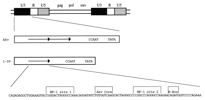
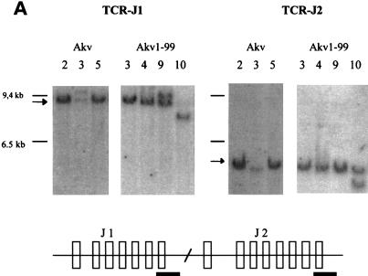
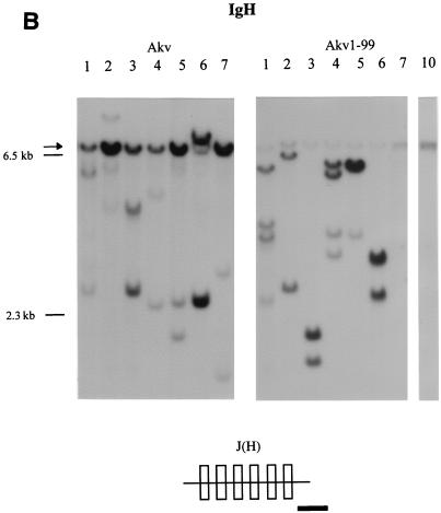
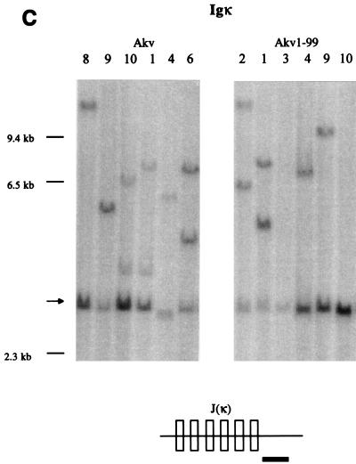
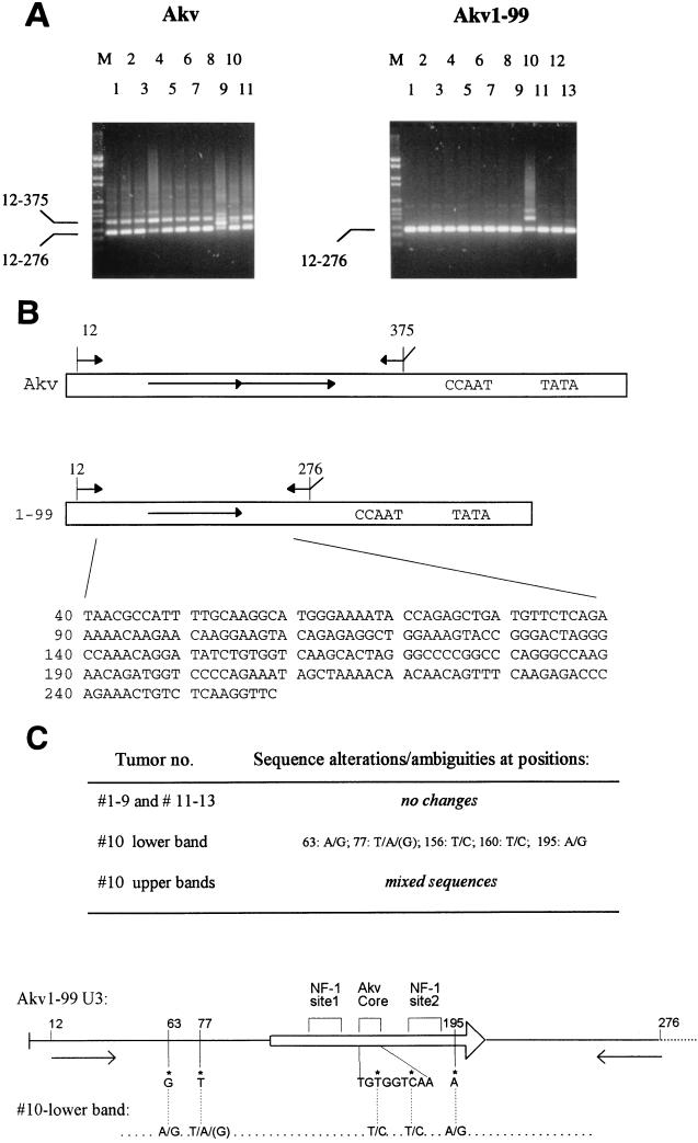
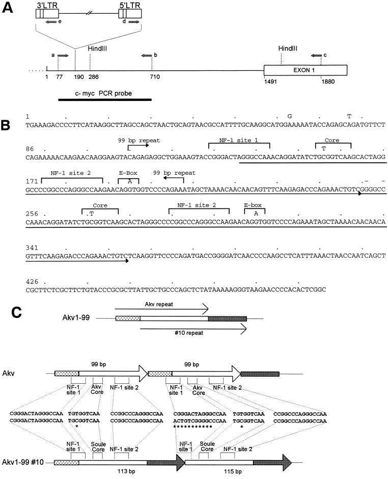
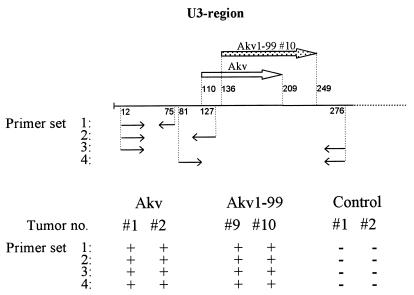
Akv is an endogenous, ecotropic murine leukemia virus (MuLV) of the AKR strain. It has served as a prototype nonpathogenic or weakly pathogenic reference virus for studies of closely related potent lymphomagenic viruses such as the T-lymphomagenic SL3-3. We here report that Akv and an Akv mutant (Akv1-99) with only one copy of the 99-bp transcriptional enhancer induce malignant lymphomas with nearly 100% incidence and mean latency periods of 12 months after injection into newborn NMRI mice. Molecular analysis of tumor DNA showed that the majority of the tumors were of the B-cell type. Sequence analysis of proviral transcriptional enhancers in DNA of B-cell lymphomas revealed conservation of the enhancer sequence, as well as a lack of sequence duplications of the Akv1-99 variant, while the repeat copy number in Akv was subject to fluctuations. In support of a B-cell specificity of the Akv enhancer, a murine plasmacytoma cell line was found to sustain three- to fivefold-higher transient transcriptional activity upon the Akv and Akv1-99 enhancers than upon the enhancer of the T-lymphomagenic SL3-3 MuLV. Thus, the overall picture is that Akv MuLV possesses a B- lymphomagenic potential and that the second copy of the 99-bp sequence seems to be of minor importance for this potential. However, in one animal the lymphomas induced by Akv1-99 were of the T-cell type. Among the 24 tumors analyzed only this one harbored a clonal proviral integration in the c-myc locus. This provirus had undergone a duplication of a 113-bp sequence of the enhancer region, partly overlapping with the 99-bp repeat of Akv, as well as a few single nucleotide alterations within and outside the repeats. Taken together with previous studies, our results suggest that T- versus B-lymphomagenic specificity of the enhancer is governed by more than one nucleotide difference and that alterations in binding sites for transcription factors of the AML1 and nuclear-factor-1 families may contribute to this specificity.
Figures
Similar articles
-
Mutation of all Runx (AML1/core) sites in the enhancer of T-lymphomagenic SL3-3 murine leukemia virus unmasks a significant potential for myeloid leukemia induction and favors enhancer evolution toward induction of other disease patterns.J Virol. 2004 Dec;78(23):13216-31. doi: 10.1128/JVI.78.23.13216-13231.2004. J Virol. 2004. PMID: 15542674 Free PMC article.
-
Stability of AML1 (core) site enhancer mutations in T lymphomas induced by attenuated SL3-3 murine leukemia virus mutants.J Virol. 1997 Jul;71(7):5080-7. doi: 10.1128/JVI.71.7.5080-5087.1997. J Virol. 1997. PMID: 9188573 Free PMC article.
-
Enhancer mutations of Akv murine leukemia virus inhibit the induction of mature B-cell lymphomas and shift disease specificity towards the more differentiated plasma cell stage.Virology. 2007 May 25;362(1):179-91. doi: 10.1016/j.virol.2006.12.016. Epub 2007 Jan 29. Virology. 2007. PMID: 17258785
-
Increased lymphomagenicity and restored disease specificity of AML1 site (core) mutant SL3-3 murine leukemia virus by a second-site enhancer variant evolved in vivo.J Virol. 1997 Oct;71(10):7273-80. doi: 10.1128/JVI.71.10.7273-7280.1997. J Virol. 1997. PMID: 9311802 Free PMC article.
-
Enhancing B-Cell Malignancies-On Repurposing Enhancer Activity towards Cancer.Cancers (Basel). 2021 Jun 29;13(13):3270. doi: 10.3390/cancers13133270. Cancers (Basel). 2021. PMID: 34210001 Free PMC article. Review.
Cited by
-
Control of pathogenicity and disease specificity of a T-lymphomagenic gammaretrovirus by E-box motifs but not by an overlapping glucocorticoid response element.J Virol. 2009 Jan;83(1):336-46. doi: 10.1128/JVI.01368-08. Epub 2008 Oct 22. J Virol. 2009. PMID: 18945767 Free PMC article.
-
Selection of reversions and suppressors of a mutation in the CBF binding site of a lymphomagenic retrovirus.J Virol. 1999 Sep;73(9):7599-606. doi: 10.1128/JVI.73.9.7599-7606.1999. J Virol. 1999. PMID: 10438850 Free PMC article.
-
Mutation of all Runx (AML1/core) sites in the enhancer of T-lymphomagenic SL3-3 murine leukemia virus unmasks a significant potential for myeloid leukemia induction and favors enhancer evolution toward induction of other disease patterns.J Virol. 2004 Dec;78(23):13216-31. doi: 10.1128/JVI.78.23.13216-13231.2004. J Virol. 2004. PMID: 15542674 Free PMC article.
-
Pvt1-encoded microRNAs in oncogenesis.Retrovirology. 2008 Jan 14;5:4. doi: 10.1186/1742-4690-5-4. Retrovirology. 2008. PMID: 18194563 Free PMC article.
-
Forward and Reverse Genetics of B Cell Malignancies: From Insertional Mutagenesis to CRISPR-Cas.Front Immunol. 2021 Aug 13;12:670280. doi: 10.3389/fimmu.2021.670280. eCollection 2021. Front Immunol. 2021. PMID: 34484175 Free PMC article. Review.
References
-
- Bergeron D, Poliquin L, Houde J, Barbeau B, Rassart E. Analysis of proviruses integrated in Fli-1 and Evi-1 regions in Cas-Br-E MuLV-induced non-T, non-B-cell leukemias. Virology. 1992;191:661–669. - PubMed
-
- Bonven B J, Nielsen A L, Nørby P L, Pedersen F S, Jørgensen P. E-box variants direct formation of distinct complexes with the basic helix-loop-helix protein ALF1. J Mol Biol. 1995;249:564–575. - PubMed
-
- Celander D, Haseltine W A. Tissue-specific transcription preference as a determinant of cell tropism and leukaemogenic potential of murine retroviruses. Nature. 1984;312:159–162. - PubMed
Publication types
MeSH terms
Substances
LinkOut - more resources
Full Text Sources
Other Literature Sources
Molecular Biology Databases
