. 1998 Jul;72(7):5762-8.
doi: 10.1128/JVI.72.7.5762-5768.1998.

Determination of the nucleotide sequence of Bombyx mori cytoplasmic polyhedrosis virus segment 9 and its expression in BmN4 cells

Affiliations

Determination of the nucleotide sequence of Bombyx mori cytoplasmic polyhedrosis virus segment 9 and its expression in BmN4 cells

K Hagiwara et al. J Virol. 1998 Jul.

Abstract

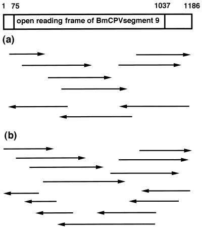
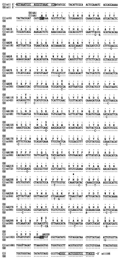
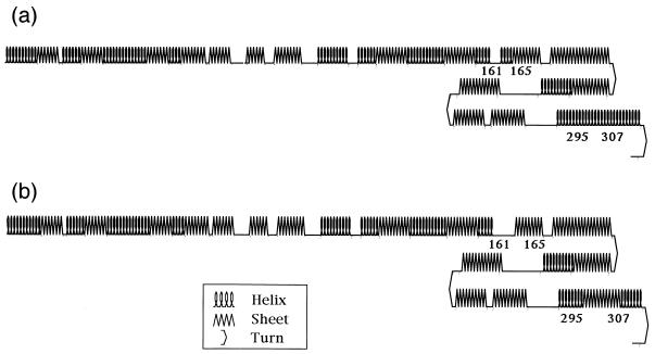
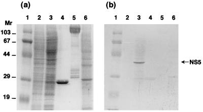
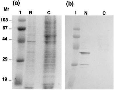
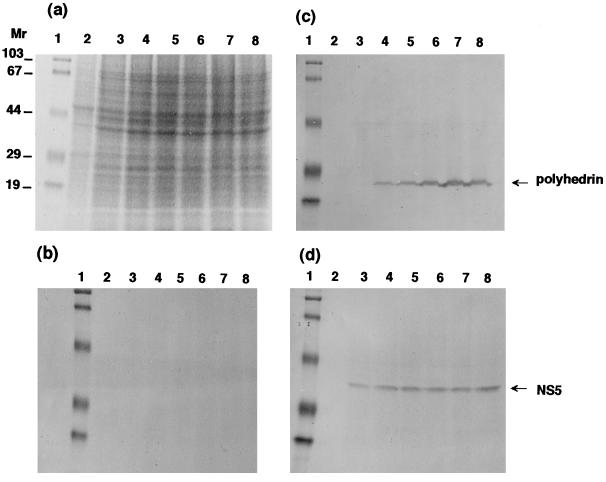
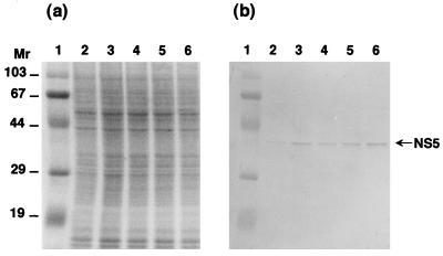
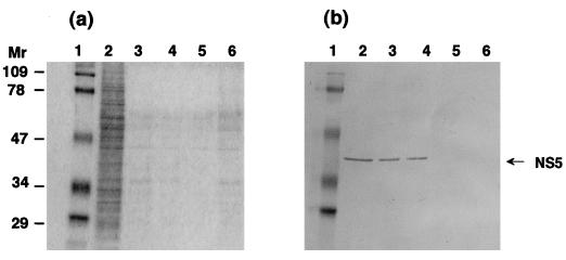
Cloning and sequencing of segment 9 of Bombyx mori cytoplasmic polyhedrosis virus (BmCPV) strains H and I were performed. The segment consisted of 1,186 bp harboring 5' and 3' noncoding regions and an open reading frame from positions 75 to 1037, encoding a protein with 320 amino acids, termed NS5. Comparison of the nucleotide sequences of NS5 for the two strains indicated 37 point differences resulting in only six amino acid replacements. Homology search showed that NS5 has localized similarities to human poliovirus RNA-dependent RNA polymerase and human rotavirus NS26. By Western blot analysis, NS5 was found in BmCPV-infected midgut cells, but not in polyhedra or virus virions, and was mainly detectable in the nucleus in BmCPV-infected BmN4 cells. Immunoblot analysis with anti-NS5 and antipolyhedrin antibodies displayed marked differences in the period of expression of NS5 and polyhedrin: the polyhedrin molecule was first detected 2 or 3 days after infection with BmCPV, whereas the expression of NS5 was initiated within a few hours. In addition, the level of polyhedrin increased as the infection developed, whereas the amount of NS5 remained essentially constant. When segment 9 was expressed with a baculovirus expression system, the resulting NS5 protein possessed the ability to bind to the double-stranded RNA genome. These results suggest that NS5 is expressed in early stages of infection and contributes to regulation of genomic RNA function.


Figures

FIG. 1
FIG. 1
Strategy for sequencing segment 9 of BmCPV strain H (a) and strain I (b). Arrows pointing to the right indicate the plus strand of segment 9 of the viral genome, and those pointing to the left indicate the minus strand. Single clones from the H and I strains were sequenced.
FIG. 2
FIG. 2
Nucleotide and deduced amino acid sequences of segment 9 from BmCPV strains H and I. Closed boxes show the initiation and termination codons, and open boxes indicate the 5′ and 3′ primer sequences. Asterisks indicate the putative N glycosylation sites. Single dots represent identical nucleotides (nt) and amino acids (AA) in the two strains.
FIG. 3
FIG. 3
Secondary structures of NS5 of BmCPV strain H (a) and strain I (b), predicted according to the method of Chou and Fasman (3, 4). The numbers represent boundary amino acid residues at a position where the secondary structure of the protein differs between the two strains.
FIG. 4
FIG. 4
Immunoblot analysis of NS5 with anti-NS5 antibody. Fifth-instar and 2-day-old larvae were infected with BmCPV strain I, and the infected midguts were collected 5 days after infection. They were homogenized with PBS, and the supernatant was collected after sedimenting cell debris by centrifugation. Uninfected midguts were prepared by the same procedure. BmNPV-infected BmN4 cells were also collected 5 days after infection, and the homogenates of 5 × 10⁵ cells were used for further analysis. BmCPV polyhedra and virions were purified as described in Materials and Methods. Each sample was applied to an SDS–12% polyacrylamide gel and subjected to electrophoresis, and protein was detected by Coomassie brilliant blue staining (a) and Western blot analysis (b). Lanes 1, molecular weight markers; lanes 2, uninfected midgut (control); lanes 3, BmCPV strain I-infected midgut; lanes 4, BmCPV strain I polyhedra (2.5 μg); lanes 5, BmCPV strain I virion (2.5 μg); lanes 6, BmNPV-infected BmN4 cells. Molecular weights are in thousands.
FIG. 5
FIG. 5
Localization of NS5 in BmN4 cells. BmCPV strain I-infected BmN4 cells were collected 5 days after infection, separated into cytosolic and nuclear fractions as described in Materials and Methods, and subjected to SDS-PAGE and detection by Coomassie brilliant blue staining (a) and Western blot analysis with anti-NS5 antibody (b). Lanes 1, N, and C show molecular weight markers and the nuclear and cytosolic fractions, respectively. Molecular weights are in thousands.
FIG. 6
FIG. 6
Time course of expression of NS5 and polyhedrin in BmCPV strain I-infected BmN4 cells. Infected BmN4 cells were collected 1 to 6 days after infection and subjected to SDS-PAGE and detection by Coomassie brilliant blue staining (a) and Western blot analysis with normal mouse serum (b), antipolyhedrin (c), and anti-NS5 (d) antibodies. Lanes 1 show molecular weight markers. Lanes 2 illustrate the results for uninfected control BmN4 cells. Lanes 3 to 8 show results obtained 1, 2, 3, 4, 5, and 6 days after infection, respectively. Homogenates of 5 × 10⁵ cells were used for analysis. Molecular weights are in thousands.
FIG. 7
FIG. 7
Time course of expression of NS5 in BmCPV strain I-infected BmN4 cells. Infected BmN4 cells were collected at 1 to 18 h after infection and subjected to SDS-PAGE and detection by Coomassie brilliant blue staining (a) and Western blot analysis with anti-NS5 antibody (b). Lanes 1 show molecular weight markers. Lanes 2 to 6 show results obtained 1, 3, 6, 12, and 18 h after infection, respectively. Homogenates of 5 × 10⁵ cells were used for analysis. Molecular weights are in thousands.
FIG. 8
FIG. 8
Assay for binding of NS5 to poly(rI) · poly(rC)-agarose. BmN4 cells were infected with recombinant BmNPV in the presence of benzamidine and collected 5 days after infection. Then the whole-cell extracts (107 cells) were mixed with poly(rI) · poly(rC)-agarose, and the proteins bound to the poly(rI) · poly(rC)-agarose were analyzed by Coomassie brilliant blue staining (a) and Western blotting (b) after SDS-PAGE. For competition assays, cell extracts were pretreated for 60 min at 4°C with 5 or 50 μg of viral dsRNA before being mixed with poly(rI) · poly(rC)-agarose. Lanes 1 show molecular weight markers. Lanes 2 show the whole-cell extracts infected by recombinant BmNPV. Lanes 4 and 5 show proteins bound to the poly(rI) · poly(rC)-agarose in the presence of 5 and 50 μg of viral dsRNA, respectively, and lanes 3 show protein binding in its absence. Lanes 6 show the proteins bound to agarose without poly(rI) · poly(rC) ligand.

Similar articles

Cited by

References

    1. Arella M, Lavallee C, Belloncik S, Furuichi Y. Molecular cloning and characterization of cytoplasmic polyhedrosis virus polyhedrin and a viable deletion mutant gene. J Virol. 1988;62:211–217. - PMC - PubMed
    1. Bruenn J A. Relationships among the positive strand and double-strand RNA viruses as viewed through their RNA-dependent RNA polymerase. Nucleic Acids Res. 1991;19:217–226. - PMC - PubMed
    1. Chou P Y, Fasman G D. Prediction of protein conformation. Biochemistry. 1974;13:222–245. - PubMed
    1. Chou P Y, Fasman G D. Empirical predictions of protein conformation. Annu Rev Biochem. 1978;47:251–276. - PubMed
    1. Dignam J D, Lebovitz R M, Roeder R G. Accurate transcription initiation by RNA polymerase II in a soluble extract from isolated mammalian nuclei. Nucleic Acids Res. 1983;11:1475–1489. - PMC - PubMed

Associated data