. 1998 Jul;72(7):5769-80.
doi: 10.1128/JVI.72.7.5769-5780.1998.

DNA packaging mutant: repression of the vaccinia virus A32 gene results in noninfectious, DNA-deficient, spherical, enveloped particles

Affiliations

DNA packaging mutant: repression of the vaccinia virus A32 gene results in noninfectious, DNA-deficient, spherical, enveloped particles

M C Cassetti et al. J Virol. 1998 Jul.

Abstract

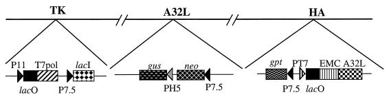
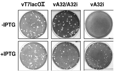
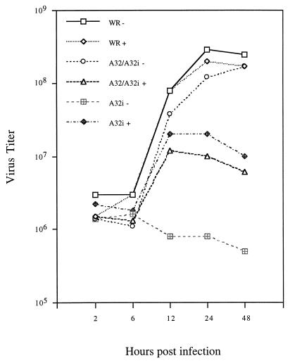
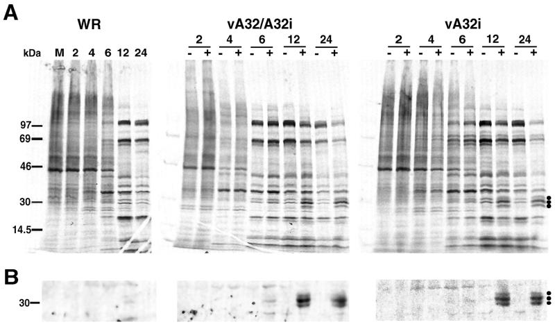
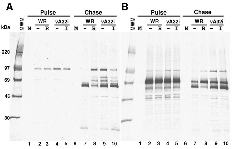
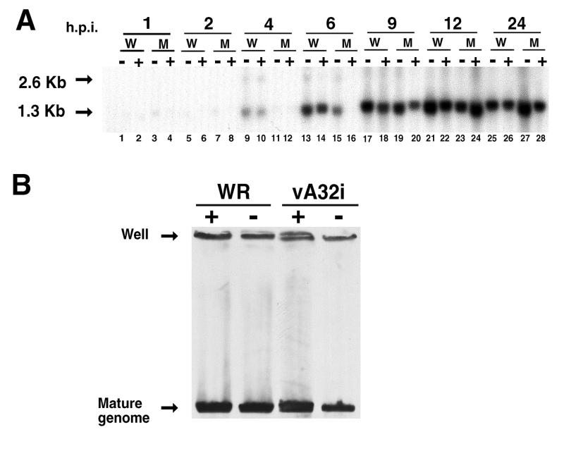
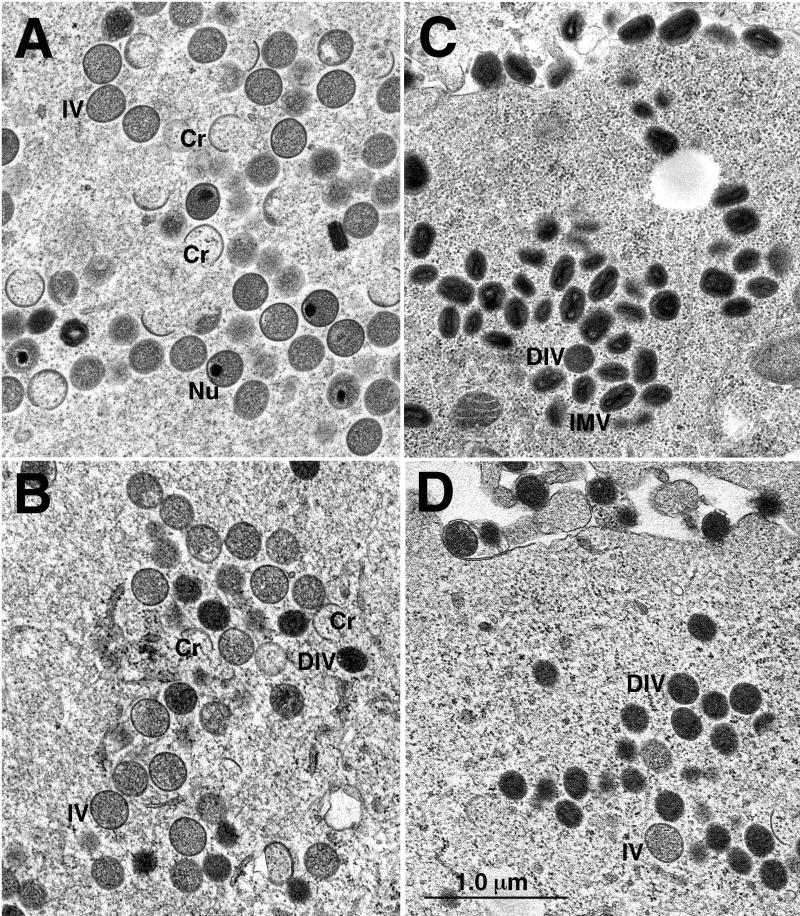
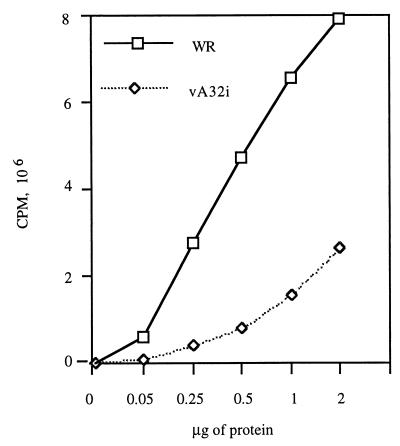
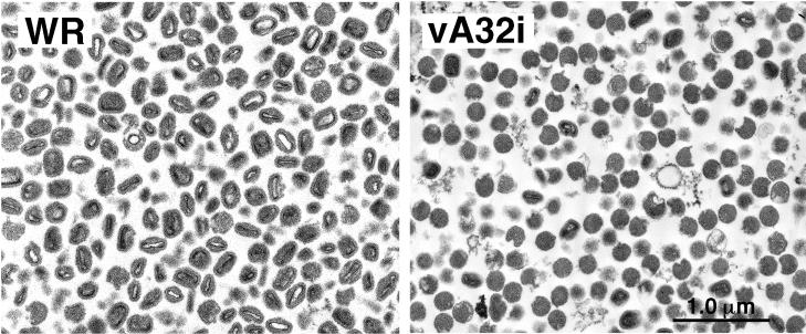
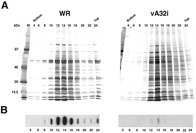
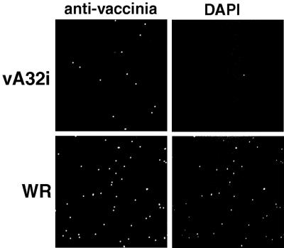
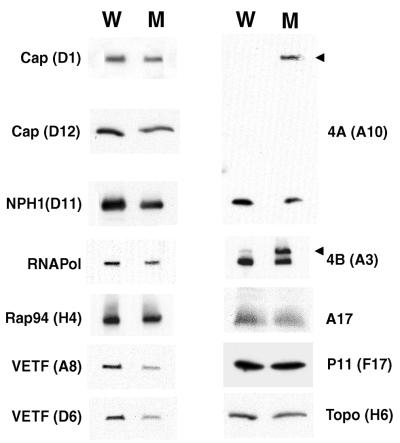
The vaccinia virus A32 open reading frame was predicted to encode a protein with a nucleoside triphosphate-binding motif and a mass of 34 kDa. To investigate the role of this protein, we constructed a mutant in which the original A32 gene was replaced by an inducible copy. The recombinant virus, vA32i, has a conditional lethal phenotype: infectious virus formation was dependent on isopropyl-beta-D-thiogalactopyranoside (IPTG). Under nonpermissive conditions, the mutant synthesized early- and late-stage viral proteins, as well as viral DNA that was processed into unit-length genomes. Electron microscopy of cells infected in the absence of IPTG revealed normal-appearing crescents and immature virus particles but very few with nucleoids. Instead of brick-shaped mature particles with defined core structures, there were numerous electron-dense, spherical particles. Some of these spherical particles were wrapped with cisternal membranes, analogous to intracellular and extracellular enveloped virions. Mutant viral particles, purified by sucrose density gradient centrifugation, had low infectivity and transcriptional activity, and the majority were spherical and lacked DNA. Nevertheless, the particle preparation contained representative membrane proteins, cleaved and uncleaved core proteins, the viral RNA polymerase, the early transcription factor and several enzymes, suggesting that incorporation of these components is not strictly coupled to DNA packaging.


Figures

FIG. 1
FIG. 1
Repression of the A32 gene. Portions of the genome of the VV mutant vA32i are represented. DNA insertions have been made into the VV thymidine kinase (TK), A32L, and hemagglutinin (HA) genes. Abbreviations: P7.5, P11, and PH5 are VV promoters; PT7 and T7pol are a bacteriophage T7 promoter and the RNA polymerase gene, respectively; EMC is a cDNA copy of the untranslated RNA leader of encephalomyocarditis virus which provides cap-independent translation; lacI and lacO are the E. coli lac repressor gene and the lac operator, respectively; gus is a color marker gene; neo and gpt are antibiotic selection genes.
FIG. 2
FIG. 2
Inducer dependence of plaque formation. BS-C-1 monolayers were infected with the mutant VVs vT7LacOI, vA32/A32i, or vA32i in the presence (+) or absence (−) of 50 μM IPTG. After 2 days, the cells were stained with crystal violet and photographed.
FIG. 3
FIG. 3
Inducer-dependent formation of infectious virus. BS-C-1 monolayers were infected with the viruses indicated at a multiplicity of 5 PFU per cell in the presence (+) or absence (−) of 50 μM IPTG. At intervals of up to 48 h, the cells were harvested and the virus titers were determined by plaque assay. For A32i, 50 μM IPTG was included in the plaque medium.
FIG. 4
FIG. 4
Protein synthesis in cells infected with wild-type and mutant viruses. BS-C-1 cells were infected with WR, vA32/A32i, or vA32i at a multiplicity of 15 PFU per cell in the presence (+) or absence (−) of 50 μM IPTG. At the indicated hours after infection, the cells were labeled for 30 min with [35S]methionine. (A) Cell lysates were analyzed by SDS-PAGE. The dots on the right indicate the bands of approximately 30 kDa that were increased in the presence of IPTG. (B) The labeled proteins that bound to beads containing antibody to the A32 protein were analyzed by SDS-PAGE. Only the portion of the autoradiograph containing proteins of approximately 30 kDa is shown.
FIG. 5
FIG. 5
Proteolytic processing of core precursors. BS-C-1 cells were infected with WR in the presence or absence of 100 μg of rifampin per ml or with vA32i in the presence or absence of 15 μM IPTG. At 9 h after infection, the cells were labeled with [35S]methionine for 30 min, washed, and incubated with medium containing excess methionine for 12 h. Cell lysates were immunoprecipitated with antibody to 4A (panel A) or 4B (panel B) and analyzed by SDS-PAGE. Autoradiographs are shown. MWM, molecular weight markers; M, mock-infected cells; WR, wild-type VV-infected cells in the absence (−) or presence (R) of rifampin; vA32i, mutant VV-infected cells in the absence (−) or presence (I) of IPTG.
FIG. 6
FIG. 6
Synthesis and processing of VV DNA. (A) At the indicated hours postinfection (h.p.i.) in the presence (+) or absence (−) of IPTG, total DNA was purified, digested with the restriction endonuclease BstEII, electrophoresed through agarose, transferred to a nylon membrane (Hybond-N+; Amersham), and probed with a radiolabeled oligonucleotide corresponding to the repeat sequence near the ends of the genome. W and M, wild-type and mutant vA32i, respectively. The arrows at 1.3 and 2.6 kb point to the fragments corresponding to the ends of mature genomes and the bridge between units of concatemeric DNA molecules, respectively. (B) Total DNA from cells infected with WR or vA32i was resolved by pulse-field gel electrophoresis and analyzed by Southern blotting. + and −, presence or absence of IPTG during infection.
FIG. 7
FIG. 7
Morphogenesis of mutant viruses. BS-C-1 cells were infected with vA32/A32i (A and C) or vA32i (B and D) in the absence of IPTG. After 24 h, the cells were fixed in glutaraldehyde and embedded in Epon, and then ultrathin sections were prepared for electron microscopy. Cr, crescents; Nu, nucleoids; IMV, intracellular mature virions; IV, immature virions; DIV, dense immature virions.
FIG. 8
FIG. 8
Transcriptional activity of purified VV particles. Sucrose gradient-purified particles from HeLa cells infected with WR or vA32i in the absence of IPTG were adjusted to equal protein concentrations and used for in vitro transcription. The incorporation of [α-32P]UMP was measured. The same sucrose gradient-purified preparation was used for the experiments depicted in Fig. 8 through 12.
FIG. 9
FIG. 9
Electron microscopy of purified virus particles. Particles purified by sucrose gradient centrifugation from HeLa cells infected with WR or vA32i in the absence of IPTG were diluted, collected by high-speed centrifugation, fixed in glutaraldehyde, and embedded in Epon. Ultrathin sections were examined by electron microscopy.
FIG. 10
FIG. 10
Protein and DNA content of purified virus particles. Particles were purified from HeLa cells infected with WR or vA32i in the absence of IPTG. Aliquots of alternate numbered fractions of the sucrose gradients were analyzed by SDS-PAGE and silver staining (A) or by slot blot hybridization with a VV DNA probe (B). M, markers with masses shown on left. Bottom and Top refer to the sucrose gradient tube.
FIG. 11
FIG. 11
Laser scanning confocal microscopy of purified viral particles. Sucrose gradient-purified particles from HeLa cells infected with WR or vA32i in the absence of IPTG were mounted on fibronectin-coated coverslips and stained with DAPI and polyclonal antibody to VV.
FIG. 12
FIG. 12
Western blot analysis of purified viral particles. Purified viral particles from HeLa cells infected with WR or vA32i in the absence of IPTG were adjusted to similar protein concentrations and analyzed by SDS-PAGE. The proteins were transferred to a polyvinylidene difluoride membrane (Immobilon-P; Millipore) and probed with antiserum to the indicated viral proteins followed by 125I-labeled protein A. W, wild-type virus particles; M, vA32i particles; arrowheads, uncleaved precursor proteins. Autoradiographs are shown.

Similar articles

Cited by

References

    1. Alexander W A, Moss B, Fuerst T R. Regulated expression of foreign genes in vaccinia virus under the control of bacteriophage T7 RNA polymerase and the Escherichia coli lac repressor. J Virol. 1992;66:2934–2942. - PMC - PubMed
    1. Baroudy B M, Venkatesan S, Moss B. Incompletely base-paired flip-flop terminal loops link the two DNA strands of the vaccinia virus genome into one uninterrupted polynucleotide chain. Cell. 1982;28:315–324. - PubMed
    1. Baroudy B M, Venkatesan S, Moss B. Structure and replication of vaccinia virus telomeres. Cold Spring Harbor Symp Quant Biol. 1982;47:723–729. - PubMed
    1. Broyles S S, Yuen L, Shuman S, Moss B. Purification of a factor required for transcription of vaccinia virus early genes. J Biol Chem. 1988;263:10754–10760. - PubMed
    1. Carroll M, Moss B. Host range and cytopathogenicity of the highly attenuated MVA strain of vaccinia virus: propagation and generation of recombinant viruses in a nonhuman mammalian cell line. Virology. 1997;238:198–211. - PubMed

LinkOut - more resources