. 1998 Jul;72(7):5877-85.
doi: 10.1128/JVI.72.7.5877-5885.1998.

Nonreciprocal packaging of human immunodeficiency virus type 1 and type 2 RNA: a possible role for the p2 domain of Gag in RNA encapsidation

Affiliations

Nonreciprocal packaging of human immunodeficiency virus type 1 and type 2 RNA: a possible role for the p2 domain of Gag in RNA encapsidation

J F Kaye et al. J Virol. 1998 Jul.

Abstract

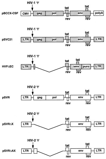
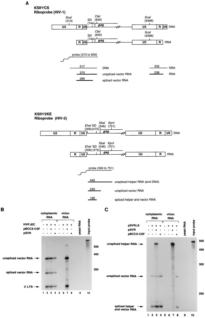
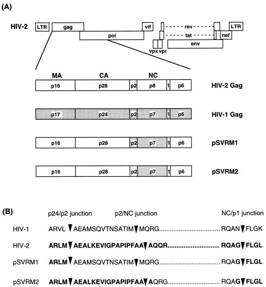
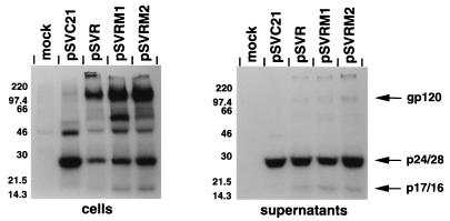
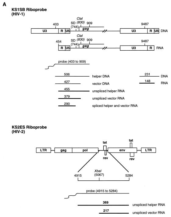
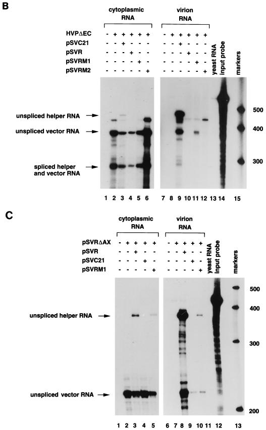
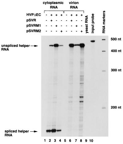
The ability of human immunodeficiency virus types 1 (HIV-1) and 2 (HIV-2) to cross-package each other's RNA was investigated by cotransfecting helper virus constructs with vectors derived from both viruses from which the gag and pol sequences had been removed. HIV-1 was able to package both HIV-1 and HIV-2 vector RNA. The unspliced HIV-1 vector RNA was packaged preferentially over spliced RNA; however, unspliced and spliced HIV-2 vector RNA were packaged in proportion to their cytoplasmic concentrations. The HIV-2 helper virus was unable to package the HIV-1 vector RNA, indicating a nonreciprocal RNA packaging relationship between these two lentiviruses. Chimeric proviruses based on HIV-2 were constructed to identify the regions of the HIV-1 Gag protein conferring RNA-packaging specificity for the HIV-1 packaging signal. Two chimeric viruses were constructed in which domains within the HIV-2 gag gene were replaced by the corresponding domains in HIV-1, and the ability of the chimeric proviruses to encapsidate an HIV-1-based vector was studied. Wild-type HIV-2 was unable to package the HIV-1-based vector; however, replacement of the HIV-2 nucleocapsid by that of HIV-1 generated a virus with normal protein processing which could package the HIV-1-based vector. The chimeric viruses retained the ability to package HIV-2 genomic RNA, providing further evidence for a lack of reciprocity in RNA-packaging ability between the HIV-1 and HIV-2 nucleocapsid proteins. Inclusion of the p2 domain of HIV-1 Gag in the chimera significantly enhanced packaging.


Figures

FIG. 1
FIG. 1
Schematic representation of helper virus and vector constructs. HIV-1 sequences are shaded. pBCCX-CSF is an HIV-1 particle producer which uses the human cytomegalovirus (CMV) immediate-early promoter to drive the expression of HIV-1 genes. pSVC21 is an HIV-1 proviral construct. HVPΔEC is an HIV-1-based vector with the sequences between ClaI (nt 830) and BalI (nt 2689) deleted and a puromycin resistance gene (puro) inserted in the nef position. pSVR is an HIV-2 proviral construct. pSVRΔX was derived from pSVR by deletion of sequences between an introduced XbaI site at nt 553 and XbaI at nt 5067. pSVRΔAX was derived from pSVR by deletion of sequences between AccI (nt 912) and XbaI (nt 5067). polyA, polyadenylation sequences; Ψ, encapsidation sequences; LTR, long terminal repeat.
FIG. 2
FIG. 2
Nonreciprocal RNA packaging by HIV-1 and HIV-2. HIV-1- and HIV-2-based vectors, HVPΔEC and pSVRΔX, were cotransfected into COS-1 cells with the HIV-1 or HIV-2 helper virus constructs pBCCX-CSF or pSVR, respectively, or alone. RNA isolated from the cytoplasm of the transfected cells or from virions was subjected to RNase protection analysis. (A) Predicted sizes of the protected fragments for the HIV-1- and HIV-2-specific riboprobes, KSIIΨCS and KSIIΨ2KE, respectively. SD, splice donor. (B) RNase protection analysis with the HIV-1-specific riboprobe, KSIIΨCS. Diagnostic bands for HIV-1 vector RNA are 375 nt (unspliced RNA), 289 nt (spliced vector RNA), and 238 nt (3′ LTR). The relative levels of unspliced and spliced vector RNA are 1.9 in lane 3 and is 65.6 in lane 7. (C) RNase protection analysis with the HIV-2 specific riboprobe, KSIIΨ2KE. Diagnostic bands for unspliced HIV-2 helper virus and vector RNA are 445 and 240 nt, respectively, and a band of 166 nt is protected for spliced RNA from both constructs. The relative levels of unspliced and spliced helper RNA are 0.7 in lane 3 and 19.8 in lane 7. The relative levels of unspliced and spliced vector are 0.4 in lane 4 and 0.4 in lane 8. Protection with control RNA (yeast RNA) and with the riboprobe without RNase treatment (input probe) is shown. The positions of RNA size markers are shown in nucleotides to the right of the panels B and C.
FIG. 3
FIG. 3
Schematic representation of chimeric constructs used in this study. (A) Gag region of wild-type HIV-1 and HIV-2 and chimeric constructs, pSVRM1 and pSVRM2, showing the subdomains. HIV-1 sequences are shaded. MA, matrix; CA, capsid. (B) Junctions between the HIV-1 and HIV-2 sequences in the chimeric constructs. Amino acid residues derived from HIV-2 sequences are shown in boldface type. The dotted regions indicate NC residues. The predicted protease cleavage sites are indicated by arrowheads.
FIG. 4
FIG. 4
Immunoprecipitation analysis of chimeric constructs. Wild-type proviral constructs pSVC21 (HIV-1) and pSVR (HIV-2) and chimeric constructs pSVRM1 and pSVRM2 were transfected into COS-1 cells. The cells were labelled with [35S]methionine from 44 to 48 h after transfection, and cell and virion lysates were subjected to immunoprecipitation analysis with pooled human sera from a panel of HIV-2-infected individuals. The molecular mass markers are shown to the left in kilodaltons. The predicted positions of viral proteins gp120, p24/28, and p17/16 are indicated.
FIG. 5
FIG. 5
RNase protection analysis of chimeric proviruses. (A) Predicted sizes of the protected fragments for the HIV-1- and HIV-2-specific riboprobes, KS1SB and KS2ES, respectively. SD, splice donor. (B) COS-1 cells were transfected with the HIV-1 vector HVPΔEC alone (lanes 2 and 8) or with pSVC21 (lanes 3 and 9), pSVR (lanes 4 and 10), pSVRM1 (lanes 5 and 11), or pSVRM2 (lanes 6 and 12) or mock transfected (lanes 1 and 7). Cytoplasmic RNA (lanes 1 to 6) and virion RNA (lanes 7 to 12) were subjected to RNase protection analysis with an HIV-1 specific riboprobe, KS1SB. The positions of unspliced helper virus RNA (455 nt), unspliced vector RNA (379 nt), and spliced helper and vector RNA (290 nt) are indicated by arrows. The relative levels of unspliced vector packaged by the various helper constructs compared to HIV-1 helper virus (given an arbitrary value of 1) are 0.31 in lane 11 and 0.03 in lane 12. Protection with yeast RNA (lane 13) and the riboprobe without RNase treatment (lane 14) is shown. The RNA molecular size markers (in nucleotides) are shown (lane 15). (C) COS-1 cells were transfected with the HIV-2 vector pSVRΔAX alone (lanes 2 and 7), or with pSVR (lanes 3 and 8), pSVC21 (lanes 4 and 9), or pSVRM1 (lanes 5 and 10) or mock transfected (lanes 1 and 6). Cytoplasmic RNA (lanes 1 to 5) and virion RNA (lanes 6 to 10) were subjected to RNase protection analysis with an HIV-2-specific riboprobe, KS2ES. The positions of unspliced HIV-2 helper virus RNA (369 nt) and unspliced vector RNA (217 nt) are indicated by arrows. The relative levels of unspliced vector and unspliced helper RNA are 3.2 in lane 3, 17.4 in lane 5, 0.9 in lane 8, and 1.1 in lane 10. Protection with yeast RNA (lane 11) and the riboprobe without RNase treatment (lane 12) are shown. The RNA molecular size markers (in nucleotides) are shown (lane 13).
FIG. 5
FIG. 5
RNase protection analysis of chimeric proviruses. (A) Predicted sizes of the protected fragments for the HIV-1- and HIV-2-specific riboprobes, KS1SB and KS2ES, respectively. SD, splice donor. (B) COS-1 cells were transfected with the HIV-1 vector HVPΔEC alone (lanes 2 and 8) or with pSVC21 (lanes 3 and 9), pSVR (lanes 4 and 10), pSVRM1 (lanes 5 and 11), or pSVRM2 (lanes 6 and 12) or mock transfected (lanes 1 and 7). Cytoplasmic RNA (lanes 1 to 6) and virion RNA (lanes 7 to 12) were subjected to RNase protection analysis with an HIV-1 specific riboprobe, KS1SB. The positions of unspliced helper virus RNA (455 nt), unspliced vector RNA (379 nt), and spliced helper and vector RNA (290 nt) are indicated by arrows. The relative levels of unspliced vector packaged by the various helper constructs compared to HIV-1 helper virus (given an arbitrary value of 1) are 0.31 in lane 11 and 0.03 in lane 12. Protection with yeast RNA (lane 13) and the riboprobe without RNase treatment (lane 14) is shown. The RNA molecular size markers (in nucleotides) are shown (lane 15). (C) COS-1 cells were transfected with the HIV-2 vector pSVRΔAX alone (lanes 2 and 7), or with pSVR (lanes 3 and 8), pSVC21 (lanes 4 and 9), or pSVRM1 (lanes 5 and 10) or mock transfected (lanes 1 and 6). Cytoplasmic RNA (lanes 1 to 5) and virion RNA (lanes 6 to 10) were subjected to RNase protection analysis with an HIV-2-specific riboprobe, KS2ES. The positions of unspliced HIV-2 helper virus RNA (369 nt) and unspliced vector RNA (217 nt) are indicated by arrows. The relative levels of unspliced vector and unspliced helper RNA are 3.2 in lane 3, 17.4 in lane 5, 0.9 in lane 8, and 1.1 in lane 10. Protection with yeast RNA (lane 11) and the riboprobe without RNase treatment (lane 12) are shown. The RNA molecular size markers (in nucleotides) are shown (lane 13).
FIG. 6
FIG. 6
RNA encapsidation in cis by chimeric proviruses. COS-1 cells were cotransfected with the HIV-1 vector HVPΔEC and pSVR (lanes 2 and 6), pSVRM1 (lanes 3 and 7), or pSVRM2 (lanes 4 and 8) or mock transfected (lanes 1 and 5). Cytoplasmic RNA (lanes 1 to 4) and virion RNA (lanes 5 to 8) was subjected to RNase protection analysis with an HIV-2-specific riboprobe, KSIIΨ2KE. The positions of unspliced HIV-2 helper virus RNA (445 nt) and spliced RNA (240 nt) are indicated by arrows. The relative levels of unspliced helper RNA and spliced RNA are 0.6 in lane 2, 1.0 in lane 3, 0.6 in lane 4, 47.0 in lane 6, 57.2 in lane 7, and 23.4 in lane 8. The relative levels of packaging of unspliced helper RNA compared to wild-type HIV-2 (given an arbitrary value of 1) are 0.26 for pSVRM1 and 1.57 for pSVRM2. Protection with yeast RNA (lane 9) and with the riboprobe without RNase treatment (lane 10) is shown. RNA size markers are shown to the right (sizes in nucleotides are indicated).

Similar articles

Cited by

References

    1. Aldovini A, Young R A. Mutations of RNA and protein sequences involved in human immunodeficiency virus type 1 packaging result in production of noninfectious virus. J Virol. 1990;64:1920–1926. - PMC - PubMed
    1. Arya S K, Gallo R C. Human immunodeficiency virus (HIV) type 2-mediated inhibition of HIV type 1: a new approach to gene therapy of HIV-infection. Proc Natl Acad Sci USA. 1996;93:4486–4491. - PMC - PubMed
    1. Arya S K, Gallo R C. Human immunodeficiency virus type 2 long terminal repeat: analysis of regulatory elements. Proc Natl Acad Sci USA. 1988;85:9753–9757. - PMC - PubMed
    1. Berkhout B. Structure and function of the human immunodeficiency virus leader RNA. Prog Nucleic Acid Res Mol Biol. 1996;54:1–34. - PubMed
    1. Berkhout B, Gatignol A, Silver J, Jeang K T. Efficient trans-activation by the HIV-2 Tat protein requires a duplicated TAR RNA structure. Nucleic Acids Res. 1990;18:1839–1846. - PMC - PubMed

Publication types

LinkOut - more resources