. 1998 Jul;72(7):5984-93.
doi: 10.1128/JVI.72.7.5984-5993.1998.

The various Sendai virus C proteins are not functionally equivalent and exert both positive and negative effects on viral RNA accumulation during the course of infection

Affiliations

The various Sendai virus C proteins are not functionally equivalent and exert both positive and negative effects on viral RNA accumulation during the course of infection

P Latorre et al. J Virol. 1998 Jul.

Abstract

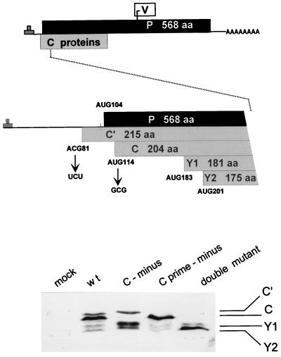
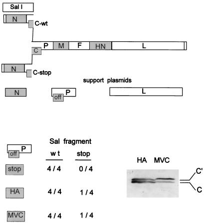
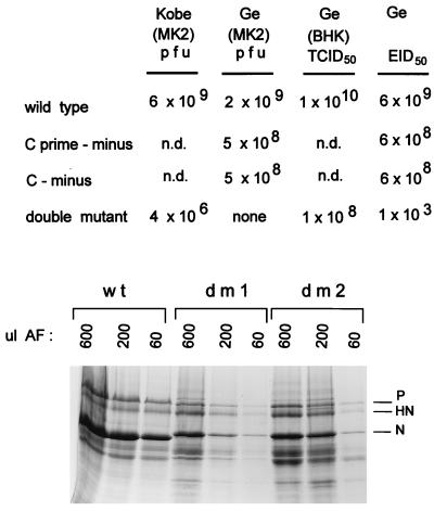
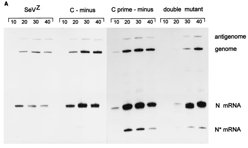
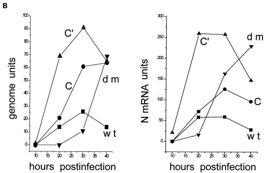
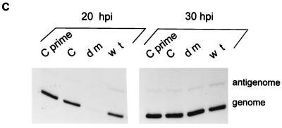
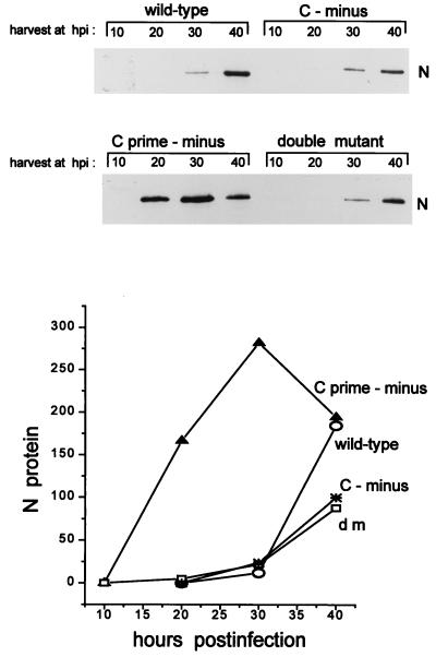
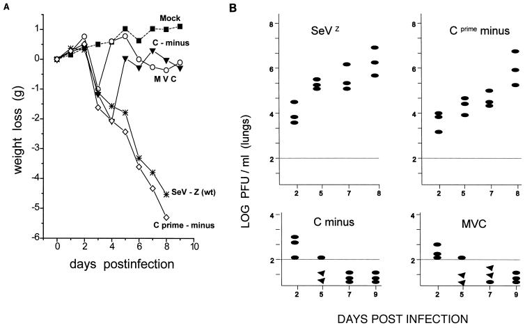
Recombinant Sendai viruses were prepared which cannot express their Cprime, C, or Cprime plus C proteins due to mutation of their respective start codons ([Cprime-minus], [C-minus] and [double mutant], respectively). The [Cprime-minus] and [C-minus] stocks were similar to that of wild-type (wt) virus in virus titer and plaque formation, whereas the double-mutant stock had a much-reduced PFU or 50% egg infective dose/particle ratio and produced very small plaques. Relative to the wt virus infection, the [Cprime-minus] and [C-minus] infections of BHK cells resulted in significantly greater accumulation of viral RNAs, consistent with the known inhibitory effects of the Cprime and C proteins. The double-mutant infection, in contrast, was delayed in its accumulation of viral RNAs; however, once accumulation started, overaccumulation quickly occurred, as in the single-mutant infections. Our results suggest that the Cprime and C proteins both provide a common positive function early in infection, so that only the double mutant undergoes delayed RNA accumulation and exhibits the highly debilitated phenotype. Later in infection, the same proteins appear to act as inhibitors of RNA accumulation. In infections of mice, [Cprime-minus] was found to be as virulent as wt virus whereas [C-minus] was highly attenuated. These results suggest that the Cprime and C proteins cannot be functionally equivalent, since C can replace Cprime for virulence in mice whereas Cprime cannot replace C.


Figures

FIG. 1
FIG. 1
ORF organization and expression of the SeV P gene. The three ORFs expressed as proteins (P, C, and V) are shown as horizontal boxes, drawn roughly to scale (top). A blow-up of the 5′ end of the mRNA is shown in the middle, and the five ribosomal start sites in this region are indicated. The mutations used to eliminate expression from the Cprime and C protein start codons are shown. The effects of these mutations on C protein expression in BHK cells infected with a 1:5 dilution of the wt (SeVZ), [C-minus], [Cprime-minus], and double-mutant virus stocks (see the text) were examined by immunoblotting with a polyclonal rabbit anti-C antiserum (bottom). aa, amino acids.
FIG. 2
FIG. 2
Inability to recovery a SeV strain which cannot express any of its C proteins. The reconstruction of full-length SeV DNA in which the C ORF remains open (C-wt) or is closed just after AUG201/Y2 (C-stop) by exchanging SalI fragments is shown schematically at the top. The three pGEM support plasmids expressing N, P, and L, needed to recover infectious virus from the full-length DNA, are shown just below; “off” in the box representing the C ORF indicates that C either is not expressed or is not completely functional (see the text). A summary of the results of the virus recoveries obtained with both SalI fragments and three different pGEM-P support plasmids (stop, HA, and MVC [see the text]) is shown at the bottom. When virus was recovered from full-length DNA containing the C-stop mutation, this virus was used to infect BHK cells, and the presence or absence of C expression was examined by immunoblotting (bottom right). Y1 or Y2 protein expression was not detected in this infection.
FIG. 3
FIG. 3
Titer determination of the various SeV stocks. Egg allantoic fluid stocks of the various mutant SeV strains and wt virus (SeVZ) were subjected to titer determination by (i) plaque formation on MK2 cells with stocks prepared in Kobe and Geneva (Ge), (ii) TCID50 determination on BHK cells, and (iii) EID50 determination in hen eggs (top). All values listed are per milliliter. Relative amounts of virus proteins present in 600, 200, and 60 μl of allantoic fluid (AF) were determined by SDS-PAGE and staining with Coomassie brilliant blue (bottom). dm1 and dm2 are independently recovered double-mutant viruses (dm2 was subjected to titer determination [above]). The N proteins of the mutant viruses migrate slightly slower than that of SeVZ, as expected, because these viruses were prepared in the rSeVZ-NH background to mark them as recombinants.
FIG. 4
FIG. 4
Plaque formation of recombinant wt and double-mutant virus. Serial dilutions of wt (rSeVZ-NH) and dm2Kobe stocks were allowed to form plaques for 9 days at 33°C on LLC-MK2 cells in the presence of trysin and the absence of serum. An example of the plaques formed by each virus and its dilution is shown.
FIG. 5
FIG. 5
Accumulation of viral RNAs in the mutant and wt virus-infected BHK cells. Parallel 4-cm-diameter BHK cultures (ca. 107 cells) were infected with 2 (MK2) PFU or 20 (BHK) TCID50 of wt, [Cprime-minus], or [C-minus] per cell and 2 (BHK) TCID50 of dm2Ge per cell. Infected cultures were harvested at various times after infection, as indicated above the lanes. The relative amounts of viral genomes, antigenomes, and N mRNAs present in equal samples of the total RNA (from one-quarter of each dish) were estimated by duplex primer extension with NP126, whose 5′ end is 126 and 70 nucleotides from that of the antigenome and N mRNA, respectively, and L15,270, whose 5′ end is 114 nucleotides from that of the genome. (A) The extended primers were separated on an 8% sequencing gel. N* mRNA is presumably a degradation product of the N mRNA. (B) The intensities of the various bands were determined in a PhosphorImager, and their values are plotted in terms of genomes and N and N* mRNA. (C) Accumulation of genomes and antigenomes in cultures infected individually with 2 (BHK) TCID50 of each virus per cell (or 0.2 PFU of wt, [Cprime-minus] or [C-minus] per cell), as above. hpi, hours p.i.
FIG. 5
FIG. 5
Accumulation of viral RNAs in the mutant and wt virus-infected BHK cells. Parallel 4-cm-diameter BHK cultures (ca. 107 cells) were infected with 2 (MK2) PFU or 20 (BHK) TCID50 of wt, [Cprime-minus], or [C-minus] per cell and 2 (BHK) TCID50 of dm2Ge per cell. Infected cultures were harvested at various times after infection, as indicated above the lanes. The relative amounts of viral genomes, antigenomes, and N mRNAs present in equal samples of the total RNA (from one-quarter of each dish) were estimated by duplex primer extension with NP126, whose 5′ end is 126 and 70 nucleotides from that of the antigenome and N mRNA, respectively, and L15,270, whose 5′ end is 114 nucleotides from that of the genome. (A) The extended primers were separated on an 8% sequencing gel. N* mRNA is presumably a degradation product of the N mRNA. (B) The intensities of the various bands were determined in a PhosphorImager, and their values are plotted in terms of genomes and N and N* mRNA. (C) Accumulation of genomes and antigenomes in cultures infected individually with 2 (BHK) TCID50 of each virus per cell (or 0.2 PFU of wt, [Cprime-minus] or [C-minus] per cell), as above. hpi, hours p.i.
FIG. 6
FIG. 6
Accumulation of viral N protein in the culture supernatants. The supernatants of the infected cultures in Fig. 5A and B were harvested at the times indicated. Virus was pelleted through a glycerol cushion (see Materials and Methods), dissolved in sample buffer, separated by SDS-PAGE (10% polyacrylamide), and immunoblotted with an anti-N monoclonal antibody. The intensities of the various bands were determined in a densitometer, and their values are plotted below the gels. hpi, hours p.i.
FIG. 7
FIG. 7
Infections of C57BL/6 mice. Groups of 7-week-old C57BL/6 mice (weighing ca. 20 g) were infected intranasally with 106 PFU of either SeVZ (wt), SeVMVC, or the recombinant [C-minus] or [Cprime-minus]. Phosphate-buffered saline was used for the mock-infected mice. (A) The body weights of the mice were measured daily and are given as weight loss relative to the weight on day 0. Three mice from each of the infected groups were sacrificed at the times indicated, and the PFU present in 10% lung homogenates were determined on LLC-MK2 cells. (B) The titers for each of the three mice are shown individually. Triangles (below the limit of detection by this method [102 PFU/lobe]) indicate that injecting these homogenates into eggs and passaging twice does lead to detectable virus; ovals indicate that virus could not be detected by this procedure.

Similar articles

Cited by

References

    1. Alkhatib G, Massie B, Briedis D J. Expression of bicistronic measles virus P/C mRNA by using hybrid adenoviruses: levels of C protein synthesized in vivo are unaffected by the presence or absence of the upstream P initiator colon. J Virol. 1988;62:4059–4069. - PMC - PubMed
    1. Barr J, Chambers P, Harriott P, Pringle C R, Easton A J. Sequence of the phosphoprotein gene of pneumonia virus of mice: expression of multiple proteins from two overlapping reading frames. J Virol. 1994;68:5330–5334. - PMC - PubMed
    1. Bellini W J, Englund G, Rozenblatt S, Arnheiter H, Richardson C D. Measles virus P gene codes for two proteins. J Virol. 1985;53:908–919. - PMC - PubMed
    1. Billeter M A, Cattaneo R, Spielhofer P, Kaelin K, Huber M, Schmid A, Baczko K, ter Meulen V. Generation and properties of measles virus mutations typically associated with subacute sclerosing panencephalitis. Ann N Y Acad Sci. 1994;724:367–377. - PubMed
    1. Boeck R, Kolakofsky D. Positions +5 and +6 can be major determinants of the efficiency of non-AUG initiation codons for protein synthesis. EMBO J. 1994;13:3608–3617. - PMC - PubMed

LinkOut - more resources