. 1998 Jul;72(7):6004-13.
doi: 10.1128/JVI.72.7.6004-6013.1998.

Interaction of the human immunodeficiency virus type 1 Vpr protein with the nuclear pore complex

Affiliations

Interaction of the human immunodeficiency virus type 1 Vpr protein with the nuclear pore complex

R A Fouchier et al. J Virol. 1998 Jul.

Abstract

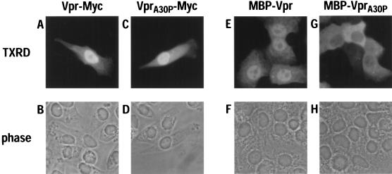
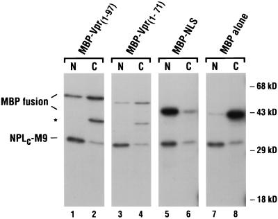
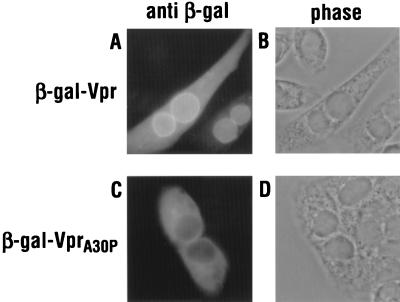
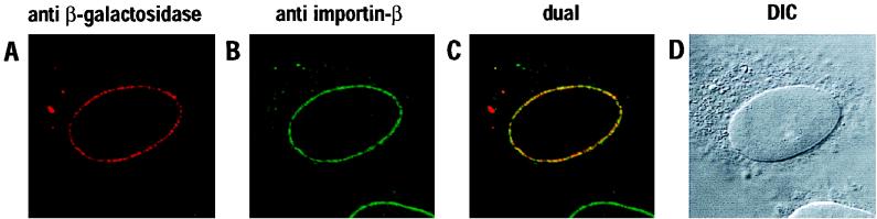
The Vpr protein of human immunodeficiency virus type 1 (HIV-1) performs a number of functions that are associated with the nucleus. Vpr enhances the nuclear import of postentry viral nucleoprotein complexes, arrests proliferating cells in the G2 phase of the cell cycle, and acts as a modest transcriptional activator. For this paper, we have investigated the nuclear import of Vpr. Although Vpr does not encode a sequence that is recognizable as a nuclear localization signal (NLS), Vpr functions as a transferable NLS both in somatic cells and in Xenopus laevis oocytes. In certain contexts, Vpr also mediates substantial accumulation at the nuclear envelope and, in particular, at nuclear pore complexes (NPCs). Consistent with this, Vpr is shown to interact specifically with nucleoporin phenylalanine-glycine (FG)-repeat regions. These findings not only demonstrate that Vpr harbors a bona fide NLS but also raise the possibility that one (or more) of Vpr's functions may take place at the NPC.


Figures

FIG. 1
FIG. 1
Subcellular localization of HIV-1 Vpr in HeLa cells. Monolayer cultures were transfected with vectors that expressed wild-type Vpr (A, B, E, and F) or the VprA30P mutant (C, D, G, and H) as either Myc-tagged proteins (A to D) or fusions to MBP (E to H). Expressed proteins were detected by indirect immunofluorescence with either the Myc-specific monoclonal antibody (A and C) or an MBP-specific antiserum (E and G), TXRD-conjugated secondary antibodies, and epifluorescence. The corresponding phase-contrast analyses are also shown (B, D, F, and H).
FIG. 2
FIG. 2
Nuclear import of MBP fusion proteins in microinjected X. laevis oocytes. The indicated MBP fusion proteins and NPLC-M9 were radiolabeled in vitro and coinjected into the cytoplasms of 10 to 20 stage VI oocytes. At 12 h, the oocytes were separated into nuclear (N) and cytoplasmic (C) fractions, and the soluble proteins were analyzed on an SDS-polyacrylamide gel. The bands corresponding to the MBP fusions and NPLC-M9 are indicated; the faster-migrating, and presumably truncated, protein present in the MBP-Vpr1–97 (lane 2) and MBP-Vpr1–71 (lane 4) samples is indicated by an asterisk.
FIG. 3
FIG. 3
Subcellular localization of β-gal–Vpr fusion proteins in HeLa cells. Monolayers were transfected with wild-type (A) or A30P mutant (C) expression vectors and analyzed by indirect immunofluorescence with a β-galactosidase-specific antiserum. The phase-contrast analyses are also shown (B and D).
FIG. 4
FIG. 4
Colocalization of wild-type β-gal–Vpr and importin-β at NPCs. Transfected HeLa cells were subjected to double-label immunofluorescence with primary antibodies specific for β-galactosidase (A) or β-importin (B) and analyzed by laser-scanning confocal microscopy. The superimposed images are shown (dual [C]) together with the corresponding differential interference contrast image (DIC [D]).
FIG. 5
FIG. 5
Interaction of Vpr with nucleoporin FG-repeat regions in vitro. Radiolabeled MBP-Vpr or MBP alone was incubated with GST, GST-Nsp1p, GST-Nup100p, or GST-Pom121 that had been prebound to glutathione-Sepharose beads. After washing, bound proteins were eluted and analyzed on an SDS-polyacrylamide gel (lanes 1 to 8) together with the input proteins (lanes 9 and 10).
FIG. 6
FIG. 6
Replication of wild-type (squares) and vpr-deficient (triangles) HIV-1YU-2 in spreading infections of nondividing and dividing cells. PBMCs (A [PBMC-1] and B [PBMC-2]), MDMs (D [MDM-1], E [MDM-2], and F [MDM-3]), primary MG (G [MG-4] and H [MG-5]), and CEM-CCR5 cells (C) were challenged with normalized virus stocks produced by transfection of 293T cells. Virus production, and hence replication, was measured as the expression of soluble p24gag in the culture supernatants.

Similar articles

Cited by

References

    1. Adam S A, Marr R S, Gerace L. Nuclear protein import in permeabilized mammalian cells requires soluble cytoplasmic factors. J Cell Biol. 1990;111:807–816. - PMC - PubMed
    1. Agostini I, Navarro J-M, Rey F, Bouhamdan M, Spire B, Vigne R, Sire J. The human immunodeficiency virus type 1 Vpr transactivator: cooperation with promoter-bound activator domains and binding to TFIIB. J Mol Biol. 1996;261:599–606. - PubMed
    1. Aitchison J D, Blobel G, Rout M P. Kap104p: a karyopherin involved in the nuclear transport of messenger RNA binding proteins. Science. 1996;274:624–627. - PubMed
    1. Ayyavoo V, Mahboubi A, Mahalingam S, Ramalingam R, Kudchodkar S, Williams W V, Green D R, Weiner D B. HIV-1 Vpr suppresses immune activation and apoptosis through regulation of nuclear factor κB. Nat Med. 1997;3:1117–1123. - PubMed
    1. Balliet J W, Kolson D L, Eiger G, Kim F M, McGann K A, Srinivasan A, Collman R. Distinct effects in primary macrophages and lymphocytes of the human immunodeficiency virus type 1 accessory genes vpr, vpu, and nef: mutational analysis of a primary HIV-1 isolate. Virology. 1994;200:623–631. - PubMed

Publication types

Substances

LinkOut - more resources