. 1998 Jul;72(7):6024-33.
doi: 10.1128/JVI.72.7.6024-6033.1998.

Particle polymorphism caused by deletion of a peptide molecular switch in a quasiequivalent icosahedral virus

Affiliations

Particle polymorphism caused by deletion of a peptide molecular switch in a quasiequivalent icosahedral virus

X F Dong et al. J Virol. 1998 Jul.

Abstract

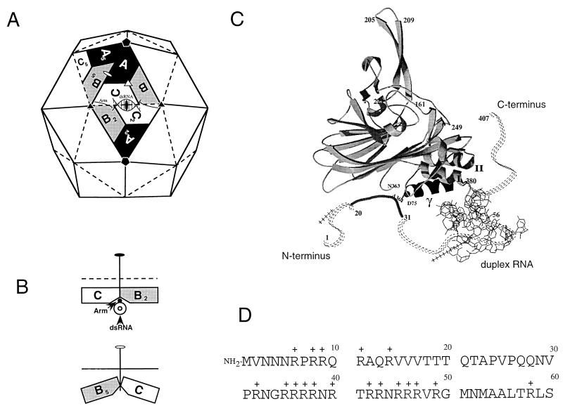
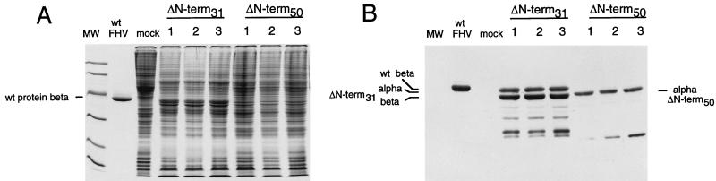
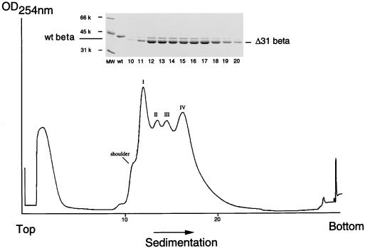
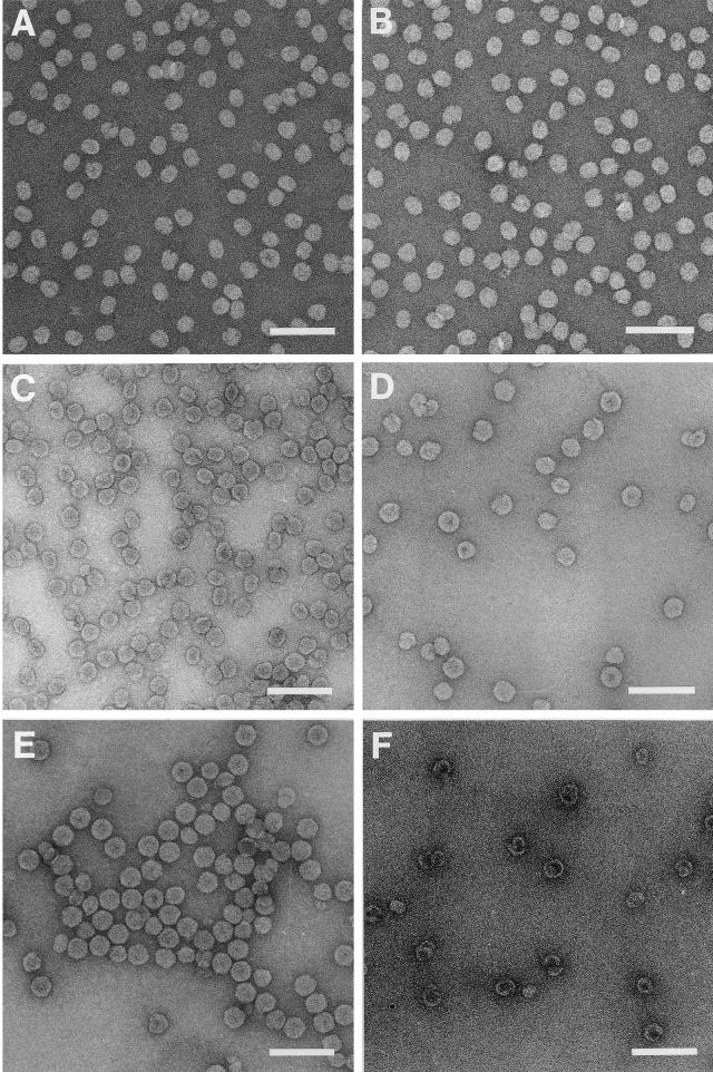
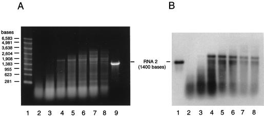
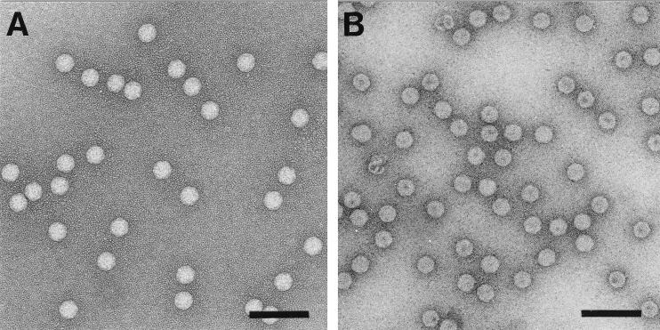
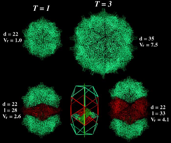
The capsid of flock house virus is composed of 180 copies of a single type of coat protein which forms a T=3 icosahedral shell. High-resolution structural analysis has shown that the protein subunits, although chemically identical, form different contacts across the twofold axes of the virus particle. Subunits that are related by icosahedral twofold symmetry form flat contacts, whereas subunits that are related by quasi-twofold symmetry form bent contacts. The flat contacts are due to the presence of ordered genomic RNA and an ordered peptide arm which is inserted in the groove between the subunits and prevents them from forming the dihedral angle observed at the bent quasi-twofold contacts. We hypothesized that by deleting the residues that constitute the ordered peptide arm, formation of flat contacts should be impossible and therefore result in assembly of particles with only bent contacts. Such particles would have T=1 symmetry. To test this hypothesis we generated two deletion mutants in which either 50 or 31 residues were eliminated from the N terminus of the coat protein. We found that in the absence of residues 1 to 50, assembly was completely inhibited, presumably because the mutation removed a cluster of positively charged amino acids required for neutralization of encapsidated RNA. When the deletion was restricted to residues 1 to 31, assembly occurred, but the products were highly heterogeneous. Small bacilliform-like structures and irregular structures as well as wild-type-like T=3 particles were detected. The anticipated T=1 particles, on the other hand, were not observed. We conclude that residues 20 to 30 are not critical for formation of flat protein contacts and formation of T=3 particles. However, the N terminus of the coat protein appears to play an essential role in regulating assembly such that only one product, T=3 particles, is synthesized.


Figures

FIG. 1
FIG. 1
Subunit interactions as seen in the structure of FHV and the N-terminal sequence of the coat protein. (A) Schematic representation of the FHV capsid as a rhombic triacontahedron. Each trapezoid represents a protein subunit which consists of 407 amino acids. The labels A, B, and C represent the three subunits in each of the 60 icosahedral asymmetric units in the T=3 particle. Although A, B, and C represent identical gene products, they are not related by strict symmetry and they are structurally slightly different. The icosahedral twofold, threefold, and fivefold axes are represented by the filled oval, triangle, and pentagon, respectively. The quasi-twofold and quasi-threefold axes are represented by the open oval and triangle. Quasisymmetry axes relate subunits only locally outside the context of the entire shell. For example, the quasi-twofold axis shown between the central triangle and its left neighbor relates subunits A, B, and C to B5, C5, and A5, respectively, thus bringing subunits clustered around a fivefold axis into coincidence with those clustered around a threefold axis. dsRNA, double-stranded RNA. (B) Side view of the joints between asymmetric units. The top diagram shows a joint at the base of the central triangle viewed along the line connecting the two threefold-symmetry axes. The dihedral angle between these two triangular surfaces is 180°C, i.e., they are coplanar. The bottom diagram shows a joint at the left of the central triangle viewed along the line connecting a threefold axis to a fivefold axis. The dihedral angle between these two surfaces is 144°. For proper assembly to occur, the trimer interfaces must be able to “hinge” with a molecular switch that determines the dihedral angle between asymmetric units. In FHV, the switch is a 10-bp RNA duplex and an 11-amino-acid peptide (residues 20 to 30) that are ordered only in the C subunits. The RNA and peptide arm form a wedge that prevents the bending of the joint at the bottom of the central triangle. (C) Ribbon diagram showing the tertiary structure of the C subunit of FHV. The subunit is oriented such that the virus exterior is at the top and the virus interior is at the bottom. The ordered peptide arm (residues 20 to 30) is represented as a solid line, whereas the disordered portions are represented as dashed lines. The duplex RNA is shown as a line diagram. (D) Amino-terminal sequence of the FHV coat protein. Note the presence of 17 positively charged amino acids, all arginines, within the first 50 residues.
FIG. 2
FIG. 2
Protein gel electrophoresis and immunoblot analysis of Sf21 cell lysates infected with three independent plaque isolates of the recombinant baculoviruses ΔNterm-31(bac) and ΔNterm-50(bac). (A) Monolayers (5 × 10⁵ cells) of Sf21 cells were infected with 100 μl of a 0.5-ml solution containing individual plaque isolates of ΔNterm-31(bac) and ΔNterm-50(bac). Five days after infection, medium was removed from the dishes and cells were suspended in 1 ml of PBS, collected by centrifugation, and washed once with 1 ml of PBS. The final pellet was resuspended in 100 μl of PBS, diluted with an equal volume of electrophoresis buffer, and heated in a boiling water bath for 10 min. Aliquots of 10 μl were electrophoresed through an SDS–12% polyacrylamide gel, followed by staining with Coomassie brilliant blue. Molecular weight (MW) markers from top to bottom were 97,000, 68,000, 43,000, 29,000, 18,000, and 14,000. wt, wild-type. (B) Proteins were subjected to electrophoresis as described for panel A and transferred to a nitrocellulose membrane. The membrane was incubated with polyclonal FHV antiserum and horseradish peroxidase- conjugated secondary antibodies as described in Materials and Methods. Immune complexes were visualized by enhanced chemiluminescence and exposure to X-ray film.
FIG. 3
FIG. 3
Sedimentation profile of VLPs synthesized in ΔNterm-31(bac)-infected Sf21 cells. Monolayers of Sf21 cells (approximately 8 × 10⁶ cells per 100-mm-diameter tissue culture plate) were infected with ΔNterm-31(bac) at a multiplicity of one PFU per cell. Dishes were incubated for 4 days, followed by purification of VLPs as described in Materials and Methods. VLPs from three to four plates were pooled and sedimented through an 11-ml 10 to 40% (wt/wt) sucrose gradient. The gradient was fractionated on an ISCO gradient fractionator, with continuous absorbance at 254 nm. (Inset) Electrophoretic analysis of gradient fractions 10 through 20. An aliquot (5 μl) of each fraction (approximately 375 μl) was mixed with an equal volume of 2× electrophoresis buffer, heated to 95°C for 10 min, and electrophoresed through an SDS–12% polyacrylamide gel. The gel was fixed and stained with Coomassie brilliant blue. wt, wild type; MW, molecular weight.
FIG. 4
FIG. 4
Electron micrographs of negatively stained, gradient-purified ΔNterm-31 VLPs. (A) Particles recovered from the shoulder of peak I; (B) particles representing peak I; (C) particles representing peak II; (D) particles representing peak III; (E) particles representing peak IV; (F) assembly products observed only in T. ni cells following infection with ΔNterm-31(bac). These products sedimented more slowly than all other VLPs and were usually detected midway between the top of the gradient and peak I. Bars, 100 nm.
FIG. 5
FIG. 5
Agarose gel electrophoresis and Northern blot analysis of RNAs encapsidated by ΔNterm-31 VLPs. (A) RNA was phenol-chloroform extracted from successive sucrose gradient fractions spanning the four peaks of a sedimentation profile similar to that shown in Fig. 3. An aliquot (4 μg) of each sample was electrophoresed through a 1% agarose-formaldehyde gel, and RNAs were visualized by staining with ethidium bromide. Lane 1, RNA size markers; lanes 2 to 8, RNAs extracted from successive gradient fractions spanning peaks I to IV (left to right); lane 9, in vitro-synthesized full-length transcript of FHV RNA 2 (1,400 bases). (B) Northern blot analysis of RNA samples shown in panel A. RNA (65 ng) was size fractionated on a 1% agarose-formaldehyde gel and transferred to a nylon membrane. Following UV cross-linking, the immobilized RNAs were hybridized to a digoxigenin-labeled probe complementary to bases 600 to 888 of FHV RNA 2. Lane 1, full-length in vitro-synthesized RNA 2 transcript (1,400 bases); lanes 2 to 8, same as lanes 2 to 8 in panel A.
FIG. 6
FIG. 6
Electron micrographs of negatively stained FHV particles. (A) Native FHV particles; (B) ΔNterm-31 (peak IV) particles used for crystallization and X-ray analysis. Bars, 100 nm.
FIG. 7
FIG. 7
A comparison of the native FHV capsid with hypothetical structures built with Cα coordinates of the coat protein subunit by applying suitable symmetry operations. In the top row at the left is a T=1 particle built from 60 subunits that form 20 trimeric units. Each trimer interacts with its three neighbors via bent contacts. Note that a T=1 particle is significantly smaller than the native T=3 particle shown to the right. Its interior volume (4.35 × 1053) can accommodate approximately 660 nt. The maximal diameter (d) of the particle is given in nanometers. Vr represents the relative interior volumes of the various particles. It was set to 1 for the T=1 particle. In the top row at right is the Cα representation of the native T=3 structure of FHV as determined by X-ray crystallography. This particle, which is shown diagramatically in Fig. 1A, contains 180 protein subunits. In the bottom row at left is the smallest possible bacilliform particle, which consists of one ring of hexameric coat protein subunits (shown in red) and two caps, each representing one-half of a T=1 particle (shown in blue). Note that the line of separation between the two halves of the T=1 particle zigzags and that the caps are rotated relative to each other by 120°. The smaller axis of this particle (d) is identical to the diameter of the T=1 particle shown in the top row, whereas the maximal length (l) of the particle depends on the number of hexameric coat protein rings inserted between the two caps. In the bottom row in the center is a schematic representation of a bacilliform particle containing two rings of hexamers and T=1 icosahedral caps. The organization of the subunits in the hexamer is indicated by the Cα chain trace of the FHV coat protein. Note that the cylindrical portion of the bacilliform particle has threefold rotational symmetry, as is indicated by the green vertical line. In the bottom row at right is a particle built from FHV coat protein subunits and representing the diagram shown in the center of the row.

References

    1. Dasmahapatra B, Dasgupta R, Ghosh A, Kaesberg P. Structure of the black beetle virus genome and its functional implications. J Mol Biol. 1985;182:183–189. - PMC - PubMed
    1. Davis T R, Wickham T J, McKenna K A, Granados R R, Shuler M L, Wood H A. Comparative recombinant protein production of eight insect cell lines. In Vitro Cell Dev Biol Anim. 1993;29A:388–390. - PubMed
    1. Earnshaw W, King J. Structure of phage P22 coat protein aggregates formed in the absence of the scaffolding protein. J Mol Biol. 1978;126:721–747. - PubMed
    1. Erickson J W, Rossmann M G. Assembly and crystallization of a T = 1 icosahedral particle from trypsinized southern bean mosaic virus coat protein. Virology. 1982;116:128–136. - PubMed
    1. Fisher A J, Johnson J E. Ordered duplex RNA controls capsid architecture in an icosahedral animal virus. Nature. 1993;361:176–179. - PubMed

Publication types

LinkOut - more resources