. 1998 Jul;72(7):6048-55.
doi: 10.1128/JVI.72.7.6048-6055.1998.

The native form and maturation process of hepatitis C virus core protein

Affiliations

The native form and maturation process of hepatitis C virus core protein

K Yasui et al. J Virol. 1998 Jul.

Abstract

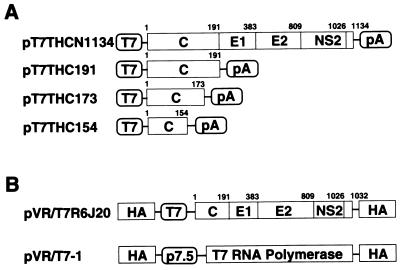
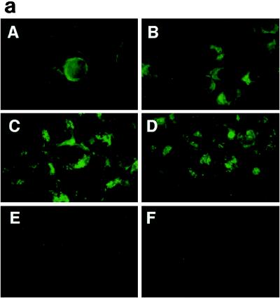
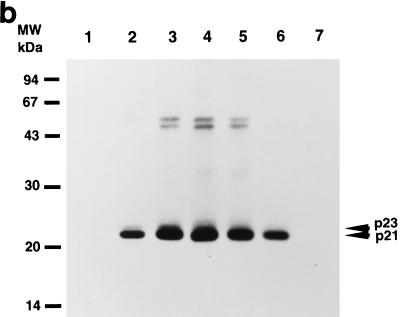
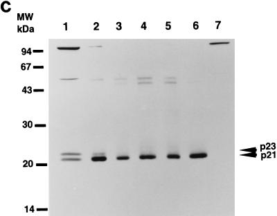
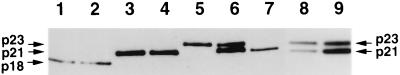
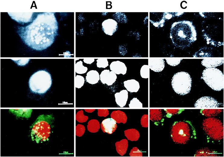
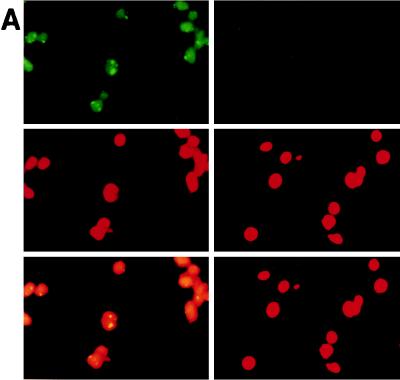
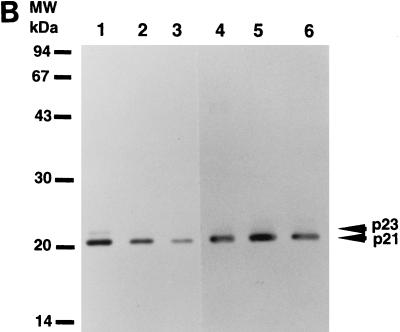
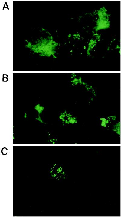
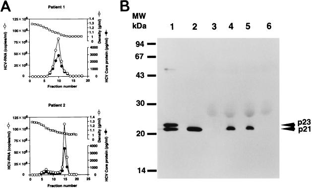
The maturation and subcellular localization of hepatitis C virus (HCV) core protein were investigated with both a vaccinia virus expression system and CHO cell lines stably transformed with HCV cDNA. Two HCV core proteins, with molecular sizes of 21 kDa (p21) and 23 kDa (p23), were identified. The C-terminal end of p23 is amino acid 191 of the HCV polyprotein, and p21 is produced as a result of processing between amino acids 174 and 191. The subcellular localization of the HCV core protein was examined by confocal laser scanning microscopy. Although HCV core protein resided predominantly in the cytoplasm, it was also found in the nucleus and had the same molecular size as p21 in both locations, as determined by subcellular fractionation. The HCV core proteins had different immunoreactivities to a panel of monoclonal antibodies. Antibody 5E3 stained core protein in both the cytoplasm and the nucleus, C7-50 stained core protein only in the cytoplasm, and 499S stained core protein only in the nucleus. These results clearly indicate that the p23 form of HCV core protein is processed to p21 in the cytoplasm and that the core protein in the nucleus has a higher-order structure different from that of p21 in the cytoplasm. HCV core protein in sera of patients with HCV infection was analyzed in order to determine the molecular size of genuinely processed HCV core protein. HCV core protein in sera was found to have exactly the same molecular weight as the p21 protein. These results suggest that p21 core protein is a component of native viral particles.


Figures

FIG. 1
FIG. 1
Construction of HCV cDNA expression vectors. (A) Structure of the HCV genome. The coding frame of the polyprotein is represented by an open box. C, core; E1 and E2, envelope 1 and 2; NS2, nonstructural protein 2. Genes were subcloned under control of the T7 promoter. (B) RVV genomes.
FIG. 2
FIG. 2
Time course of changes in expression of HCV core protein. (a) Indirect immunofluorescence microscopy. RK13 cells were infected with both RVVs, LO-R6J20 and LO-T7-1, fixed with acetone and methanol at −20°C for 10 min, and stained 3 (A), 6 (B), 12 (C), and 24 (D) h after infection. E, 24 h after infection with LO-T7-1; F, negative serum. Immunostaining was performed with monoclonal antibody 515S. (b and c) Western blot analysis. RK13 cells were infected with both LO-R6J20 RVV and LO-T7-1 RVV and harvested at various time points after infection: 3 h (lane 1), 6 h (lane 2), 12 h (lane 3), 24 h (lane 4), 48 h (lane 5), and 72 h (lane 6). As a negative control, RK13 cells were infected with only LO-T7-1 RVV and harvested at 24 h after infection (lane 7). Western blotting was performed with monoclonal antibody 515S. In panel b, the same amount (15 μg/lane) of infected RK13 cell lysate was loaded in all lanes. In panel c, he total loaded protein was 54, 6, 3, 3, 3, 6, and 54 μg in lanes 1 to 7, respectively. The two HCV core proteins, p21 and p23, are indicated on the right, and the sizes of protein molecular weight (MW) markers are indicated on the left.
FIG. 2
FIG. 2
Time course of changes in expression of HCV core protein. (a) Indirect immunofluorescence microscopy. RK13 cells were infected with both RVVs, LO-R6J20 and LO-T7-1, fixed with acetone and methanol at −20°C for 10 min, and stained 3 (A), 6 (B), 12 (C), and 24 (D) h after infection. E, 24 h after infection with LO-T7-1; F, negative serum. Immunostaining was performed with monoclonal antibody 515S. (b and c) Western blot analysis. RK13 cells were infected with both LO-R6J20 RVV and LO-T7-1 RVV and harvested at various time points after infection: 3 h (lane 1), 6 h (lane 2), 12 h (lane 3), 24 h (lane 4), 48 h (lane 5), and 72 h (lane 6). As a negative control, RK13 cells were infected with only LO-T7-1 RVV and harvested at 24 h after infection (lane 7). Western blotting was performed with monoclonal antibody 515S. In panel b, the same amount (15 μg/lane) of infected RK13 cell lysate was loaded in all lanes. In panel c, he total loaded protein was 54, 6, 3, 3, 3, 6, and 54 μg in lanes 1 to 7, respectively. The two HCV core proteins, p21 and p23, are indicated on the right, and the sizes of protein molecular weight (MW) markers are indicated on the left.
FIG. 3
FIG. 3
Determination of the origins of the two forms of HCV core proteins, p21 and p23 by Western blot analysis. Plasmids pT7THC154, pT7THC173, and pT7THC191 were transiently expressed in an in vitro translation system (lanes 1, 3, and 5, respectively) and were also transfected into RK13 cells, which were then infected with LO-T7-1 RVV and harvested 12 h after infection (lanes 2, 4, and 6, respectively). Plasmid pT7THCN1134 was also transfected into RK13 cells, which were then infected with LO-T7-1 RVV and harvested 12 h after infection (lanes 7). RK13 cells infected with both LO-T7-1 RVV and LO-R6J20 RVV were then harvested 3 h after infection (lane 8). The same sample was loaded in lanes 6 and 9.
FIG. 4
FIG. 4
Subcellular localization of HCV core protein. RK13 cells (A) and HPB-Ma cells (B) were infected with both the LO-R6J20 and the LO-T7-1 RVVs. Cells were fixed with acetone and methanol at −20°C for 10 min 12 h after infection, counterstained with FITC-conjugated anti-mouse Ig (for HCV core protein) and propidium iodide (for nuclear DNA), and viewed with a confocal laser scanning microscope. (C) CHO cells stably transformed with an HCV core protein expression vector. Upper panels, HCV core protein; middle panels, nuclear DNA; lower panels, merged image of HCV core protein (green) and nucleus (red).
FIG. 5
FIG. 5
Subcellular fractionation of HCV core protein. (A) Immunostaining of pure nuclear fraction N2. N2 fractions from RK13 cells infected with LO-R6J20 and LO-T7-1 RVVs for 12 h were separated as described in Materials and Methods, stained with FITC-conjugated anti-mouse Ig (for HCV core protein) (upper panels), and counterstained with propidium iodide (for nucleus DNA) (middle panels), and the resultant images were merged (lower panels). Fraction N2 was stained with anticore monoclonal antibody 515S (upper left) or anti-poliovirus receptor monoclonal antibody as a negative control (upper right). (B) The cytoplasmic fraction (Cy), crude nuclear fraction (N1), and pure nuclear fraction (N2) from RK13 cells infected with LO-R6J20 RVV and LO-T7-1 RVV for 6 h (lanes 1 to 3) or 12 h (lanes 4 to 6) were separated as described in Materials and Methods. The HCV core protein in each fraction was analyzed by Western blotting. Lanes 1 and 4, Cy; lanes 2 and 5, N1; lanes 3 and 6, N2.
FIG. 5
FIG. 5
Subcellular fractionation of HCV core protein. (A) Immunostaining of pure nuclear fraction N2. N2 fractions from RK13 cells infected with LO-R6J20 and LO-T7-1 RVVs for 12 h were separated as described in Materials and Methods, stained with FITC-conjugated anti-mouse Ig (for HCV core protein) (upper panels), and counterstained with propidium iodide (for nucleus DNA) (middle panels), and the resultant images were merged (lower panels). Fraction N2 was stained with anticore monoclonal antibody 515S (upper left) or anti-poliovirus receptor monoclonal antibody as a negative control (upper right). (B) The cytoplasmic fraction (Cy), crude nuclear fraction (N1), and pure nuclear fraction (N2) from RK13 cells infected with LO-R6J20 RVV and LO-T7-1 RVV for 6 h (lanes 1 to 3) or 12 h (lanes 4 to 6) were separated as described in Materials and Methods. The HCV core protein in each fraction was analyzed by Western blotting. Lanes 1 and 4, Cy; lanes 2 and 5, N1; lanes 3 and 6, N2.
FIG. 6
FIG. 6
Subcellular localization of HCV core protein determined by immunostaining with different antibodies. RK13 cells infected with both LO-R6J20 RVV and LO-T7-1 RVV were fixed with acetone and methanol at −20°C for 10 min 12 h after infection and stained with the anti-HCV core protein monoclonal antibodies 5E3 (A), C7-50 (B), and 499 (C).
FIG. 7
FIG. 7
(A) Quantification of HCV core protein and HCV RNA in fractions from sucrose density gradient centrifugation of the patient sera. (B) Western blot analysis of HCV native core protein in HCV-infected-patient sera or HCV-negative sera. Lanes 1 and 2, HCV core protein in RK13 cells harvested 3 and 24 h, respectively, after infection with both the LO-R6J20 and the LO-T7-1 RVVs; lanes 4 and 5, HCV core protein from HCV-infected-patient sera; lanes 3 and 6, HCV-negative sera. MW, molecular weight markers.

Similar articles

Cited by

References

    1. Barba B, Harper F, Harada T, Kohara M, Goulinet S, Matsuura Y, Eder G, Schaff Z, Chapman M J, Miyamura T, Brechot C. Hepatitis C virus core protein shows a cytoplasmic localization and associates to cellular lipid storage droplets. Proc Natl Acad Sci USA. 1997;94:1200–1205. - PMC - PubMed
    1. Chen C, Okayama H. High-efficiency transformation of mammalian cells by plasmid DNA. Mol Cell Biol. 1987;7:2745–2752. - PMC - PubMed
    1. Choo Q-L, Kuo G, Weiner A J, Overby L R, Bradley D W, Houghton M. Isolation of cDNA derived from a blood-borne non-A, non-B viral hepatitis genome. Science. 1989;244:359–362. - PubMed
    1. Choo Q-L, Richman K, Han J H, Berger K, Lee C, Dong C, Gallegos C, Coit D, Medina-Selby A, Barr P, Weiner A J, Bradley D W, Kuo G, Houghton M. Genetic organization and diversity of the hepatitis C virus. Proc Natl Acad Sci USA. 1991;88:2451–2455. - PMC - PubMed
    1. Esteban J I, Esteban R, Viladomiu L, Lopez-Talavera J C, Gonzalez A, Hernandez J M, Roget M, Vargas V, Genesca J, Buti M, Guardia J. Hepatitis C virus antibodies among risk groups in Spain. Lancet. 1989;ii:294–296. - PubMed

Publication types

Substances

LinkOut - more resources