. 1998 Jul;72(7):6104-12.
doi: 10.1128/JVI.72.7.6104-6112.1998.

Human herpesvirus 6 open reading frame U12 encodes a functional beta-chemokine receptor

Affiliations

Human herpesvirus 6 open reading frame U12 encodes a functional beta-chemokine receptor

Y Isegawa et al. J Virol. 1998 Jul.

Abstract

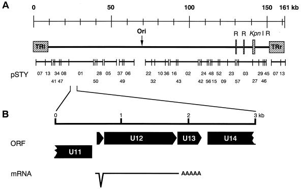
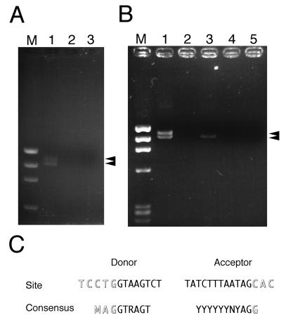
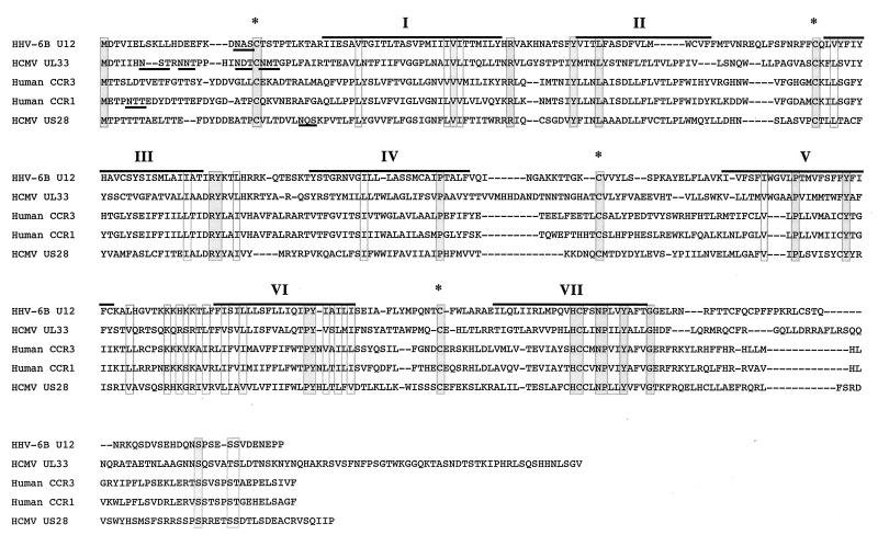
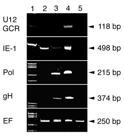
Human herpesvirus 6 (HHV- 6), which belongs to the betaherpesvirus subfamily and infects mainly T cells in vitro, causes acute and latent infections. HHV- 6 contains two genes (U12 and U51) that encode putative homologs of cellular G-protein-coupled receptors (GCR), while three other betaherpesviruses, human cytomegalovirus, murine cytomegalovirus, and human herpesvirus 7, have three, one, and two GCR-homologous genes, respectively. The U12 gene is expressed late in infection from a spliced mRNA. The U12 gene was cloned, and the protein was expressed in cells and analyzed for its biological characteristics. U12 functionally encoded a calcium-mobilizing receptor for beta-chemokines such as regulated upon activation, normal T expressed and secreted (RANTES), macrophage inflammatory proteins 1alpha and 1beta (MIP-1alpha and MIP-1beta) and monocyte chemoattractant protein 1 but not for the alpha-chemokine interleukin-8, suggesting that the chemokine selectivity of the U12 product was distinct from that of the known mammalian chemokine receptors. These findings suggested that the product of U12 may play an important role in the pathogenesis of HHV- 6 through transmembrane signaling by binding with beta-chemokines.


Figures

FIG. 1
FIG. 1
General organization of an HHV- 6 GCR-homologous gene, and structure of the HHV- 6 GCR transcript. (A) Location of a GCR-homologous gene in the HHV- 6 (HST) genome. (B) Direction of ORFs and splicing site of U12.
FIG. 2
FIG. 2
RT-PCR assay of U12 mRNA in the HST-infected cells and the U12- transfected cells. (A) U12 ORF was amplified from cDNA synthesized with total RNA purified from HST- or mock-infected CBMCs with a pair of primers, 01GCR- Met-Kpn and 01GCR-Ter-Bam, as described in Materials and Methods. Lanes: 1, HST-infected CBMCs; 2, HST-infected CBMCs without RT treatment; 3, mock-infected CBMCs; M, φX174 × HaeIII. (B) U12 ORF was amplified from cDNA synthesized with total RNA purified from U12-transfected K562 cells. Lanes: 1, genomic U12-transfected cells; 2, genomic U12-transfected cells without RT treatment; 3, U12 cDNA-transfected cells; 4, U12 cDNA-transfected cells without RT treatment; 5, pCEP4-transfected cells; M, φX174 × HaeIII. (C) Comparison of amino acid sequences between U12 splice sites and consensus donor-acceptor sites as reported by Green (26). Outlined letters are exon nucleotides, and normal letters are intron nucleotides.
FIG. 3
FIG. 3
Sequence alignment of the U12 cDNA product with the HCMV US28 and UL33 products and human CCR-1 and CCR-3. Dashes indicate gaps that were inserted to optimize the alignment. The locations of predicted membrane-spanning segments are denoted by I to VII, and overlines indicate their amino acid sequences. Underlines designated predicted sites for N-linked glycosylation. Asterisks indicate the positions where the five sequences have the conserved cysteine residues in the predicted cytoplasmic domains. Identical and homologous amino acids of the five sequences are enclosed in dark and light gray boxes, respectively.
FIG. 4
FIG. 4
RT-PCR assay of the transcripts in the HST-infected cells treated with CHX and PFA. Total RNAs purified from mock-infected (lane 5) and HST-infected (lanes 2 to 4) CBMCs, which were treated with CHX for 24 h (lane 2) or PFA for 24 h (lane 3) or left untreated (lane 4), were used for RT-PCR of the parts of HHV- 6 IE-1, Pol, gH, EF, and U12, as described in Materials and Methods. Lane 1 contains molecular size markers.
FIG. 5
FIG. 5
Expression of U12 in HHV- 6B-infected cells at various times after infection. Western blot analysis shows proteins produced by CBMCs that were infected with the HST-infected cells (lanes 6 to 10) or mock-infected cells (lanes 1 to 5). Lanes: 1 and 6, mock- and HST-infected cells at 0 h; 2 and 7, mock- and HST-infected cells at 12 h; 3 and 8, mock- and HST-infected cells at 24 h; 4 and 9, mock- and HST-infected cells at 48 h; 5 and 10, mock- and HST-infected cells at 72 h; M, molecular size markers.
FIG. 6
FIG. 6
Binding of 125I-RANTES to K562 cells stably transfected with pCEU12c. (A) Binding isotherm of 125I-RANTES. (B) Displacement of RANTES binding to U12-transfected K562 cells by unlabeled chemokines. Each point represents the mean ± standard error of the mean for triplicate determinations. The average total binding was 6,300 cpm. Nonspecific binding was 600 cpm. The binding parameters for competing unlabeled RANTES are shown at the lower left of panel A. Untransfected K562 cells did not specifically bind the radioligand. ○, RANTES; •, MCP-1; □, MIP-1α; ▪, MIP-1β; ▵, IL-8.
FIG. 7
FIG. 7
Transmembrane signaling by the product of HHV- 6 U12. (A) Kinetics. The intracellular Ca2+ concentration was monitored by measuring the relative fluorescence of Indo-1-loaded K562 cells stably transfected with pCECXCR-1, pCEU12c, and pCECCR-1, indicated at the top of each column of tracings. The cells were stimulated, at the time indicated by the arrowheads, with the chemokine indicated on the left of each row of tracings and at the concentration indicated to the right of the corresponding arrow. The tracings shown are from a single experiment representative of three separate experiments. (B) Concentration dependence. The magnitude of the peak of the calcium transient elicited by the indicated concentration of RANTES is shown.
FIG. 8
FIG. 8
Transmembrane signaling by the product of HHV- 6 U12: desensitization. The relative fluorescence was monitored from Indo-1-loaded K562 cells stably transfected with pCEU12c and during sequential addition of test substances at the times indicated by the arrowheads. The concentration and identity of each stimulus are indicated to the right of each arrow. The tracings are from a single experiment representative of two separate experiments.

Similar articles

Cited by

References

    1. Ablashi D V, Balachandran N, Josephs S F, Hung C L, Krueger G R F, Kramarsky B, Salahuddin S Z, Gallo R C. Genomic polymorphism, growth properties, and immunologic variations in human herpesvirus-6 isolates. Virology. 1991;184:545–552. - PubMed
    1. Ahuja S K, Murphy P M. Molecular piracy of mammarian interleukin-8 receptor type B by herpesvirus saimiri. J Biol Chem. 1993;268:20691–20694. - PubMed
    1. Arvanitakis L, Geras-Raake E, Varma A, Gershengorn M C, Cerarman E. Human herpesvirus KSHV encodes a constitutively active G-protein-coupled receptor linked to cell proliferation. Nature. 1997;358:347–350. - PubMed
    1. Aubin J-T, Agut H, Collandre H, Yamanishi K, Chandran B, Montagnier L, Huraux J-M. Antigenic and genetic differentiation of the two putative types of human herpesvirus 6. J Virol Methods. 1993;41:223–234. - PubMed
    1. Aubin J-T, Collandre H, Candotti D, Ingrand D, Rouzioux C, Burgard M, Richard S, Huraux J-M, Agut H. Several groups among human herpesvirus 6 strains can be distinguished by Southern blotting and polymerase chain reaction. J Clin Microbiol. 1991;29:367–372. - PMC - PubMed

Publication types