. 1998 Jul;72(7):6119-30.
doi: 10.1128/JVI.72.7.6119-6130.1998.

Heparan sulfate proteoglycan binding by herpes simplex virus type 1 glycoproteins B and C, which differ in their contributions to virus attachment, penetration, and cell-to-cell spread

Affiliations

Heparan sulfate proteoglycan binding by herpes simplex virus type 1 glycoproteins B and C, which differ in their contributions to virus attachment, penetration, and cell-to-cell spread

S Laquerre et al. J Virol. 1998 Jul.

Abstract

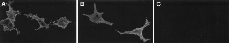
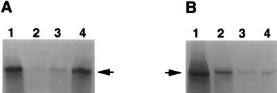
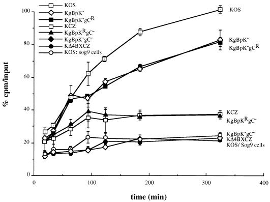
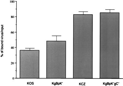
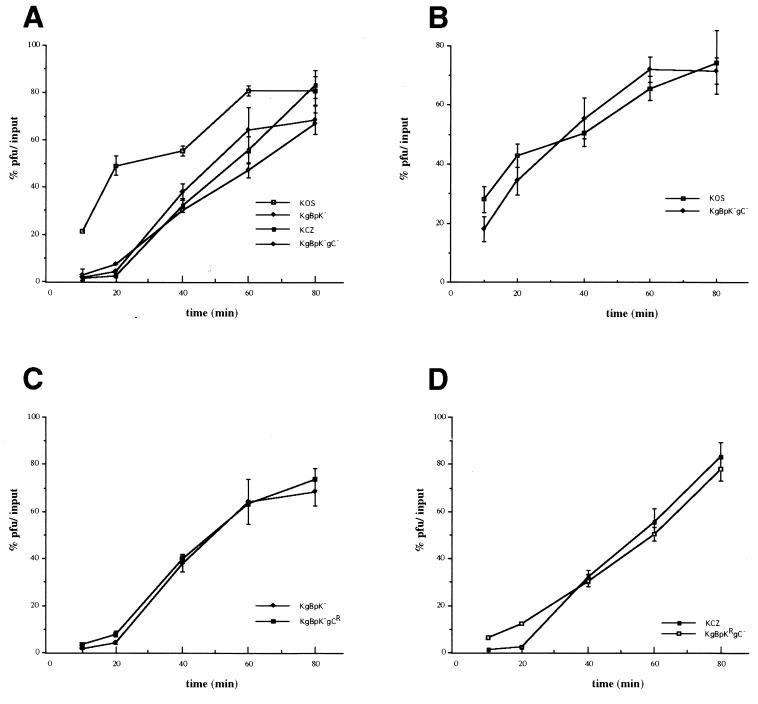
Herpes simplex virus type 1 (HSV-1) mutants defective for envelope glycoprotein C (gC) and gB are highly impaired in the ability to attach to cell surface heparan sulfate (HS) moieties of proteoglycans, the initial virus receptor. Here we report studies aimed at defining the HS binding element of HSV-1 (strain KOS) gB and determining whether this structure is functionally independent of gB's role in extracellular virus penetration or intercellular virus spread. A mutant form of gB deleted for a putative HS binding lysine-rich (pK) sequence (residues 68 to 76) was transiently expressed in Vero cells and shown to be processed normally, leading to exposure on the cell surface. Solubilized gBpK- also had substantially lower affinity for heparin-acrylic beads than did wild-type gB, confirming that the HS binding domain had been inactivated. The gBpK- gene was used to rescue a KOS gB null mutant virus to produce the replication-competent mutant KgBpK-. Compared with wild-type virus, KgBpK- showed reduced binding to mouse L cells (ca. 20%), while a gC null mutant virus in which the gC coding sequence was replaced by the lacZ gene (KCZ) was substantially more impaired (ca. 65%-reduced binding), indicating that the contribution of gC to HS binding was greater than that of gB. The effect of combining both mutations into a single virus (KgBpK-gC-) was additive (ca. 80%-reduced binding to HS) and displayed a binding activity similar to that observed for KOS virus attachment to sog9 cells, a glycosaminoglycan-deficient L-cell line. Cell-adsorbed individual and double HS mutant viruses exhibited a lower rate of virus entry following attachment, suggesting that HS binding plays a role in the process of virus penetration. Moreover, the KgBpK- mutant virus produced small plaques on Vero cells in the presence of neutralizing antibody where plaque formation depended on cell-to-cell virus spread. These studies permitted the following conclusions: (i) the pK sequence is not essential for gB processing or function in virus infection, (ii) the lysine-rich sequence of gB is responsible for HS binding, and (iii) binding to HS is cooperatively linked to the process of efficient virus entry and lateral spread but is not absolutely required for virus infectivity.


Figures

FIG. 1
FIG. 1
Cell surface detection of gBpK mutant glycoprotein. Vero cells were transfected with expression plasmids encoding the wild-type gB protein (HCMV-BXX) (A) and the pK-deleted gB protein (HCMV-gBpK) (B) or were mock transfected (C). After 30 h, unfixed transfected monolayers were incubated with a pool of monoclonal anti-gB antibody for 1 h at 4°C, washed with cold TBS, and incubated for 1 h at 4°C with a cy3 anti-mouse antibody. Monolayers were visualized with a Nikon microscope (model 211910) and photographed.
FIG. 2
FIG. 2
Heparin binding capacity of gBpK mutant glycoprotein. Vero cells were transfected with plasmids encoding wild-type gB (A) and gBpK (B). Twenty-four hours posttransfection, the cell monolayers were infected with KΔ4BX virus in the presence of [35S]methionine/cysteine, as described in Materials and Methods. Ten hours p.i., the monolayers were harvested and solubilized with 0.1% Triton X-100-containing buffer and equivalent amounts of detergent-extracted protein (lanes 1) and were incubated for 2 h at 4°C with heparin-acrylic beads. The unbound proteins (lanes 2) and proteins eluted from the heparin-acrylic beads with detergent buffer (lanes 3) or detergent buffer supplemented with 10 mg of heparin per ml (lanes 4) were immunoprecipitated with a pool of gB-specific MAbs. Each sample was analyzed by SDS-PAGE and autoradiography. The arrows indicate the positions of gB wild-type or gBpK mutant glycoproteins.
FIG. 3
FIG. 3
Southern blot characterization of the recombinant viruses. Viral DNAs from KOS (lane 1), KgBpK (lane 2), KCZ (lane 3), and KgBpKgC (lane 4), as well as viral DNAs from KgBpKgCR and KgBpKRgC, were digested with the restriction endonuclease BamHI (A) or NcoI (B and C) and subjected to Southern blot analysis. (A) A 32P-labeled gB probe hybridized to a 7,774-bp fragment of wild-type gB sequence encoded by KOS (lane 1) and KCZ (lane 3), and KgBpKRgC (lane 6) hybridized with a smaller fragment of 3,009 bp in the recombinant viruses KgBpK (lane 2), KgBpKgC (lane 4), and KgBpKgCR (lane 5) due to the introduction of a BamHI restriction endonuclease site within the gB sequence at the site of the deletion of amino acids 68 to 76. (B) A 32P-labeled gC probe (642-bp NcoI fragment of pgC1) encoding the deleted gC sequence hybridized to a 642-bp fragment containing wild-type gC in KOS (lane 7), KgBpK (lane 8), and KgBpKgCR (lane 11). (C) A different gC probe (828-bp NcoI fragment of pgC1 undeleted in all viruses) hybridized with an 11.2-kbp fragment in KCZ (lane 15), KgBpKgC (lane 16), and KgBpKRgC (lane 18), in which the gC coding sequence was deleted and replaced with the human cytomegalovirus immediate early promoter driving the lacZ gene, and it hybridized with an 828-bp fragment in KOS (lane 13), KgBpK (lane 14), and KgBpKgCR (lane 17), in which the wild-type gC coding sequence was present.
FIG. 4
FIG. 4
Comparative incorporation of glycoproteins B, C, and D into mutant virus envelopes. [35S]methionine/cysteine-labeled sucrose-purified KOS (A), KgBpK (B), KCZ (C), KgBpKgC (D), KgBpKgCR (E), and KgBpKRgC (F) viruses were solubilized with detergent and immunoprecipitated with a pool of MAbs directed against gB (lanes 1), gC (lanes 2), or gD (lanes 3). The immune complexes were captured on protein A-Sepharose, resuspended in Laemmli buffer, and separated by SDS-PAGE. Asterisks indicate the positions of the glycoproteins designated on the left, when present.
FIG. 5
FIG. 5
Cell surface binding capacity of viruses altered in HS proteoglycan binding domains. Vero cells infected with KOS, KCZ, KgBpK, KgBpKgC, KgBpKRgC, or KgBpKgCR viruses and C1 cells infected with KΔ4BXCZ virus were labeled with [35S]methionine/cysteine, and the virions from cell supernatants were subsequently purified on sucrose gradients. The binding capacity of each purified virion was determined on mouse L cells and compared to the binding of wild-type KOS virus on sog9 cells (GAG-deficient L-cell derivatives). Aliquots from the different virus preparations were incubated on the two cell lines at 4°C for up to 320 min and washed with cold TBS, and the cells were scraped, harvested, and counted for virus-associated radioactivity. The percentage of bound virus was determined as radioactive counts representing the bound fraction divided by the total counts per minute (input). The binding capacity of KOS virus on mouse L cells after 320 min was designated 100% binding. Error bars indicate results determined for triplicate wells.
FIG. 6
FIG. 6
Contribution of a viral glycoprotein(s) other than gB and gC to HS binding. Confluent monolayers of Vero cells in 12-well plates were inoculated with 100 PFU of each test virus per well for an adsorption period of 2 h at 4°C. The cells were then rinsed three times with complete medium in the absence or presence of 500 μg of heparin per ml, overlaid with medium containing methyl-cellulose, and shifted to 37°C to allow viral plaque formation. The cells were then fixed and stained with crystal violet, and plaques were counted. At each time point, the average number of plaques produced on heparin-washed monolayers was expressed as a percentage of the average number of plaques produced on complete medium-washed monolayers. Error bars indicate results determined for triplicate wells.
FIG. 7
FIG. 7
Rate of penetration of viruses altered in HS proteoglycan binding domains. Confluent monolayers of Vero cells (A, C, and D) or sog9 cells (B) in 6-well plates were inoculated with 250 PFU of each indicated virus per well for an adsorption period of 2 h at 4°C. The cells were then rinsed three times, overlaid with complete medium, and shifted to 37°C to allow virus penetration. At selected times after the temperature shift, the cells were treated with 2 ml of glycine buffer (0.1 M glycine [pH 3.0]) or TBS for 1 min. The cell monolayers were then washed three times with complete medium, overlaid with medium containing methyl-cellulose, and incubated at 37°C to allow viral plaques to form. After plaque formation, cells were fixed and stained with crystal violet, and plaques were counted. At each time point, the average number of plaques produced on glycine-treated monolayers was expressed as a percentage of the average number of plaques produced on TBS-treated monolayers. Results are averages from triplicate wells.
FIG. 8
FIG. 8
Effect of HS binding mutations on lateral mutant virus spread. Confluent monolayers of Vero and sog9 cells were infected with 300 PFU of KOS, KCZ, KgBpK, and KgBpKgC viruses per well. Twenty-four, 36, and 48 h p.i., Vero cell (A) and sog9 cell (B) monolayers were fixed with ice-cold methanol and processed for immunofluorescence with an anti-rabbit HSV-1 polyclonal antibody for 1 h at 37°C, washed with TBS, and incubated for 1 h at 37°C with a cy3 anti-rabbit antibody. Plaque sizes were determined with a Zeiss Axiophot microscope linked to a Xillix digital camera. For Vero cells, statistically significant differences (P < 0.05) in plaque sizes between viruses are marked by like symbols above the measured values; for sog9 cells, the plaque sizes were not statistically different (P > 0.39).

Similar articles

Cited by

References

    1. Allen G P, Coogle L D. Characterization of an equine herpesvirus type 1 gene encoding a glycoprotein (gp13) with homology to herpes simplex virus glycoprotein C. J Virol. 1988;62:2850–2858. - PMC - PubMed
    1. Baines J D, Roizman B. The UL10 gene of herpes simplex virus 1 encodes a novel glycoprotein, gM, which is present in the virion and in the plasma membrane of infected cells. J Virol. 1993;67:1441–1452. - PMC - PubMed
    1. Banfield B W, Leduc Y, Esford L, Schubert K, Tufaro F. Sequential isolation of proteoglycan synthesis mutants by using herpes simplex virus as a selective agent: evidence for a proteoglycan-independent virus entry pathway. J Virol. 1995;69:3290–3298. - PMC - PubMed
    1. Bergström T, Trybala E, Spillmann D. Heparan sulfate and viral tropism. Nat Med. 1997;3:1177. - PubMed
    1. Cai W, Gu B, Person S. Role of glycoprotein B of herpes simplex virus type 1 in viral entry and cell fusion. J Virol. 1988;62:2596–2604. - PMC - PubMed

Publication types

Substances

LinkOut - more resources