. 1998 Jul;72(7):6199-206.
doi: 10.1128/JVI.72.7.6199-6206.1998.

The NS5A/NS5 proteins of viruses from three genera of the family flaviviridae are phosphorylated by associated serine/threonine kinases

Affiliations

The NS5A/NS5 proteins of viruses from three genera of the family flaviviridae are phosphorylated by associated serine/threonine kinases

K E Reed et al. J Virol. 1998 Jul.

Abstract

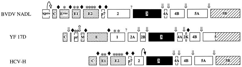
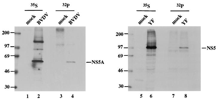
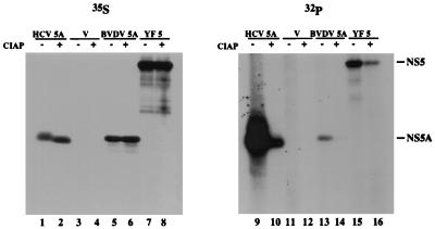
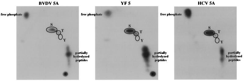
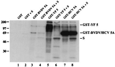
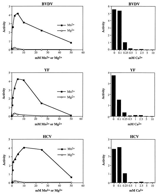
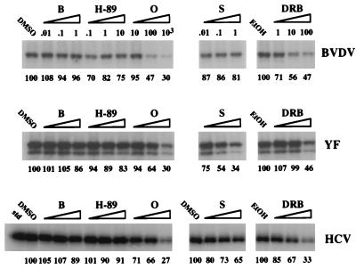
Phosphorylation of the expressed NS5A protein of hepatitis C virus (HCV), a member of the Hepacivirus genus of the family Flaviviridae, has been demonstrated in mammalian cells and in a cell-free assay by an associated kinase activity. In this report, phosphorylation is also shown for the NS5A and NS5 proteins, respectively, of bovine viral diarrhea virus (BVDV) and yellow fever virus (YF), members of the other two established genera in this family. Phosphorylation of BVDV NS5A and YF NS5 was observed in infected cells, transient expression experiments, and a cell-free assay similar to the one developed for HCV NS5A. Phosphoamino acid analyses indicated that all three proteins were phosphorylated by serine/threonine kinases. Similarities in the properties of BVDV NS5A, YF NS5, and HCV NS5A phosphorylation in vitro further suggested that closely related kinases or the same kinase may phosphorylate these viral proteins. Conservation of this trait among three quite distantly related viruses representing three separate genera suggests that phosphorylation of the NS5A/NS5 proteins or their association with cellular kinases may play an important role in the flavivirus life cycle.


Figures

FIG. 1
FIG. 1
Features of the BVDV, YF, and HCV polyproteins. C, capsid; E, E1, E2, and Erns, envelope proteins; Npro, N-terminal autoprotease; prM, membrane precursor protein; p7 and 2K, small polypeptides of unknown function; 1, 2, 2A, 2B, 3, 4A, 4B, 5, 5A, and 5B, NS proteins. Stippled boxes, structural proteins; solid boxes, NS proteins containing the serine protease and nucleoside triphosphatase/helicase activities; striped boxes, polymerase domains. Asterisks indicate glycosylation sites; solid diamonds indicate host signalase cleavage sites; straight arrows mark the Golgi furin-like protease cleavage site in YF 17D (⇕) and the viral serine protease cleavage sites (⇓). The HCV NS2-3 and BVDV N-terminal autoproteases are indicated by curved arrows with solid and open arrowheads, respectively.
FIG. 2
FIG. 2
Phosphorylation of BVDV NS5A and YF NS5 in virus-infected cells. Monolayers of Madin-Darby bovine kidney (MDBK) or SW-13 cells, a human adrenocortical carcinoma cell line, in 35-mm wells were infected, respectively, with BVDV NADL at a multiplicity of infection of 0.1 in 400 μl of phosphate-buffered saline–2% horse serum or with YF 17D at a multiplicity of infection of 5 in 200 μl of Earle’s minimal essential medium (MEM)–2% fetal bovine serum (FBS) for 1 h at 37°C. After the initial infection period, Dulbecco’s MEM–1 mM sodium pyruvate–10% horse serum or Earle’s MEM–2% FBS was added to the BVDV-infected MDBK cells or the YF-infected SW-13 cells, respectively, and incubation was continued for 20 h at 37°C. The cells were then labeled for 4 h at 37°C with MEM containing 2% of the normal methionine concentration, 3% dialyzed FBS, and 100 μCi of Expre35S35S (NEN) or with MEM lacking phosphate supplemented with 3% dialyzed FBS and 400 to 500 μCi of [32P]orthophosphate (ICN) per ml, as indicated. Cell lysates were prepared, and BVDV NS5A or YF NS5 was immunoprecipitated with region-specific antisera (3, 7) and protein A-agarose as previously described (36), except that BVDV NS5A samples were subjected to two successive rounds of IP to reduce the nonspecific background. IPs were analyzed by sodium dodecyl sulfate (SDS)–8% polyacrylamide gel electrophoresis (PAGE), followed by autoradiography. Samples from mock-infected cells are shown in lanes 1, 3, 5, and 7. The sizes of molecular size marker proteins (in kilodaltons) are indicated to the left of each panel.
FIG. 3
FIG. 3
Phosphorylation of HCV NS5A, BVDV NS5A, and YF NS5 transiently expressed in BHK-21 cells. BHK-21 cells were infected with vTF7-3 (10), transfected with pTM3 (32) (lanes 3, 4, 11, and 12), pTM3/HCV 5A (36) (lanes 1, 2, 9, and 10), pTM3/BVDV 5A (lanes 5, 6, 13, and 14), or pBRTM/YF 5 (lanes 7, 8, 15, and 16), labeled with 80 μCi of Expre35S35S (NEN) or 100 μCi of [32P]orthophosphate (ICN) per ml, and harvested; NS5A and NS5 were then immunoprecipitated with region-specific antisera (3, 7, 14) by using Pansorbin cells (Calbiochem) and analyzed by SDS–8% PAGE and autoradiography (36). IPs in the even-numbered lanes were treated with 20 U of CIAP in 100 μl of phosphatase buffer (50 mM Tris-Cl [pH 7.5], 1 mM MgCl2, 0.1 mM ZnCl2, 1 mM spermidine) for 1 h at 37°C prior to SDS-PAGE; mock phosphatase treatments were performed on samples shown in the odd-numbered lanes. pTM3/BVDV 5A was constructed by inserting the NcoI-XhoI fragment of a PCR product amplified from pTM3/BVDV 2398-3988 (50a) with primers corresponding to the N and C termini of BVDV NS5A (BRL 367 [5′-AACCATGGCGTCCGGAAATTACATT-3′] and BRL 368 [5′-AACTCGAGCTATAGCTTCATGGCATA-3′]) into the NcoI-XhoI site of pTM3 (32). pBRTM/YF 5 was constructed by inserting the NcoI-EcoRI and EcoRI-PstI fragments of pBS.YF.NS5 (6a) into pBRTM/HCV 827-3011 (14) that had been digested with NcoI and PstI to remove the HCV sequences.
FIG. 4
FIG. 4
Phosphoamino acid analyses of BVDV NS5A, YF NS5, and HCV NS5A expressed transiently in BHK-21 cells. BHK-21 cells were infected with vTF7-3 and transfected with pTM3/BVDV 5A, pBRTM/YF 5, or pTM3/HCV 5A, labeled with [32P]orthophosphate (ICN), and harvested; the NS5A and NS5 proteins were then isolated and subjected to phosphoamino acid analysis as previously described (36). The positions of comigrating unlabeled phosphoamino acid standards are indicated by the outlined ovals.
FIG. 5
FIG. 5
In vitro phosphorylation of fusion proteins consisting of GST and BVDV NS5A, YF NS5, or HCV NS5A by a kinase activity associated with the NS5A/NS5 region. pTM3/GST (36), pTM3/GST-BVDV 5A, pBRTM/GST-YF 5, or pTM3/GST-HCV 5A (36) was expressed in BHK-21 cells, and the respective fusion proteins were purified and assayed for associated kinase activity as previously described (36). A GST-truncated HCV NS5A substrate (S) produced in E. coli was added to the kinase reaction mixtures for which results are shown in lanes 2, 4, 6, and 8. The sizes of marker proteins (in kilodaltons) are on the left. pTM3/GST/BVDV 5A was constructed by inserting the BamHI-XhoI fragment of pGEX-3x/BVDV 5A into the BamHI-XhoI site of pTM3/GST. To construct pGEX-3x/BVDV 5A, pTM3/BVDV 5A was digested with BspEI and StuI, the 5′ overhang produced by BspEI was filled in with T4 DNA polymerase, and the resulting blunt-ended fragment was inserted into pGEX-3x (Pharmacia) that had been linearized with EcoRI and treated with T4 DNA polymerase. pBRTM/GST-YF 5 was subcloned by ligation of the XbaI-BsiCI fragment of pTM3/GST and the SfuI-SstII fragment of pGEX-3x/YF 5∗ into the XbaI-SstII site of pBRTM/YF 5. pGEX-3x/YF 5∗ was constructed by digesting pBRTM/YF 5 with NcoI and HincII, treating it with T4 DNA polymerase, and ligating the resulting fragment to the blunt ends of pGEX-3x digested with AvaI and treated with T4 DNA polymerase. The GST-truncated HCV NS5A substrate was purified as described elsewhere (21) from 10-ml cultures of E. coli TOPP2 (Stratagene), transformed with pGEX-3x/HCV 2179-2420 (36), and grown for 24 h at room temperature after induction with 0.1 mM isopropyl-β-d-thiogalactopyranoside (IPTG).
FIG. 6
FIG. 6
Comparison of the effects of divalent cation concentration on the in vitro phosphorylation of GST-BVDV NS5A, GST-YF NS5, and GST-HCV NS5A. pTM3/GST-BVDV 5A, pBRTM/GST-YF 5, or pTM3/GST-HCV 5A was expressed in BHK-21 cells, and the respective fusion proteins were purified and assayed for associated kinase activity as previously described (36). Clarified lysates were pooled and divided into equal aliquots prior to isolation on glutathione-agarose to ensure that all in vitro kinase reactions contained equal amounts of protein. Standard kinase wash buffers and kinase reaction buffers were used in these experiments with the following exceptions: (i) 5 mM MnCl2 was replaced with the indicated concentrations of MnCl2 in reaction mixtures for which activities are shown in panels on the left, and (ii) the indicated concentrations of CaCl2 were included, along with 5 mM MnCl2, in reaction mixtures for which activities are shown in the panels on the right. The level of phosphorylation in each reaction was determined by phosphorimager quantitation (Bio-Rad) of SDS-8% polyacrylamide gels.
FIG. 7
FIG. 7
Comparison of the effects of protein kinase inhibitors on GST-BVDV NS5A, GST-YF NS5, and GST-HCV NS5A phosphorylation in vitro. Assays were performed as previously described (36), except that the kinase reaction buffers contained various concentrations of protein kinase inhibitors, as indicated in micromolar units. Provided below each lane are the percentages of NS5A or NS5 phosphorylation, as determined by phosphorimager quantitation, relative to the appropriate solvent controls (dimethyl sulfoxide [DMSO] for bisindolylmaleimide I-HCl [B], H-89, olomoucine [O], and staurosporine [S] and ethanol [EtOH] for DRB). An additional dimethyl sulfoxide control is shown for GST-HCV NS5A phosphorylation assays containing staurosporine because these samples were analyzed on a separate SDS-8% polyacrylamide gel.

Similar articles

Cited by

References

    1. Asabe S-I, Tanji Y, Satoh S, Kaneko T, Kimura K, Shimotohno K. The N-terminal region of hepatitis C virus-encoded NS5A is important for NS4A-dependent phosphorylation. J Virol. 1997;71:790–796. - PMC - PubMed
    1. Casato M, Agnello V, Pucillo L P, Knight G B, Leoni M, Del Vecchio S, Mazzilli C, Antonelli G, Bonomo L. Predictors of long-term response to high-dose interferon therapy in type II cryoglobulinemia associated with hepatitis C virus infection. Blood. 1997;90:3865–3873. - PubMed
    1. Chambers T J, McCourt D W, Rice C M. Production of yellow fever virus proteins in infected cells: identification of discrete polyprotein species and analysis of cleavage kinetics using region-specific polyclonal antisera. Virology. 1990;177:159–174. - PubMed
    1. Chayama K, Tsubota A, Kobayashi M, Okamoto K, Hashimoto M, Miyano Y, Koike H, Kobayashi M, Koida I, Arase Y, Saitoh S, Suzuki Y, Murashima N, Ikeda K, Kumada H. Pretreatment virus load and multiple amino acid substitutions in the interferon sensitivity-determining region predict the outcome of interferon treatment in patients with chronic genotype 1b hepatitis C virus infection. Hepatology. 1997;25:745–749. - PubMed
    1. Chijiwa T, Mishima A, Hagiwara M, Sano M, Hayashi K, Inoue T, Naito K, Toshioka T, Hidaka H. Inhibition of forskolin-induced neurite outgrowth and protein phosphorylation by a newly synthesized selective inhibitor of cyclic AMP-dependent protein kinase, N-[2-(p-bromocinnamylamino)ethyl]-5-isoquinolinesulfonamide (H-89), of PC12D pheochromocytoma cells. J Biol Chem. 1990;265:5267–5272. - PubMed

Publication types

MeSH terms

Substances