. 1998 Jun;42(6):1437-46.
doi: 10.1128/AAC.42.6.1437.

Characterization of a murine monoclonal antibody to Cryptococcus neoformans polysaccharide that is a candidate for human therapeutic studies

Affiliations

Characterization of a murine monoclonal antibody to Cryptococcus neoformans polysaccharide that is a candidate for human therapeutic studies

A Casadevall et al. Antimicrob Agents Chemother. 1998 Jun.

Abstract

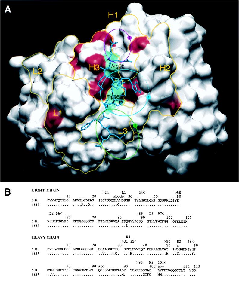
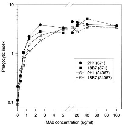
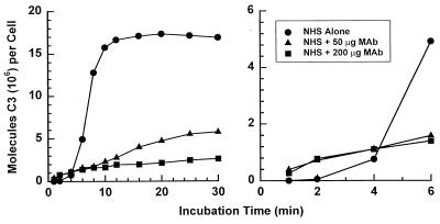
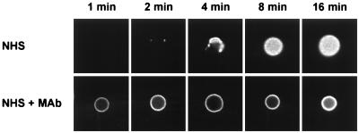
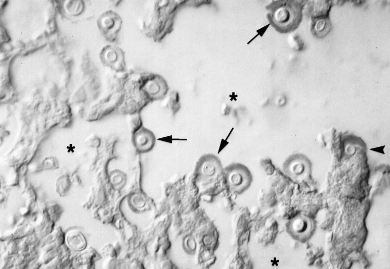
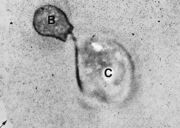
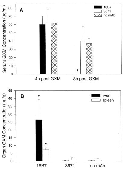
The murine monoclonal antibody (MAb) 18B7 [immunoglobulin G1(kappa)] is in preclinical development for treatment of Cryptococcus neoformans infections. In anticipation of its use in humans, we defined the serological and biological properties of MAb 18B7 in detail. Structural comparison to the related protective MAb 2H1 revealed conservation of the antigen binding site despite several amino acid differences. MAb 18B7 was shown by immunofluorescence and agglutination studies to bind to all four serotypes of C. neoformans, opsonize C. neoformans serotypes A and D, enhance human and mouse effector cell antifungal activity, and activate the complement pathway leading to deposition of complement component 3 (C3) on the cryptococcal capsule. Administration of MAb 18B7 to mice led to rapid clearance of serum cryptococcal antigen and deposition in the liver and spleen. Immunohistochemical studies revealed that MAb 18B7 bound to capsular glucuronoxylomannan in infected mouse tissues. No reactivity of MAb 18B7 with normal human, rat, or mouse tissues was detected. The results show that both the variable and constant regions of MAb 18B7 are biologically functional and support the use of this MAb in human therapeutic trials.


Figures

FIG. 1
FIG. 1
(A) Solvent-accessible surfaces of the MAb 2H1 binding site with the amino acid differences between MAbs 2H1 and 18B7 highlighted in red. L1, L2, and L3 refer to the three light chain CDRs. H1, H2, and H3 refer to the three heavy chain CDRs. Yellow lines denote the structural area associated with CDRs. The positions of arginine H95 in the VH CDR3 and tryptophan L96 in the VL CDR3 are shown in green. The position of the peptide mimotope PA1 is drawn in blue (50). The sequence of PA1 is GLQYTPSWMLVG. The blue circle labeled “1” denotes the location of the central hydrophobic pocket delimited by VH CDR1, CDR2, and CDR3 and VL CDR1 and -3. The magenta circle labeled “2” denotes the location of a small hydrophobic pocket containing arginine H95. The green circle labeled “3” denotes the location of a potential extension of the binding groove that is highly conserved in both MAbs 2H1 and 18B7. (B) Sequence comparison of heavy and light chain variable regions of MAbs 2H1 and 18B7 (numbering according to Kabat et al. [24]).
FIG. 2
FIG. 2
Spot ELISA for C. neoformans with MAb 18B7. (A) Graphic representation of the ELISA configuration. GAM, goat anti-mouse. (B and C) Light microscopy images of C. neoformans 24067 captured and detected by the assay. In this assay the C. neoformans cells stain blue. Magnification, ×200 (B) and ×400 (C). Bars, 20 μm.
FIG. 3
FIG. 3
Phagocytosis of C. neoformans 24067 and 371 by J774.16 cells in the presence and absence of MAbs 18B7 and 2H1. Each point is the average of three measurements.
FIG. 4
FIG. 4
Kinetics for activation and binding of C3 fragments to serotype A C. neoformans cells incubated with 40% NHS in the presence and absence of MAb 18B7. The binding of C3 fragments was determined by incorporation of trace amounts of 125I-labeled C3 into the reaction mixture.
FIG. 5
FIG. 5
Immunofluorescence analysis of the sites for binding of C3 to serotype A C. neoformans cells incubated for 1, 2, 4, 8, and 16 min with 40% NHS in the presence (50 μg/ml) or absence of MAb 18B7. Sites of C3 deposition were determined by the use of FITC-labeled antiserum to C3. All images were collected under identical conditions.
FIG. 6
FIG. 6
Immunohistochemistry for 18B7 by light microscopy shows antibody binding to yeast cell capsules in a mouse lung during experimental infection. Arrows point to several immunostained cryptococci. The asterisks denote air spaces. The arrowhead points to shed polysaccharide inside an alveolar macrophage. In this assay the polysaccharide stains red. Magnification, ×400.
FIG. 7
FIG. 7
Immunogold labeling shows that the 18B7 epitope is evenly distributed throughout the C. neoformans capsule in a mouse lung during experimental infection. C, cryptococcus cell body; B, bud. The arrow points to the edge of the capsule. No immunogold labeling was apparent in mouse tissues or alveolar spaces (not shown). Magnification, ×10,000.
FIG. 8
FIG. 8
Effect of MAb administration on serum GXM level and organ deposition of GXM. Mice were given 50 μg of strain 24067 GXM i.v. followed by one of the following: 1 mg of 18B7, 1 mg of 3671 (isotype-matched irrelevant MAb), or an equivalent volume of PBS. The bars denote the average GXM concentrations (n = 4). The error bars denote standard deviations. The asterisks denote P values of <0.05 compared with conditions where MAb 3671 or PBS was administered.

References

    1. Belay T, Cherniak R, Kozel T R, Casadevall A. Reactivity patterns and epitope specificities of anti-Cryptococcus neoformans monoclonal antibodies by enzyme-linked immunosorbent assay and dot enzyme assay. Infect Immun. 1997;65:718–728. - PMC - PubMed
    1. Casadevall A, DeShaw M, Fan M, Dromer F, Kozel T R, Pirofski L-A. Molecular and idiotypic analysis of antibodies to Cryptococcus neoformans glucuronoxylomannan. Infect Immun. 1994;62:3864–3872. - PMC - PubMed
    1. Casadevall A, Mukherjee J, Devi S J N, Schneerson R, Robbins J B, Scharff M D. Antibodies elicited by a Cryptococcus neoformans glucuronoxylomannan-tetanus toxoid conjugate vaccine have the same specificity as those elicited in infection. J Infect Dis. 1992;65:1086–1093. - PubMed
    1. Cherniak R, Morris L C, Belay T, Spitzer E D, Casadevall A. Variation in the structure of glucuronoxylomannan in isolates from patients with recurrent cryptococcal meningitis. Infect Immun. 1995;63:1899–1905. - PMC - PubMed
    1. Cherniak R, Reiss E, Turner S. A galactoxylomannan antigen of Cryptococcus neoformans serotype A. Carbohydr Res. 1982;103:239–250.

Publication types

MeSH terms

LinkOut - more resources