Temporal sequence of cell wall disassembly in rapidly ripening melon fruit
- PMID: 9625688
- PMCID: PMC34955
- DOI: 10.1104/pp.117.2.345
Temporal sequence of cell wall disassembly in rapidly ripening melon fruit
Abstract
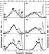
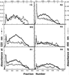
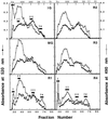
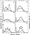
The Charentais variety of melon (Cucumis melo cv Reticulatus F1 Alpha) was observed to undergo very rapid ripening, with the transition from the preripe to overripe stage occurring within 24 to 48 h. During this time, the flesh first softened and then exhibited substantial disintegration, suggesting that Charentais may represent a useful model system to examine the temporal sequence of changes in cell wall composition that typically take place in softening fruit. The total amount of pectin in the cell wall showed little reduction during ripening but its solubility changed substantially. Initial changes in pectin solubility coincided with a loss of galactose from tightly bound pectins, but preceded the expression of polygalacturonase (PG) mRNAs, suggesting early, PG-independent modification of pectin structure. Depolymerization of polyuronides occurred predominantly in the later ripening stages, and after the appearance of PG mRNAs, suggesting the existence of PG-dependent pectin degradation in later stages. Depolymerization of hemicelluloses was observed throughout ripening, and degradation of a tightly bound xyloglucan fraction was detected at the early onset of softening. Thus, metabolism of xyloglucan that may be closely associated with cellulose microfibrils may contribute to the initial stages of fruit softening. A model is presented of the temporal sequence of cell wall changes during cell wall disassembly in ripening Charentais melon.
Figures



Similar articles
-
Polygalacturonase gene expression in ripe melon fruit supports a role for polygalacturonase in ripening-associated pectin disassembly.Plant Physiol. 1998 Jun;117(2):363-73. doi: 10.1104/pp.117.2.363. Plant Physiol. 1998. PMID: 9625689 Free PMC article.
-
Ethylene regulation of fruit softening and cell wall disassembly in Charentais melon.J Exp Bot. 2007;58(6):1281-90. doi: 10.1093/jxb/erl283. Epub 2007 Feb 17. J Exp Bot. 2007. PMID: 17308329
-
Cell wall metabolism during maturation, ripening and senescence of peach fruit.J Exp Bot. 2004 Sep;55(405):2029-39. doi: 10.1093/jxb/erh227. Epub 2004 Jul 30. J Exp Bot. 2004. PMID: 15286150
-
Fruit ripening phenomena--an overview.Crit Rev Food Sci Nutr. 2007;47(1):1-19. doi: 10.1080/10408390600976841. Crit Rev Food Sci Nutr. 2007. PMID: 17364693 Review.
-
Fruit Calcium: Transport and Physiology.Front Plant Sci. 2016 Apr 29;7:569. doi: 10.3389/fpls.2016.00569. eCollection 2016. Front Plant Sci. 2016. PMID: 27200042 Free PMC article. Review.
Cited by
-
Functional copy number variation of CsSHINE1 is associated with fruit skin netting intensity in cucumber, Cucumis sativus.Theor Appl Genet. 2022 Jun;135(6):2101-2119. doi: 10.1007/s00122-022-04100-4. Epub 2022 May 7. Theor Appl Genet. 2022. PMID: 35524817
-
Changes in cell wall polysaccharides of green bean pods during development.Plant Physiol. 1999 Oct;121(2):363-72. doi: 10.1104/pp.121.2.363. Plant Physiol. 1999. PMID: 10517827 Free PMC article.
-
Deciphering the genetic control of fruit texture in apple by multiple family-based analysis and genome-wide association.J Exp Bot. 2017 Mar 1;68(7):1451-1466. doi: 10.1093/jxb/erx017. J Exp Bot. 2017. PMID: 28338805 Free PMC article.
-
Fruit Characteristics, Peel Nutritional Compositions, and Their Relationships with Mango Peel Pectin Quality.Plants (Basel). 2021 Jun 4;10(6):1148. doi: 10.3390/plants10061148. Plants (Basel). 2021. PMID: 34200110 Free PMC article.
-
Quantitative Proteomics Analysis of Herbaceous Peony in Response to Paclobutrazol Inhibition of Lateral Branching.Int J Mol Sci. 2015 Oct 14;16(10):24332-52. doi: 10.3390/ijms161024332. Int J Mol Sci. 2015. PMID: 26473855 Free PMC article.
References
-
- Ahmed AER, Labavitch JM. A simplified method for accurate determination of cell wall uronide content. J Food Biochem. 1977;1:361–365.
-
- Blakeney AB, Harris PJ, Henry RJH, Stone BA. A simple and rapid preparation of alditol acetates for monosaccharide analysis. Carbohydr Res. 1983;11:3291–3299.
-
- Blumenkrantz N, Asboe-Hansen G. New method for quantitative determination of uronic acids. Anal Biochem. 1973;54:484–489. - PubMed
-
- Brady CJ. Fruit ripening. Annu Rev Plant Physiol. 1987;38:155–178.
-
- Brady CJ, Macalpine G, McGlasson WB, Ueda Y. Polygalacturonase in tomato fruits and the induction of ripening. Aust J Plant Physiol. 1982;9:171–178.
LinkOut - more resources
Full Text Sources
Other Literature Sources

