. 1998 Jun;117(2):687-93.
doi: 10.1104/pp.117.2.687.

Wound signaling in tomato plants. Evidence that aba is not a primary signal for defense gene activation

Affiliations

Wound signaling in tomato plants. Evidence that aba is not a primary signal for defense gene activation

GF Birkenmeier et al. Plant Physiol. 1998 Jun.

Abstract

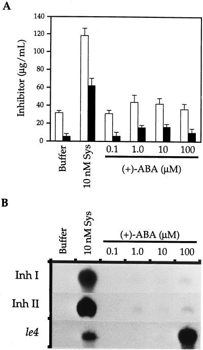
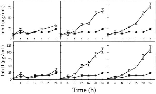
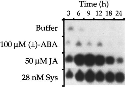
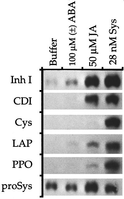
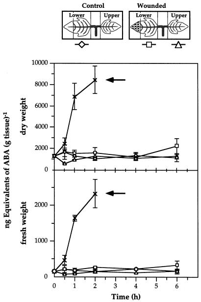
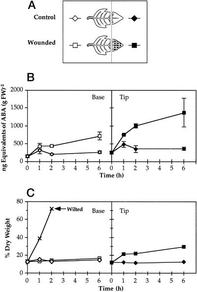
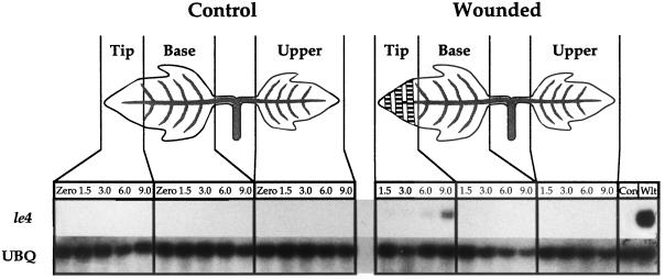
The effects of abscisic acid (ABA) on the accumulation of proteinase inhibitors I (Inh I) and II (Inh II) in young, excised tomato (Lycopersicon esculentum L.) plants were investigated. When supplied to excised plants through the cut stems, 100 &mgr;m ABA induced the activation of the ABA-responsive le4 gene. However, under the same conditions of assay, ABA at concentrations of up to 100 &mgr;m induced only low levels of proteinase-inhibitor proteins or mRNAs, compared with levels induced by systemin or jasmonic acid over the 24 h following treatment. In addition, ABA only weakly induced the accumulation of mRNAs of several other wound-response proteins. Assays of the ABA concentrations in leaves following wounding indicated that the ABA levels increased preferentially near the wound site, suggesting that ABA may have accumulated because of desiccation. The evidence suggests that ABA is not a component of the wound-inducible signal transduction pathway leading to defense gene activation but is likely involved in the general maintenance of a healthy plant physiology that facilitates a normal wound response.


Figures

Figure 1
Figure 1
The effects of systemin (Sys) and ABA on the accumulation of Inh I and II mRNAs and proteins in young tomato plants. Excised plants were continuously supplied with buffer or with buffer plus systemin or ABA as indicated. A, Expressed leaf juice was assayed for Inh I (white bars) and II (black bars) protein content by radial immunodiffusion after 24 h of treatment with systemin and ABA. The means ± se of six plants per treatment were plotted. B, After 6 h, leaves from six plants per treatment were frozen for mRNA analysis by northern blotting and hybridization with radiolabeled probes prepared from cDNAs encoding Inh I, Inh II, and le4.
Figure 2
Figure 2
Time course of Inh I and II protein accumulation in young tomato plants supplied with buffer (♦) or with buffer plus ABA (100 μm, ▵), JA (40 μm, □), or systemin (28 nm, ○). At 4-h intervals, leaf juice was expressed and assayed for proteinase inhibitors by radial immunodiffusion. Each data point represents the average of 14 plants ± se.
Figure 3
Figure 3
Time course of Inh II mRNA accumulation in young tomato plants in response to ABA, JA, and systemin (Sys). The plants were supplied with buffer or with buffer plus ABA, JA, or systemin as shown. At the indicated times, leaves from six plants per treatment were frozen for RNA extraction and assayed by northern blotting. Three independent experiments were performed and results from one representative experiment are shown.
Figure 4
Figure 4
Induction of genes encoding several swrps in young tomato plants in response to buffer or to buffer plus ABA, JA, or systemin (Sys). After 12 h, leaves from six plants per treatment were frozen for RNA extraction. mRNA detection was achieved by northern blotting, and hybridization was achieved with radiolabeled probes prepared from cDNAs encoding Inh I, cathepsin D inhibitor (CDI), Cys proteinase inhibitor (Cys), Leu amino peptidase (LAP), polyphenol oxidase (PPO), or prosystemin (proSys). Three independent experiments were performed, which were in full agreement. Results from one representative experiment are shown.
Figure 5
Figure 5
Time course of the accumulation of ABA in young tomato plants in response to wounding and wilting with respect to tissue dry (top) and fresh (bottom) weight. Leaves or leaf subsections were collected at the indicated times as illustrated in the sketch. At each interval, the leaves of four plants per treatment were individually assayed for ABA content by an indirect ELISA method and each assay was performed in triplicate. The means ± se are plotted. Wilting was performed by exposing excised lower leaves to air at 28°C and 80% RH. The hatch marks on the wounded leaflet represent the locations of hemostat-crushed tissue. Arrows indicate wilted leaves.
Figure 6
Figure 6
Time course of the accumulation of ABA and corresponding water loss in leaves of young tomato plants in response to wounding. A, The illustration shows the location of tissues tested with respect to the site of injury. The hatch marks on the wounded leaflet represent the locations of hemostat-crushed tissue. B, At each interval, leaves from six plants per treatment were assayed in pairs for ABA content by an indirect ELISA method, and each assay was performed in triplicate. The means ± se are plotted. C, Water loss from tissues used in B was monitored by plotting the dry weight percentage of fresh weight (FW) over time. Wilting was performed as described in Figure 5.
Figure 7
Figure 7
Time-course accumulation of le4 mRNA upon wounding. Six leaves per treatment were collected at the indicated times and divided into sections as illustrated. Sectioned tissue was frozen for RNA analysis by northern blotting and hybridization with radiolabeled probes prepared from cDNAs encoding le4 and ubiquitin (UBQ). Two independent experiments were performed and results from one representative experiment are shown. The hatch marks on the wounded leaflet represent the locations of hemostat-crushed tissue. Con, Leaves of intact, untreated plants; Wlt, excised leaves collected 4 h after being allowed to wilt to 88% of their original fresh weight.

Similar articles

Cited by

References

    1. Bergey DR, Howe GA, Ryan CA. Polypeptide signaling for plant defensive genes exhibits analogies to defense signaling in animals. Proc Natl Acad Sci USA. 1996;93:12053–12058. - PMC - PubMed
    1. Bishop PD, Pearce G, Bryant JE, Ryan CA. Isolation and characterization of the proteinase inhibitor-inducing factor from tomato leaves: identity and activity of poly- and oligogalacturonide fragments. J Biol Chem. 1984;259:13172–13176. - PubMed
    1. Cohen A, Bray EA. Characterization of three mRNAs that accumulate in wilted tomato leaves in response to elevated levels of endogenous abscisic acid. Planta. 1990;182:27–33. - PubMed
    1. Conconi A, Miquel M, Browse JA, Ryan CA. Intracellular levels of free linolenic and linoleic acids increase in tomato leaves in response to wounding. Plant Physiol. 1996;111:797–803. - PMC - PubMed
    1. Constabel CP, Bergey DR, Ryan CA. Systemin activates synthesis of wound-inducible tomato leaf polyphenol oxidase via the octadecanoid defense signaling pathway. Proc Natl Acad Sci USA. 1995;92:407–411. - PMC - PubMed

LinkOut - more resources