. 1998 Jun 15;187(12):1965-75.
doi: 10.1084/jem.187.12.1965.

Identification of podocalyxin-like protein as a high endothelial venule ligand for L-selectin: parallels to CD34

Affiliations

Identification of podocalyxin-like protein as a high endothelial venule ligand for L-selectin: parallels to CD34

C Sassetti et al. J Exp Med. .

Abstract

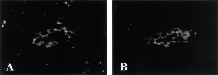
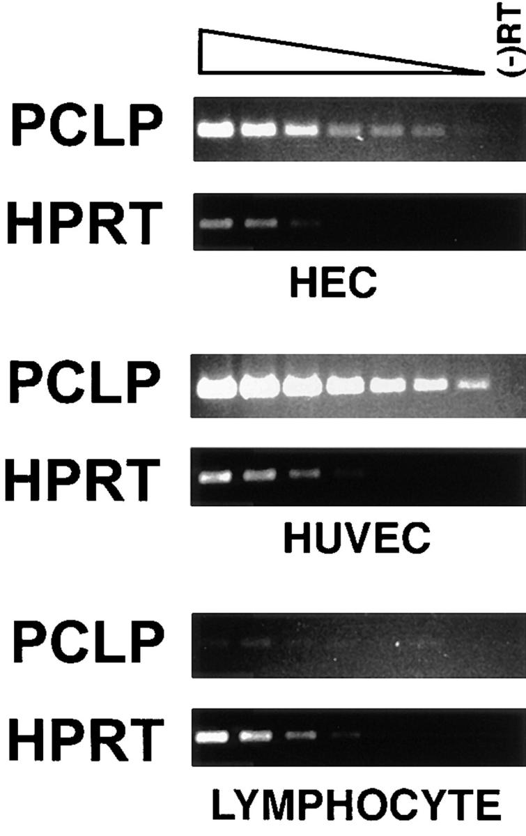
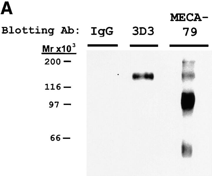
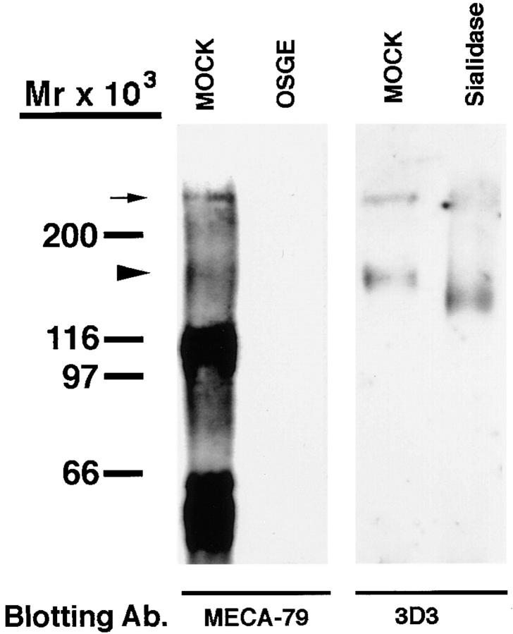
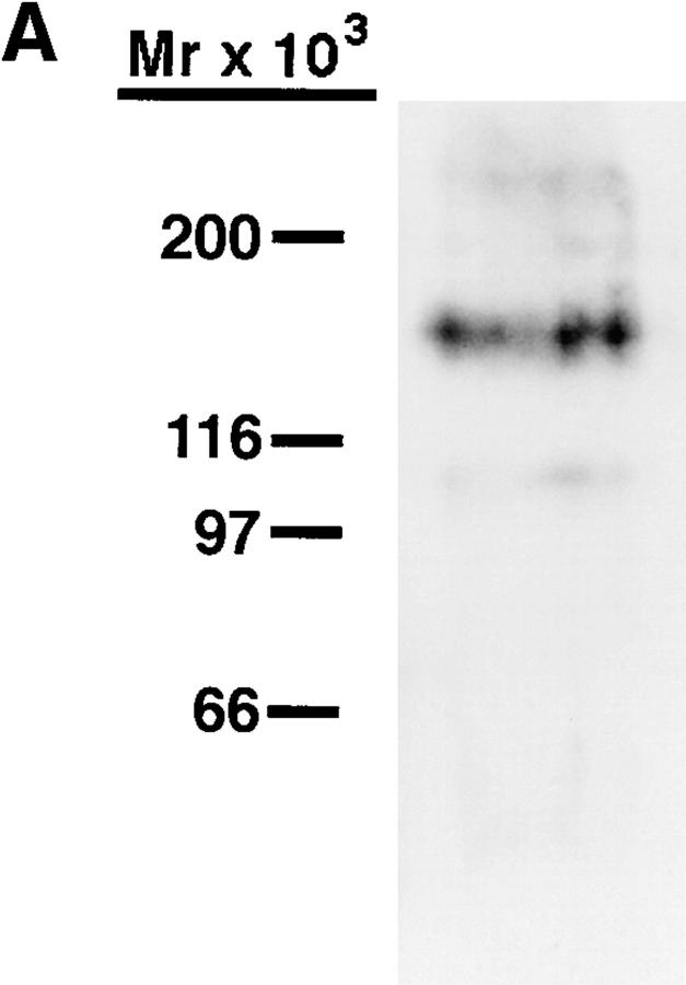
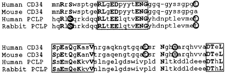
The leukocyte adhesion molecule, L-selectin, mediates the recruitment of lymphocytes to secondary lymphoid organs via interactions with specific ligands presented on high endothelial venules (HEV). Although the HEV-derived ligands for L-selectin are still incompletely defined, they share a common sialomucin-like structure which is thought to present clustered oligosaccharides to the lectin domain of L-selectin. Podocalyxin-like protein (PCLP) is a transmembrane sialomucin that is similar in structure to the well-characterized L-selectin ligand CD34. PCLP has been shown previously to be expressed on the foot processes of podocytes in the kidney glomerulus as well as on vascular endothelium at some sites. We have determined that PCLP is present on HEV, where it binds to both recombinant L-selectin and the HEV-specific monoclonal antibody MECA-79. Furthermore, purified HEV-derived PCLP is able to support the tethering and rolling of lymphocytes under physiological flow conditions in vitro. These results suggest a novel function for PCLP as an adhesion molecule and allow the definition of conserved structural features in PCLP and CD34, which may be important for L-selectin ligand function.


Figures

Figure 1
Figure 1
Coexpression of MECA-79 and PCLP in HEV of tonsil. Frozen sections were stained simultaneously with the PCLP mAb 4F10 (A) and MECA-79 (B). A and B show the same HEV viewed with an FITC filter (A) or a Texas red filter (B).
Figure 2
Figure 2
Expression of PCLP in secondary lymphoid organs. Frozen sections of tonsil and associated connective tissue (A and D) and paraffin-embedded sections of tonsil (E and F), appendix (B), and lymph node (C) were stained with the anti-PCLP mAb 2A4. Arrows, HEV. Arrowheads, PCLP-expressing capillaries. Bars, 200 μm (A), 100 μm (B, D, E, and F), and 50 μm (C). Data are representative of two different samples of tonsil and lymph node, and three samples of appendix.
Figure 3
Figure 3
Detection of PCLP mRNA by PCR. Fragments of the PCLP and HPRT sequences were amplified by PCR from serial dilutions of cDNA from purified HEC, tonsillar lymphocytes, and HUVEC. (-)RT, PCR reactions using samples from which the reverse transcriptase was omitted. Note that a faint PCLP band is barely visible in the lowest dilution of lymphocyte cDNA.
Figure 4
Figure 4
Western blotting and immunoprecipitation of PNAd with PCLP mAb. (A) PNAd was electrophoresed on 7.5% SDS gels and blotted with either MECA-79, the PCLP mAb 3D3, or control mouse IgG. (B) Samples of PNAd were immunoprecipitated (I.P.) with protein A–Sepharose loaded with either 3D3 (PCLP mAb) or control mouse IgG. The unbound supernatant (SUP) as well as the fraction bound to the antibody (PEL) were analyzed by MECA-79 Western blotting after 7.5% SDS-PAGE.
Figure 4
Figure 4
Western blotting and immunoprecipitation of PNAd with PCLP mAb. (A) PNAd was electrophoresed on 7.5% SDS gels and blotted with either MECA-79, the PCLP mAb 3D3, or control mouse IgG. (B) Samples of PNAd were immunoprecipitated (I.P.) with protein A–Sepharose loaded with either 3D3 (PCLP mAb) or control mouse IgG. The unbound supernatant (SUP) as well as the fraction bound to the antibody (PEL) were analyzed by MECA-79 Western blotting after 7.5% SDS-PAGE.
Figure 5
Figure 5
Binding of PNAd to L-selectin. Samples of PNAd were precipitated with protein A beads loaded with either L-selectin–IgG (L-IgG) or with CD4/ IgG chimeras (CD4-IgG). The unbound supernatants (SUP) and EDTA elutions of bound material (EDTA) were electrophoresed on a 7.5% SDS gel and Western blotted with MECA-79. Arrowhead, The 160-kD band representing PCLP.
Figure 6
Figure 6
OSGE and sialidase digestion of PCLP. PNAd was either treated with OSGE and Western blotted with MECA-79, or treated with sialidase and blotted with the PCLP mAb 3D3. Mock treatments were identical except the enzyme was omitted. Arrowhead, The 160-kD PCLP band. Small arrow, The presumed PCLP multimer.
Figure 7
Figure 7
Tethering of lymphocytes to purified PCLP under flow. (A) MECA-79 Western blotting of the purified PCLP, which was used in the flow chamber experiments, showing a major band at 160 kD. (B) Peripheral blood T lymphocytes or Jurkat T lymphoma cells were infused into the flow chamber at a constant wall shear stress, and the cell tethering rate to the PCLP-coated surface was determined. (C) Immobilized PCLP was treated with OSGE, sialidase, MECA-79, or PCLP mAb (3D3) before infusion of Jurkat cells. Jurkat cells were treated with L-selectin mAb (LAM1-3) or fucoidin before infusion. Error bars represent SD of measurements from at least three separate fields of view.
Figure 7
Figure 7
Tethering of lymphocytes to purified PCLP under flow. (A) MECA-79 Western blotting of the purified PCLP, which was used in the flow chamber experiments, showing a major band at 160 kD. (B) Peripheral blood T lymphocytes or Jurkat T lymphoma cells were infused into the flow chamber at a constant wall shear stress, and the cell tethering rate to the PCLP-coated surface was determined. (C) Immobilized PCLP was treated with OSGE, sialidase, MECA-79, or PCLP mAb (3D3) before infusion of Jurkat cells. Jurkat cells were treated with L-selectin mAb (LAM1-3) or fucoidin before infusion. Error bars represent SD of measurements from at least three separate fields of view.
Figure 8
Figure 8
Alignment of PCLP and CD34 cytoplasmic tails. Regions depicted encompass residues 281–354 of human CD34, 309–382 of mouse CD34, 453–528 of human PCLP, and 477–551 of rabbit PCLP, and begin immediately after the predicted transmembrane domain of each protein. Regions of >50% homology are boxed. Potential protein kinase C (S/T-X-R/K) and casein kinase II (S/T-XX-D/E) phosphorylation sites are circled.

References

    1. Gowans JL, Knight EJ. The route of recirculation of lymphocytes in the rat. Proc R Soc Lond Ser B. 1964;159:257–282. - PubMed
    1. Butcher EC, Picker LJ. Lymphocyte homing and homeostasis. Science. 1996;272:60–66. - PubMed
    1. Girard J-P, Springer TA. High endothelial venules (HEV): specialized endothelium for lymphocyte migration. Immunol Today. 1995;16:449–457. - PubMed
    1. Kikuta A, Rosen SD. Localization of ligands for L-selectin in mouse peripheral lymph node high endothelial cells by colloidal gold conjugates. Blood. 1994;84:3766–3775. - PubMed
    1. Lawrence MB, Berg EL, Butcher EC, Springer TA. Rolling of lymphocytes and neutrophils on peripheral node addressin and subsequent arrest on ICAM-1 in shear flow. Eur J Immunol. 1995;25:1025–1031. - PubMed

Publication types

MeSH terms