. 1998 Jun 15;187(12):1985-93.
doi: 10.1084/jem.187.12.1985.

The role of lymphocyte subsets in accelerated diabetes in nonobese diabetic-rat insulin promoter-B7-1 (NOD-RIP-B7-1) mice

Affiliations

The role of lymphocyte subsets in accelerated diabetes in nonobese diabetic-rat insulin promoter-B7-1 (NOD-RIP-B7-1) mice

F S Wong et al. J Exp Med. .

Abstract

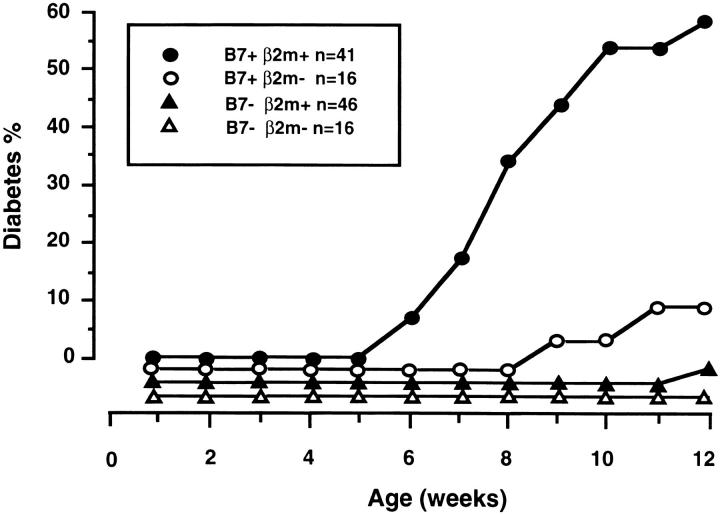
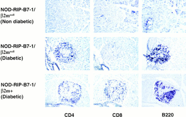
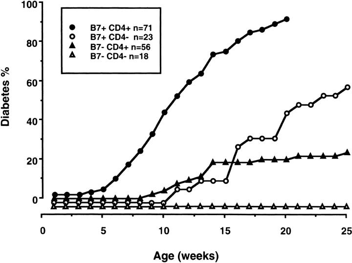
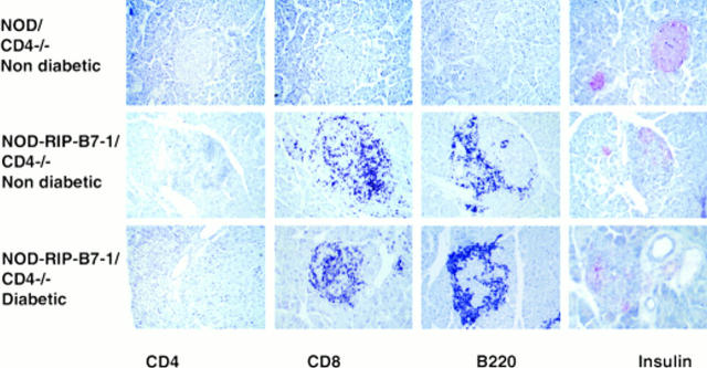
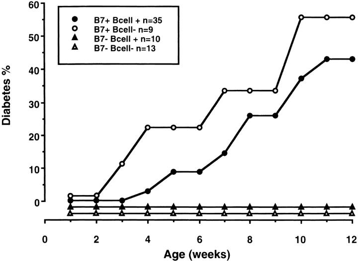
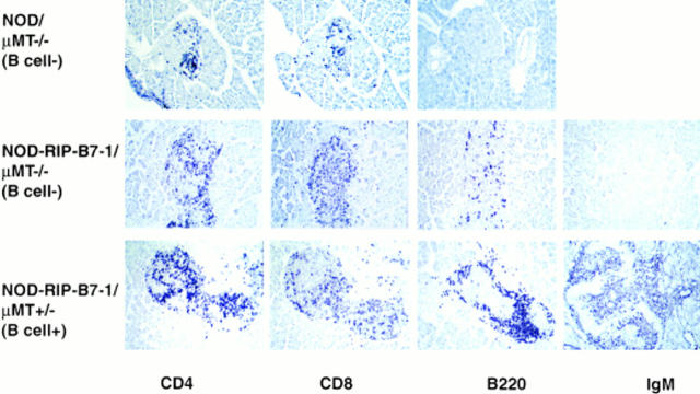
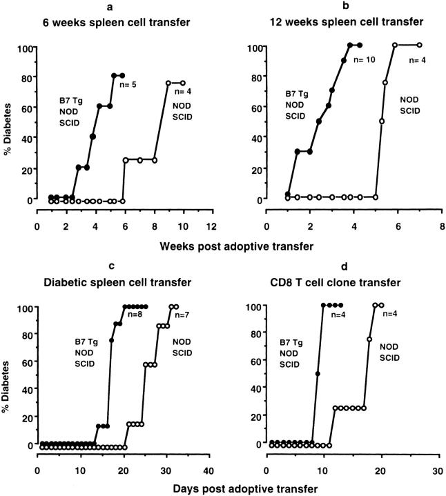
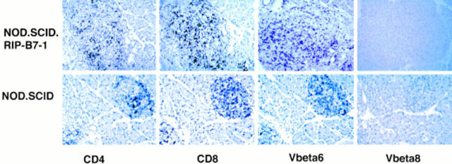
B7-1 transgene expression on the pancreatic islets in nonobese diabetic (NOD) mice leads to accelerated diabetes, with >50% of animals developing diabetes before 12 wk of age. The expression of B7-1 directly on the pancreatic beta cells, which do not normally express costimulator molecules, converts the cells into effective antigen-presenting cells leading to an intensified autoimmune attack. The pancreatic islet infiltrate in diabetic mice consists of CD8 T cells, CD4 T cells, and B cells, similar to diabetic nontransgenic NOD mice. To elucidate the relative importance of each of the subsets of cells, the NOD-rat insulin promoter (RIP)-B7-1 animals were crossed with NOD.beta2microglobulin -/- mice which lack major histocompatibility complex class I molecules and are deficient in peripheral CD8 T cells, NOD.CD4 -/- mice which lack T cells expressing CD4, and NOD.muMT -/- mice which lack B220-positive B cells. These experiments showed that both CD4 and CD8 T cells were necessary for the accelerated onset of diabetes, but that B cells, which are needed for diabetes to occur in normal NOD mice, are not required. It is possible that B lymphocytes play an important role in the provision of costimulation in NOD mice which is unnecessary in the NOD-RIP-B7-1 transgenic mice.


Figures

Figure 1
Figure 1
Mice were observed and screened for glycosuria. All mice which had glycosuria had a blood glucose measurement taken using One Touch test strips (LifeScan, Inc., Milpitas, CA), and diabetes was diagnosed if the blood glucose was >250 mg/dl. The graph shows the percentage of diabetes in mice that were RIP-B7-1 transgene positive and β2m sufficient (filled circles), n = 41; RIP-B7-1 transgene positive and β2m deficient (open circles), n = 16; RIP-B7-1 transgene negative and β2m sufficient (filled triangles), n = 46; and RIP-B7-1 transgene negative and β2m deficient (open triangles), n = 16. The numbers indicate the total population of both male and female mice.
Figure 2
Figure 2
Pancreatic section from an NOD-RIP-B7-1/β2mnull mouse that was nondiabetic is shown (top), illustrating staining with anti-CD4, anti-CD8, and anti-B220 antibodies at 24 wk, and showing that when diabetes did not occur, there was no insulitis. (Middle) Staining from one of the two NOD-RIP-B7-1/β2mnull mice that became diabetic, showing the presence of CD4 T cells and B cells. (Bottom) Staining of a pancreatic section from a diabetic NOD-RIP-B7-1/β2m-sufficient mouse.
Figure 3
Figure 3
The graph shows the percentage of diabetes in mice that were RIP-B7-1 transgene positive and CD4 sufficient (filled circles), n = 71; RIP-B7-1 transgene positive and CD4 deficient (open circles) n = 23; RIP-B7-1 transgene negative and CD4 sufficient (filled triangles), n = 56; and RIP-B7-1 transgene negative and CD4 deficient (open triangles), n = 18. The numbers indicate the total population of both male and female mice.
Figure 4
Figure 4
(Top) Pancreatic sections from a non–RIP-B7-1 transgenic mouse that was CD4 deficient. All these mice were nondiabetic, and there was no insulitis seen, as illustrated by staining with anti-CD4, anti-CD8, and anti-B220 antibodies at 24 wk. The islets are intact, as shown by staining with insulin (red). (Middle) Staining from an NOD-RIP-B7-1/CD4 −/− mouse that did not become diabetic. (Bottom) Staining of a pancreatic section from a diabetic NOD-RIP-B7-1/CD4–sufficient mouse.
Figure 5
Figure 5
The graph shows the percentage of diabetes in mice that were RIP-B7-1 transgene positive and B cell sufficient (filled circles), n = 35; RIP-B7-1 transgene positive and B cell deficient (open circles), n = 9; RIP-B7-1 transgene negative and B cell sufficient (filled triangles), n = 10; and RIP-B7-1 transgene negative and B cell deficient (open triangles), n = 13. The numbers indicate the total population of both male and female mice.
Figure 6
Figure 6
(Top) Pancreatic sections from a nontransgenic mouse that was B cell deficient taken at 30 wk. Mild insulitis is seen, as illustrated by staining with anti-CD4, anti-CD8, and anti-B220 antibodies. (Middle) Staining from an NOD-RIP-B7-1/μMT −/− mouse that became diabetic, showing intense insulitis. There are a few B220-positive cells seen, but these are negative for staining with anti-Ig antibody and are likely to be T cells. (Bottom) Staining of a pancreatic section from a diabetic NOD-RIP-B7-1, B cell-sufficient mouse.
Figure 7
Figure 7
Incidence of diabetes after adoptive transfer of 6-wk-old NOD spleen cells (a), 12-wk-old NOD spleen cells (b), diabetic spleen cells (c), and CD8 cloned T cells (d) into NOD.SCID-RIP-B7-1 mice (filled circles) and NOD.SCID mice (open circles).
Figure 8
Figure 8
Immunohistochemistry showing staining with anti-CD4, anti-CD8, anti-Vβ6, and anti-Vβ8 in NOD.SCID-RIP-B7-1 mice (top) and NOD.SCID mice (bottom) that have become diabetic after adoptive transfer of spleen cells from 12-wk-old NOD mice.

Similar articles

Cited by

References

    1. Miller BJ, Appel MC, O'Neil JJ, Wicker LS. Both the Lyt-2+ and L3T4+ T cell subsets are required for the transfer of diabetes in nonobese diabetic mice. J Immunol. 1988;140:52–58. - PubMed
    1. Bendelac A, Carnaud C, Boitard C, Bach J-F. Syngeneic transfer of autoimmune diabetes from diabetic NOD mice to healthy neonates: requirement for both L3T4+ and Lyt2+ T cells. J Exp Med. 1987;166:823–832. - PMC - PubMed
    1. Serreze DV, Leiter EH, Christianson GJ, Greiner D, Roopenian DC. Major histocompatibility complex class I-deficient NOD-B2mnull mice are diabetes and insulitis resistant. Diabetes. 1994;43:505–509. - PubMed
    1. Wicker LS, Leiter EH, Todd JA, Renjilian RJ, Peterson E, Fischer PA, Podolin PL, Zijlstra M, Jaenisch R, Peterson LB. Beta 2-microglobulin-deficient NOD mice do not develop insulitis or diabetes. Diabetes. 1994;43:500–504. - PubMed
    1. Katz J, Benoist C, Mathis D. Major histocompatibility complex class I molecules are required for the development of insulitis in non-obese diabetic mice. Eur J Immunol. 1993;23:3358–3360. - PubMed

Publication types

MeSH terms