. 1998 Jun 15;187(12):2037-44.
doi: 10.1084/jem.187.12.2037.

Extrathymic T cell deletion and allogeneic stem cell engraftment induced with costimulatory blockade is followed by central T cell tolerance

Affiliations

Extrathymic T cell deletion and allogeneic stem cell engraftment induced with costimulatory blockade is followed by central T cell tolerance

T Wekerle et al. J Exp Med. .

Abstract

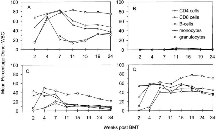
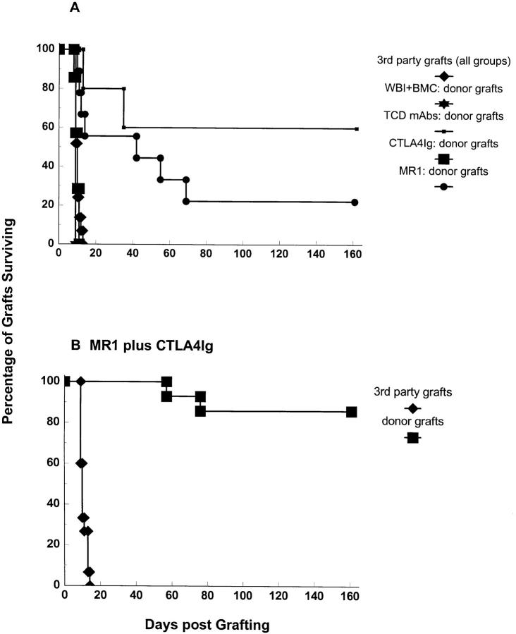
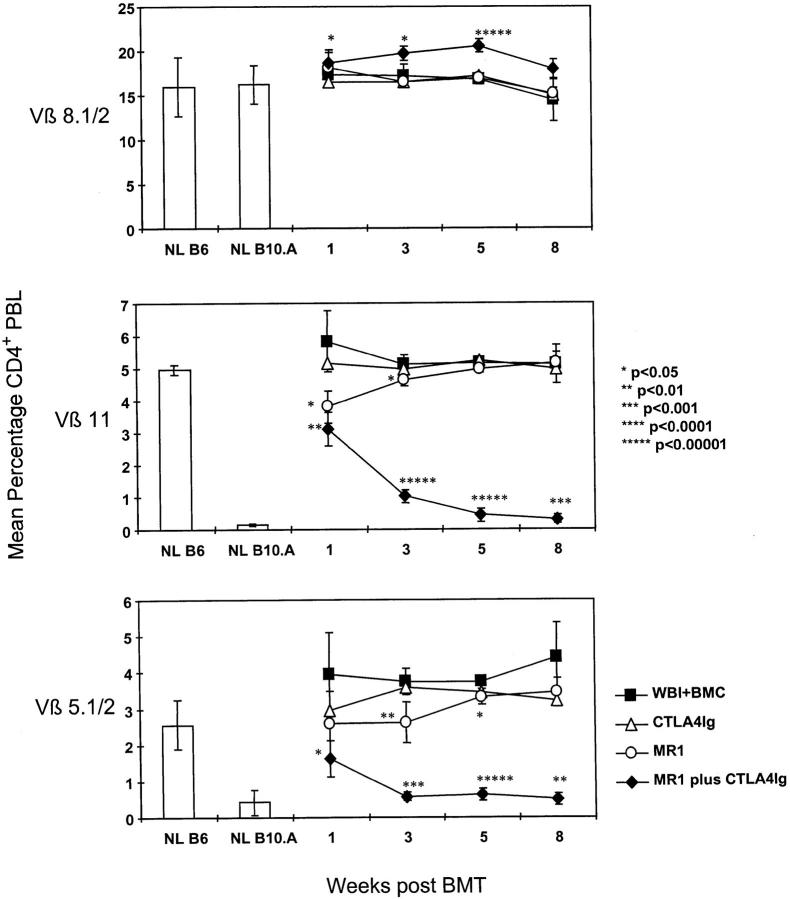
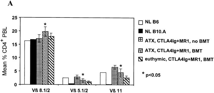
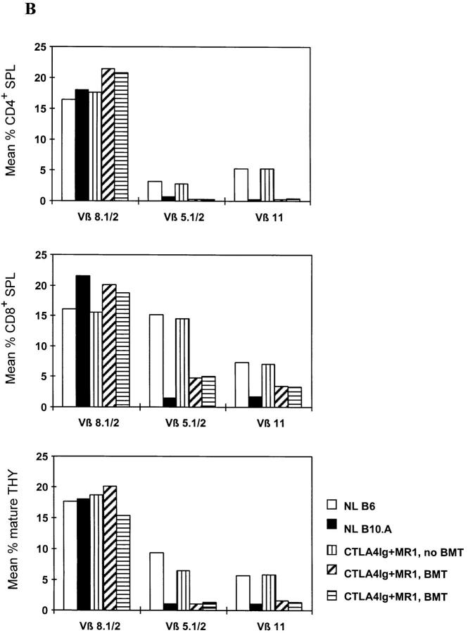
A reliable, nontoxic method of inducing transplantation tolerance is needed to overcome the problems of chronic organ graft rejection and immunosuppression-related toxicity. Treatment of mice with single injections of an anti-CD40 ligand antibody and CTLA4Ig, a low dose (3 Gy) of whole body irradiation, plus fully major histocompatibility complex-mismatched allogeneic bone marrow transplantation (BMT) reliably induced high levels (>40%) of stable (>8 mo) multilineage donor hematopoiesis. Chimeric mice permanently accepted donor skin grafts (>100 d), and rapidly rejected third party grafts. Progressive deletion of donor-reactive host T cells occurred among peripheral CD4(+) lymphocytes, beginning as early as 1 wk after bone marrow transplantation. Early deletion of peripheral donor-reactive host CD4 cells also occurred in thymectomized, similarly treated marrow recipients, demonstrating a role for peripheral clonal deletion of donor-reactive T cells after allogeneic BMT in the presence of costimulatory blockade. Central intrathymic deletion of newly developing T cells ensued after donor stem cell engraftment had occurred. Thus, we have shown that high levels of chimerism and systemic T cell tolerance can be reliably achieved without myeloablation or T cell depletion of the host. Chronic immunosuppression and rejection are avoided with this powerful, nontoxic approach to inducing tolerance.


Figures

Figure 1
Figure 1
High levels of multilineage donor chimerism in peripheral blood for 34 wk after BMT. Results from one of two similar experiments are shown as group averages. All animals received 3 Gy WBI and 1.5 × 10⁷ allogeneic BM cells on day 0. Only MR1 plus CTLA4Ig together (D) allowed the reliable induction of stable chimerism (n = 5), with high levels of donor cells in all lineages throughout the follow-up. Administration of MR1 alone (C) led to significant levels of chimerism, but chimerism declined over time (n = 5). When CTLA4Ig was given alone (B), no chimerism was detectable by FCM (n = 4). A control group (n = 5), receiving depleting doses of anti-CD4 and anti-CD8 mAbs on day −5 and day −1 (A), showed substantial levels of donor chimerism, with long-term levels of T cell chimerism being significantly lower than B cell, granulocyte, and monocyte chimerism.
Figure 2
Figure 2
Permanent survival of donor-specific skin grafts in chimeras prepared with 3 Gy WBI and allogeneic (B10.A) BM cells and treatment with MR1 plus CTLA4Ig. Combined results from two experiments are shown. Recipients were grafted with donor-specific (B10.A) and third party (A.SW) skin grafts at 3, 6, or 10 wk after BMT. Mice receiving the full treatment of BMT and MR1 plus CTLA4Ig accepted donor skin grafts (B) permanently (12 out of 14), with the exception of 2 animals that rejected their grafts at days 57 and 76, respectively. 9 grafts have been accepted in perfect condition for >110 d, and 5 grafts for >140 d. Third-party skin grafts (B) were rejected in the expected time frame (MST = 10 d). MR1 alone (A) led to prolongation of donor-specific skin graft survival (MST = 42 d), but only 2 out of 9 grafts survived >100 d. CTLA4Ig alone (A) failed to improve skin graft survival (n = 7, MST = 10 d). Control mice treated with 3 Gy WBI plus BMC (n = 4) and mice receiving 3 Gy WBI and MR1 plus CTLA4Ig alone (without BMT, data not shown) rejected donor skin within 2 wk. In a control group prepared with T cell–depleting mAbs on day −5 and day −1 plus BMT (plus 3 Gy WBI) (n = 5), donor skin grafts were permanently accepted in 60% of mice. Third party grafts were rejected within 2 wk in all groups.
Figure 3
Figure 3
Specific deletion of donor-reactive peripheral T cells in recipients of BMT and MR1 plus CTLA4Ig. Results from one of two similar experiments are shown. FCM was performed at indicated time points, with the percentage of Vβ-positive cells being determined among gated CD4+ PBLs. The mean percentage of CD4+ lymphocytes expressing Vβ5.1/2 or Vβ11 was significantly lower in mice receiving BM cells (BMC) (plus 3 Gy WBI) with MR1 plus CTLA4Ig (n = 10) than in recipients of BMC (plus 3 Gy WBI) alone (n = 4), as early as 1 wk after BMT (P <0.01 for Vβ11, P <0.05 for Vβ5). The specific deletion gradually became more complete at 3, 5, and 8 wk after BMT and was sustained for the length of follow-up. The percentage of Vβ8.1/2+ CD4 cells remained similar in all groups, demonstrating specificity of the Vβ5.1/2 or Vβ11 deletion in mixed chimeras. Mice receiving BMC (plus 3 Gy WBI) alone or in addition to CTLA4Ig (n = 4) did not show any deletion, nor did control mice treated with MR1 plus CTLA4Ig alone (without BMC; data not shown). MR1 alone led only to a slight and transient deletion in this experiment (n = 5). Error bars indicate standard deviation. P values are shown for comparison with the control group receiving 3 Gy WBI plus BMC. NL B6 denotes naive C57BL/6 control; NL B10.A denotes naive B10.A control.
Figure 4
Figure 4
Extrathymic clonal deletion after BMT and costimulatory blockade with CTLA4Ig and MR1. (A) ATX recipients (n = 6) showed specific, partial deletion of Vβ5.1/2+ and Vβ11+ CD4 cells in PBLs, 1 wk after BMT plus CTLA4Ig and MR1 and 3 Gy WBI. A similar degree of deletion was observed in euthymic controls (n = 5) prepared with the same conditioning. CD4 cells in ATX controls receiving CTLA4Ig plus MR1 and WBI without BMT (n = 4) did not show deletion of these Vβ. The percentage of Vβ+ cells was determined by FCM analysis of gated CD4+ PBLs. P values are shown for comparison between ATX BMT recipients receiving CTLA4Ig plus MR1 and ATX non-BMT controls. (B) In two euthymic chimeras killed 20 wk after BMT (under cover of 3 Gy WBI, plus treatment with CTLA4Ig plus MR1), Vβ5.1/2+ and Vβ11+CD4+ splenocytes (SPL) were deleted to the same extent as in naive B10.A controls (top). In contrast, the percentage of Vβ5.1/2+ and Vβ11+CD8+ splenocytes was reduced compared to naive B6 mice, but was substantially higher than in naive B10.A mice (middle). Mature Vβ5.1/2+ and Vβ11+ thymocytes (THY) showed deletion comparable to B10.A at the same time (bottom). A control mouse receiving WBI and CTLA4Ig plus MR1 (but no BMT) showed no deletion in either splenocytes or thymocytes. The percentage of Vβ+ cells was determined by FCM analysis in gated CD4+ (or CD8+) 34-2-12–negative splenocytes, and in gated KH-95high (i.e., Db high)-thymocytes (or Dd high in the case of BIO.A controls; see Materials and Methods). NL B6 denotes naive C57BL/6 control; NL B10.A denotes naive B10.A control.
Figure 4
Figure 4
Extrathymic clonal deletion after BMT and costimulatory blockade with CTLA4Ig and MR1. (A) ATX recipients (n = 6) showed specific, partial deletion of Vβ5.1/2+ and Vβ11+ CD4 cells in PBLs, 1 wk after BMT plus CTLA4Ig and MR1 and 3 Gy WBI. A similar degree of deletion was observed in euthymic controls (n = 5) prepared with the same conditioning. CD4 cells in ATX controls receiving CTLA4Ig plus MR1 and WBI without BMT (n = 4) did not show deletion of these Vβ. The percentage of Vβ+ cells was determined by FCM analysis of gated CD4+ PBLs. P values are shown for comparison between ATX BMT recipients receiving CTLA4Ig plus MR1 and ATX non-BMT controls. (B) In two euthymic chimeras killed 20 wk after BMT (under cover of 3 Gy WBI, plus treatment with CTLA4Ig plus MR1), Vβ5.1/2+ and Vβ11+CD4+ splenocytes (SPL) were deleted to the same extent as in naive B10.A controls (top). In contrast, the percentage of Vβ5.1/2+ and Vβ11+CD8+ splenocytes was reduced compared to naive B6 mice, but was substantially higher than in naive B10.A mice (middle). Mature Vβ5.1/2+ and Vβ11+ thymocytes (THY) showed deletion comparable to B10.A at the same time (bottom). A control mouse receiving WBI and CTLA4Ig plus MR1 (but no BMT) showed no deletion in either splenocytes or thymocytes. The percentage of Vβ+ cells was determined by FCM analysis in gated CD4+ (or CD8+) 34-2-12–negative splenocytes, and in gated KH-95high (i.e., Db high)-thymocytes (or Dd high in the case of BIO.A controls; see Materials and Methods). NL B6 denotes naive C57BL/6 control; NL B10.A denotes naive B10.A control.

References

    1. Gjertson, D.W. 1991. Survival trends in long-term first cadaver-donor kidney transplants. In Clinical Transplants. P.I. Terasaki and J.M. Cecka, editors. UCLA Tissue Typing Laboratory, Los Angeles. 225–235. - PubMed
    1. Linsley PS, Ledbetter JA. The role of the CD28 receptor during T cell responses to antigen. Annu Rev Immunol. 1993;11:191–212. - PubMed
    1. Sayegh MH, Akalin E, Hancock WW, Russell ME, Carpenter CB, Linsley PS, Turka LA. CD28-B7 blockade after alloantigenic challenge in vivo inhibits Th1 cytokines but spares Th2. J Exp Med. 1995;181:1869–1874. - PMC - PubMed
    1. Foy TM, Aruffo A, Bajorath J, Buhlmann JE, Noelle RJ. Immune regulation by CD40 and its ligand gp39. Annu Rev Immunol. 1996;14:591–617. - PubMed
    1. Hancock WW, Sayegh MH, Peach R, Linsley PS, Turka LA. Costimulatory function of CD40L, CD80 and CD86 in vascularized murine cardiac allograft rejection. Proc Natl Acad Sci USA. 1996;93:13967–13972. - PMC - PubMed

Publication types