Comparative Study
. 1998 Jun 15;187(12):2045-53.
doi: 10.1084/jem.187.12.2045.

Dermal mast cells determine susceptibility to ultraviolet B-induced systemic suppression of contact hypersensitivity responses in mice

Affiliations
Comparative Study

Dermal mast cells determine susceptibility to ultraviolet B-induced systemic suppression of contact hypersensitivity responses in mice

P H Hart et al. J Exp Med. .

Abstract

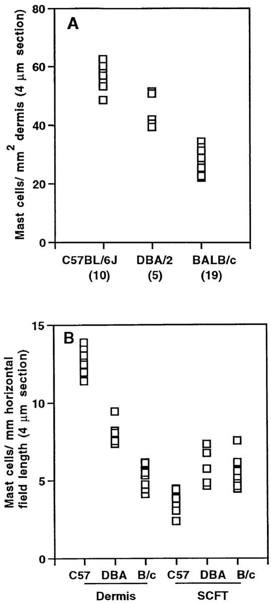
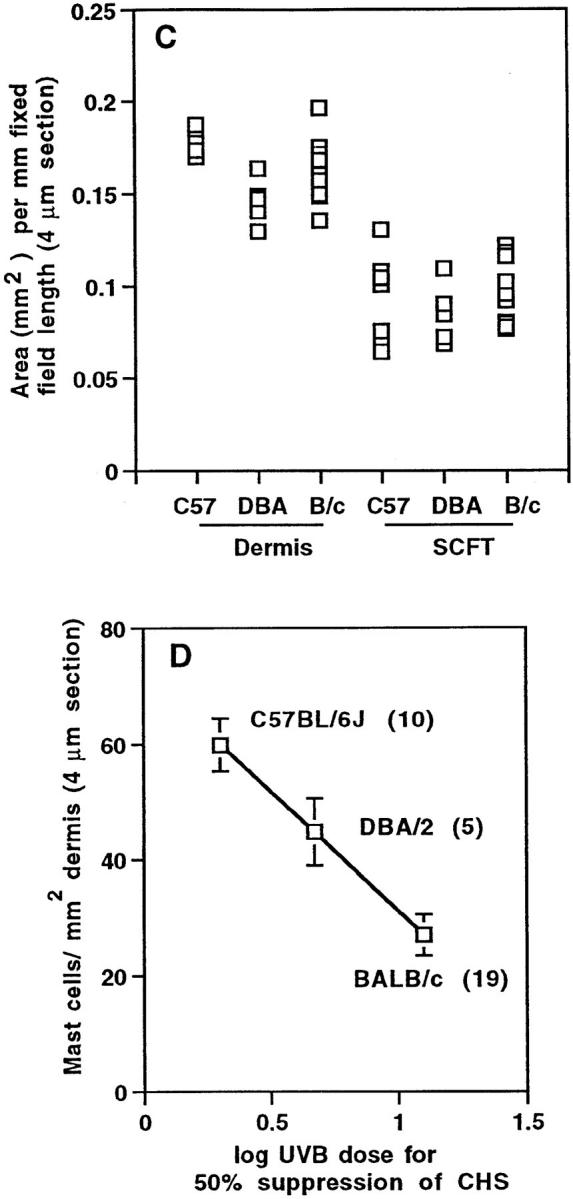
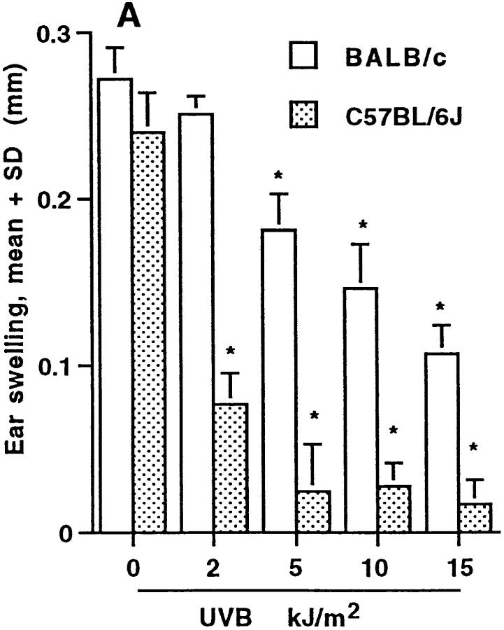
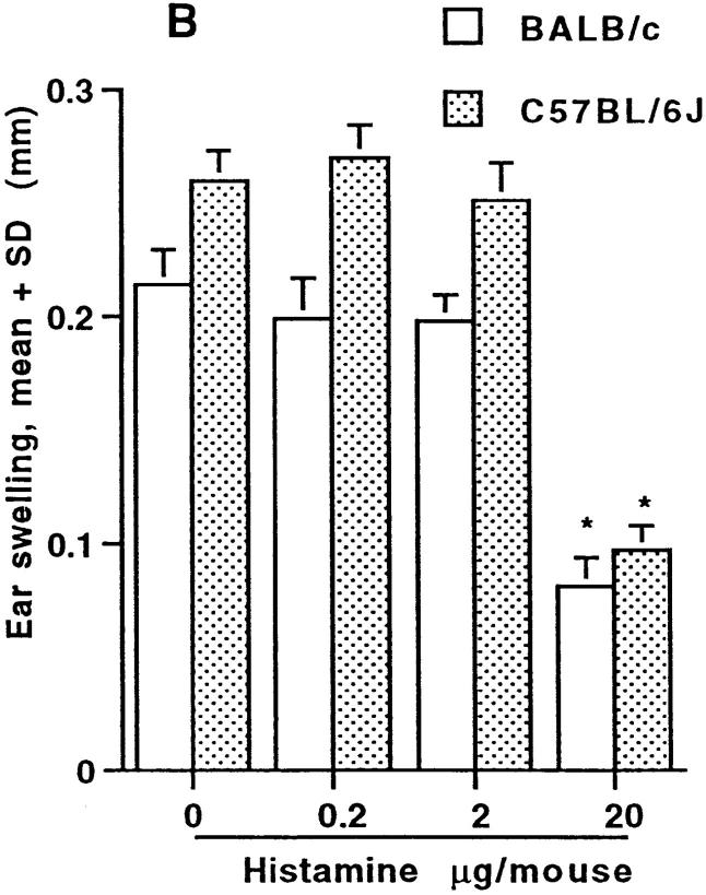
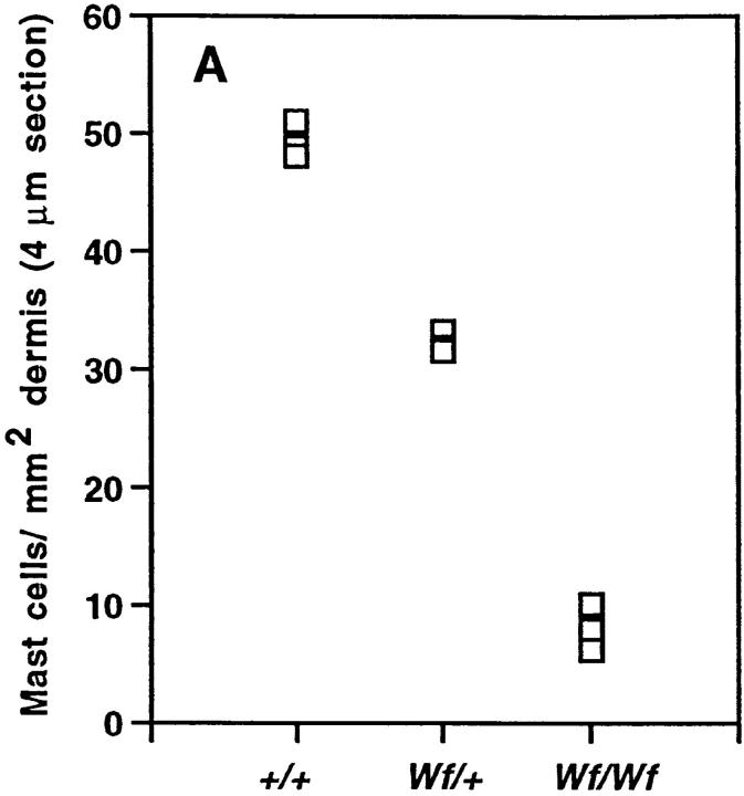
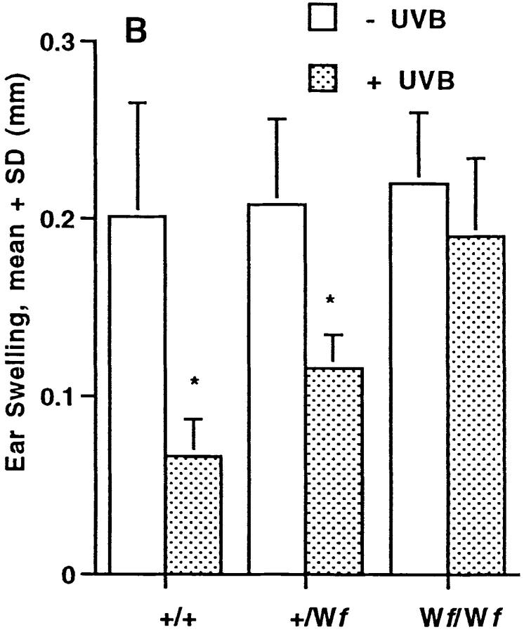
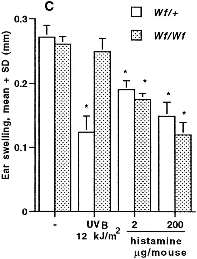
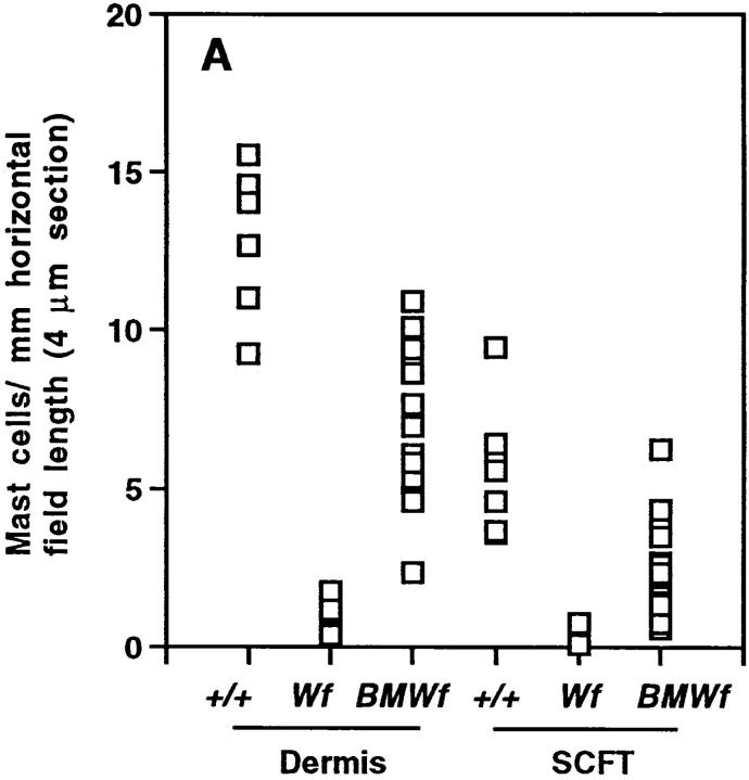
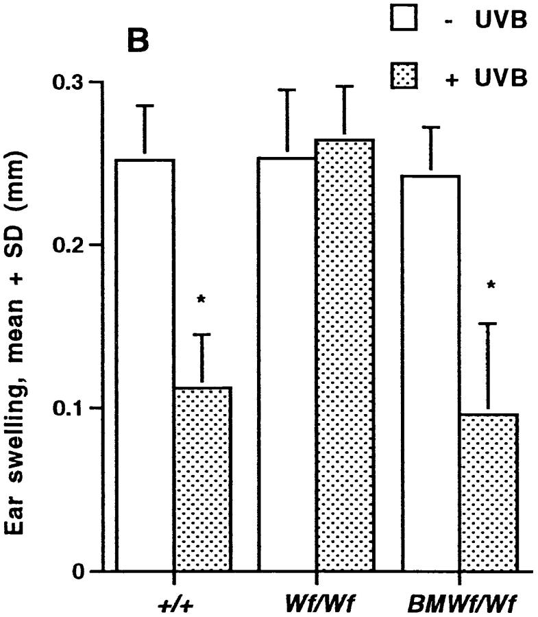
Different strains of mice have varying susceptibilities to ultraviolet radiation (UV) of wavelength 280-320 nm (UVB) for 50% suppression of systemic contact hypersensitivity (CHS) responses. Prevalence of histamine-staining dermal mast cells in different strains of mice (C57BL/ 6J, DBA/2, BALB/c) correlated directly with their susceptibility to UVB-induced systemic immunosuppression. BALB/c mice carrying Uvs1, a major locus for susceptibility to UV-induced immunosuppression, contained greater numbers of dermal mast cells than BALB/c mice of the same parental origin. Strains of mice that were differentiated on their susceptibility to UVB-induced downregulation of systemic CHS responses were similar in their susceptibility to histamine-induced immunomodulation. Histamine, but not UVB irradiation, decreased systemic CHS responses in mast cell-depleted mice (W f/W f). Reconstitution of the dorsal skin of W f/W f mice with bone marrow-derived mast cell precursors from nonmutant mice rendered the mice susceptible to UVB irradiation for systemic suppression of CHS responses. UVB irradiation did not suppress delayed type hypersensitivity responses to allogeneic spleen cells in W f/W f mice. In contrast, UV irradiation suppressed CHS responses in W f/W f mice when hapten was applied to the irradiated site. This study demonstrates that dermal mast cells are necessary for the induction of systemic suppression of CHS responses by UVB radiation, and suggests that mast cell- derived histamine is one component of this UVB-induced systemic immunosuppression.


Figures

Figure 1
Figure 1
Mast cells in the dorsal skin of C57BL/6J, DBA/2, and BALB/c mice. (A) Mast cells per mm2 dermal area. (B) Mast cells per mm horizontal field length in the dermis and SCFT of C57BL/6J (C57; n = 10), DBA/2 (DBA; n = 5), and BALB/c (B/c; n = 11) mice. (C) The area (mm2) of the dermis and SCFT per mm of fixed field length for the same mice as shown in B. (D) The correlation between mast cell prevalence in the dermis (A) and susceptibility to UVB radiation for systemic suppression of CHS responses. Each point in A and B represents the mean mast cell number (expressed per area and fixed field length, respectively), and in C, area per fixed field length for 30–60 measurements/mouse. In A and D, the number of mice per group is shown in brackets. In D, the mean mast cell number per mm2 dermis for 4-μm sections (±SD) was plotted against the log of the UV dose (as documented by others [5]) required for 50% suppression of CHS responses.
Figure 1
Figure 1
Mast cells in the dorsal skin of C57BL/6J, DBA/2, and BALB/c mice. (A) Mast cells per mm2 dermal area. (B) Mast cells per mm horizontal field length in the dermis and SCFT of C57BL/6J (C57; n = 10), DBA/2 (DBA; n = 5), and BALB/c (B/c; n = 11) mice. (C) The area (mm2) of the dermis and SCFT per mm of fixed field length for the same mice as shown in B. (D) The correlation between mast cell prevalence in the dermis (A) and susceptibility to UVB radiation for systemic suppression of CHS responses. Each point in A and B represents the mean mast cell number (expressed per area and fixed field length, respectively), and in C, area per fixed field length for 30–60 measurements/mouse. In A and D, the number of mice per group is shown in brackets. In D, the mean mast cell number per mm2 dermis for 4-μm sections (±SD) was plotted against the log of the UV dose (as documented by others [5]) required for 50% suppression of CHS responses.
Figure 4
Figure 4
Mast cells in the dorsal skin of BALB/c and BALB/c-Uvs1 mice. (A) Mast cells per mm2 dermal area. (B) Mast cells per mm horizontal field length in the dermis and SCFT of BALB/c (–, n = 9) and BALB/c-Uvs1 (Uvs1, n = 11) mice. (C) The area (mm2) of the dermis per mm of fixed field length for the same mice as shown in B. Each point in A and B represents the mean mast cell number (expressed per area and fixed field length, respectively), and in C, area per fixed field length, for 30–60 measurements/mouse. In A and C, the number of mice per group is shown in brackets.
Figure 4
Figure 4
Mast cells in the dorsal skin of BALB/c and BALB/c-Uvs1 mice. (A) Mast cells per mm2 dermal area. (B) Mast cells per mm horizontal field length in the dermis and SCFT of BALB/c (–, n = 9) and BALB/c-Uvs1 (Uvs1, n = 11) mice. (C) The area (mm2) of the dermis per mm of fixed field length for the same mice as shown in B. Each point in A and B represents the mean mast cell number (expressed per area and fixed field length, respectively), and in C, area per fixed field length, for 30–60 measurements/mouse. In A and C, the number of mice per group is shown in brackets.
Figure 2
Figure 2
Histologic sections of dorsal skin from representative mice. (A) BALB/c, (B) C57BL/6J. Arrows, Mast cells.
Figure 3
Figure 3
(A) Location of 400 mast cells from the dorsal skins of each of three C57BL/6J and three BALB/c mice examined for histamine content. The proportion of mast cells (mean + SD) in the upper dermis (D), lower dermis, and SCFT is shown. (B) Histamine content of these cells as assessed by measuring the integrated optical density (mean + SD) for individual histamine-stained cells.
Figure 3
Figure 3
(A) Location of 400 mast cells from the dorsal skins of each of three C57BL/6J and three BALB/c mice examined for histamine content. The proportion of mast cells (mean + SD) in the upper dermis (D), lower dermis, and SCFT is shown. (B) Histamine content of these cells as assessed by measuring the integrated optical density (mean + SD) for individual histamine-stained cells.
Figure 5
Figure 5
The effect of (A) UVB and (B) histamine on the CHS responses of BALB/c and C57BL/6J mice to the hapten, TNCB. In A, mice were UVB irradiated on their shaved dorsal surface with increasing doses of UVB (290–320 nm) as indicated. In B, mice were injected subcutaneously on their backs with increasing doses of histamine per mouse. The mean change in ear swelling (+SD) for five mice/group is shown; the groups of mice were always coded before challenge with hapten and ear thickness measurements. *Result significantly different (P <0.05) to that of nonirradiated mice in A or control mice in B.
Figure 5
Figure 5
The effect of (A) UVB and (B) histamine on the CHS responses of BALB/c and C57BL/6J mice to the hapten, TNCB. In A, mice were UVB irradiated on their shaved dorsal surface with increasing doses of UVB (290–320 nm) as indicated. In B, mice were injected subcutaneously on their backs with increasing doses of histamine per mouse. The mean change in ear swelling (+SD) for five mice/group is shown; the groups of mice were always coded before challenge with hapten and ear thickness measurements. *Result significantly different (P <0.05) to that of nonirradiated mice in A or control mice in B.
Figure 6
Figure 6
(A) Dermal mast cells per mm2 area in dorsal skin of W  f/W  f, heterozygous W  f/+, and wild-type +/+ mice (n = three in each group). Quantification was as per Fig. 1 A. (B) The contact hypersensitivity response to TNCB by W  f/W  f mice after exposure to 12 kJ/m2 UVB. The mean result (+SD) for mice from three experiments is shown. (C) The CHS response to TNCB by W  f/W  f mice after UVB irradiation or administration of 200 and 2 μg histamine/ mouse. The mean result (+SD) for five mice/group is shown. In B and C, a result significantly different (*P <0.05) to that of nonirradiated mice of the same type.
Figure 6
Figure 6
(A) Dermal mast cells per mm2 area in dorsal skin of W  f/W  f, heterozygous W  f/+, and wild-type +/+ mice (n = three in each group). Quantification was as per Fig. 1 A. (B) The contact hypersensitivity response to TNCB by W  f/W  f mice after exposure to 12 kJ/m2 UVB. The mean result (+SD) for mice from three experiments is shown. (C) The CHS response to TNCB by W  f/W  f mice after UVB irradiation or administration of 200 and 2 μg histamine/ mouse. The mean result (+SD) for five mice/group is shown. In B and C, a result significantly different (*P <0.05) to that of nonirradiated mice of the same type.
Figure 7
Figure 7
Reconstitution of the dorsal skin of W  f/W  f mice with bone marrow–derived mast cell precursors. (A) Mast cells per mm horizontal field length in the dermis and SCFT of wild-type (+/+, n = 7), W  f/W  f (W  f; n = 7), and W  f/W  f mice reconstituted with bone marrow–derived mast cell precursors (BMWf; n = 18). (B) The effect of UVB (12 kJ/m2) on the systemic CHS response to the hapten, TNCB, by wild-type (+/+), W  f/W  f, and W  f/W  f mice reconstituted with bone marrow–derived mast cell precursors (BMW  f/W  f). The mean change in ear swelling (+SD) for 5 mice/group is shown with the exception of 12 UVB-irradiated BMW  f/ W   f mice; the groups of mice were coded before challenge with hapten and ear thickness measurements. *A result significantly different (P <0.05) to that of nonirradiated mice.
Figure 7
Figure 7
Reconstitution of the dorsal skin of W  f/W  f mice with bone marrow–derived mast cell precursors. (A) Mast cells per mm horizontal field length in the dermis and SCFT of wild-type (+/+, n = 7), W  f/W  f (W  f; n = 7), and W  f/W  f mice reconstituted with bone marrow–derived mast cell precursors (BMWf; n = 18). (B) The effect of UVB (12 kJ/m2) on the systemic CHS response to the hapten, TNCB, by wild-type (+/+), W  f/W  f, and W  f/W  f mice reconstituted with bone marrow–derived mast cell precursors (BMW  f/W  f). The mean change in ear swelling (+SD) for 5 mice/group is shown with the exception of 12 UVB-irradiated BMW  f/ W   f mice; the groups of mice were coded before challenge with hapten and ear thickness measurements. *A result significantly different (P <0.05) to that of nonirradiated mice.

Similar articles

Cited by

References

    1. Kripke ML. Immunologic mechanisms in UV-radiation carcinogenesis. Adv Cancer Res. 1981;34:69–106. - PubMed
    1. Mottram PL, Mirisklavos A, Clunie GJA, Noonan FP. A single dose of UV radiation suppresses delayed type hypersensitivity responses to alloantigens and prolongs heart allograft survival in mice. Immunol Cell Biol. 1988;66:377–385. - PubMed
    1. Gilmour JW, Vestey JP, Norval M. The effect of UV therapy on immune function in patients with psoriasis. Br J Dermatol. 1993;129:28–38. - PubMed
    1. Yoshikawa T, Streilein JW. Genetic basis of the effects of ultraviolet light B on cutaneous immunity. Evidence that polymorphism at the Tnfα and Lpsloci governs susceptibility. Immunogenetics. 1990;32:398–405. - PubMed
    1. Noonan FP, Hoffman HA. Susceptibility to immunosuppression by ultraviolet B radiation in the mouse. Immunogenetics. 1994;39:29–39. - PubMed

Publication types

MeSH terms