

beta1 integrins are critically involved in neutrophil locomotion in extravascular tissue In vivo
- PMID: 9625769
- PMCID: PMC2212368
- DOI: 10.1084/jem.187.12.2091
beta1 integrins are critically involved in neutrophil locomotion in extravascular tissue In vivo
Abstract
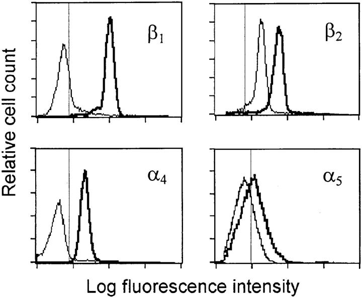
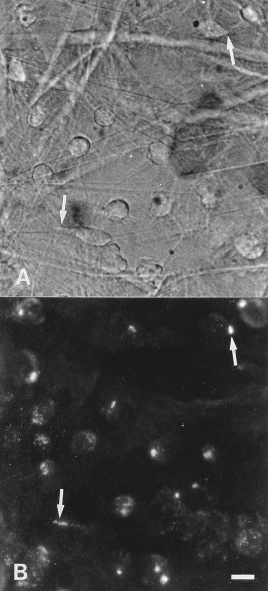
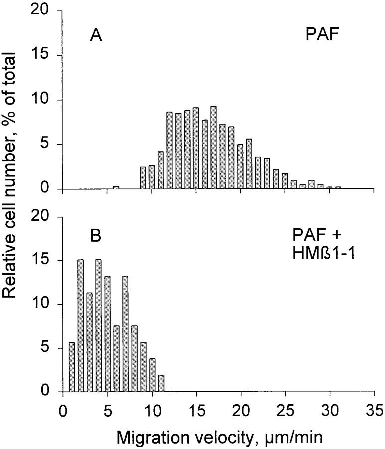
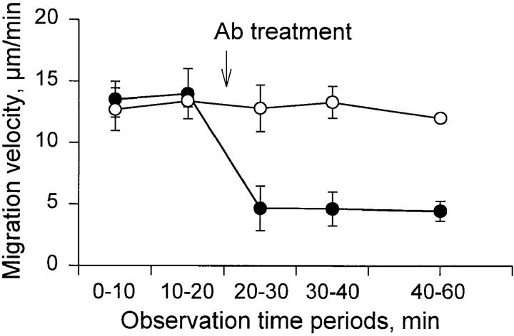
Recruitment of leukocytes from blood to tissue in inflammation requires the function of specific cell surface adhesion molecules. The objective of this study was to identify adhesion molecules that are involved in polymorphonuclear leukocyte (PMN) locomotion in extravascular tissue in vivo. Extravasation and interstitial tissue migration of PMNs was induced in the rat mesentery by chemotactic stimulation with platelet-activating factor (PAF; 10(-7) M). Intravital time-lapse videomicroscopy was used to analyze migration velocity of the activated PMNs, and the modulatory influence on locomotion of locally administered antibodies or peptides recognizing various integrin molecules was examined. Immunofluorescence flow cytometry revealed increased expression of alpha4, beta1, and beta2 integrins on extravasated PMNs compared with blood PMNs. Median migration velocity in response to PAF stimulation was 15.5 +/- 4.5 micron/min (mean +/- SD). Marked reduction (67 +/- 7%) in motility was observed after treatment with mAb blocking beta1 integrin function (VLA integrins), whereas there was little, although significant, reduction (22 +/- 13%) with beta2 integrin mAb. Antibodies or integrin-binding peptides recognizing alpha4beta1, alpha5beta1, or alphavbeta3 were ineffective in modulating migration velocity. Our data demonstrate that cell surface expression of beta1 integrins, although limited on blood PMNs, is induced in extravasated PMNs, and that members of the beta1 integrin family other than alpha4beta1 and alpha5beta1 are critically involved in the chemokinetic movement of PMNs in rat extravascular tissue in vivo.
Figures

References
-
- Carlos TM, Harlan JM. Leukocyte–endothelial adhesion molecules. Blood. 1994;84:2068–2101. - PubMed
-
- Hemler ME. VLA proteins in the integrin family: structures, functions, and their role on leukocytes. Annu Rev Immunol. 1990;8:365–400. - PubMed
-
- Gehlsen KR, Dillner L, Engvall E, Ruoslahti E. The human laminin receptor is a member of the integrin family of cell adhesion receptors. Science. 1988;241:1228–1229. - PubMed
Publication types
MeSH terms
Substances
LinkOut - more resources
Full Text Sources
Other Literature Sources
