. 1998 Jul 1;510 ( Pt 1)(Pt 1):209-23.
doi: 10.1111/j.1469-7793.1998.209bz.x.

Neuroeffector transmission in arterioles of the guinea-pig choroid

Affiliations

Neuroeffector transmission in arterioles of the guinea-pig choroid

H Hashitani et al. J Physiol. .

Abstract

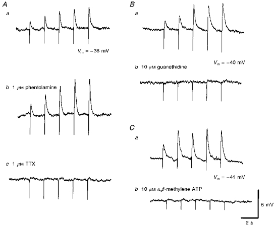
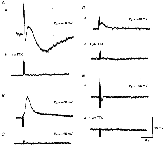
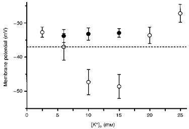
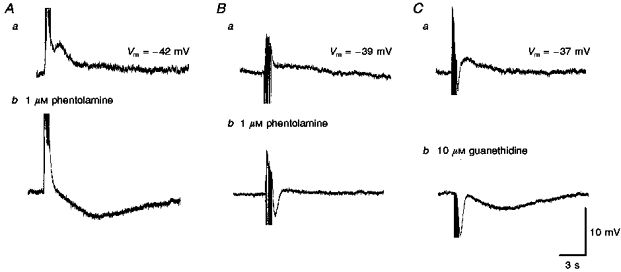
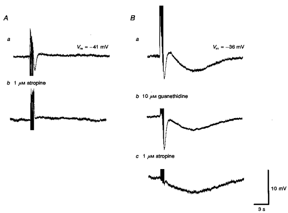
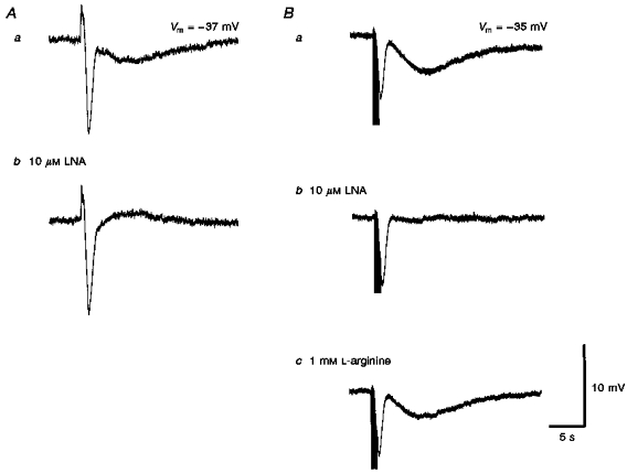
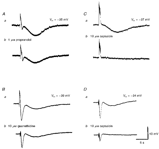
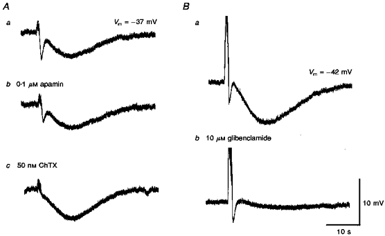
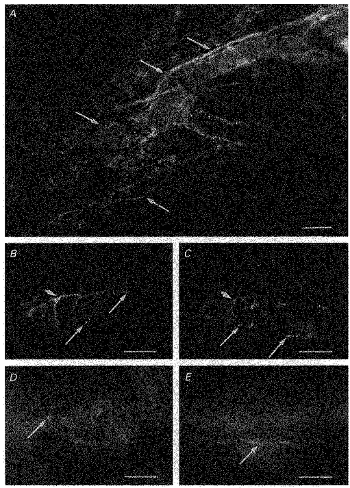
1. Using conventional microelectrode techniques, membrane potentials were recorded from smooth muscle cells of guinea-pig choroidal arterioles. 2. Transmural stimulation initiated excitatory junction potentials (EJPs) which were abolished by either guanethidine or alpha,beta-methylene-ATP but not by phentolamine, indicating that they resulted from activation of purinoceptors. 3. Trains of stimuli evoked EJPs which were followed by a slow depolarization, an inhibitory junction potential (IJP) or a biphasic membrane potential change which consisted of an IJP and a subsequent slow depolarization. 4. Slow depolarizations were abolished by either phentolamine or guanethidine, indicating that they resulted from activation of alpha-adrenoceptors. 5. IJPs were abolished by atropine but not by guanethidine, and were reduced by 50 % by apamin with the residual response being abolished by charybdotoxin, indicating that they resulted from the activation of muscarinic receptors which open two sets of Ca2+-activated K+ channels. 6. Most responses were followed by slow hyperpolarizations. These were almost abolished by L-nitroarginine, an effect which was partly overcome by L-arginine, and were abolished by glibenclamide, indicating that they resulted from the release of NO and activation of ATP-sensitive K+ channels. 7. Immunohistochemical analysis showed that arterioles were densely innervated by adrenergic nerve fibres. A population of fibres, likely to be cholinergic, was also identified. Furthermore, populations of nerve fibres immunoreactive to antibodies against either nitric oxide synthase (NOS) or substance P (SP) were also identified. 8. These findings indicate that choroidal arterioles of the guinea-pig are innervated by at least three different populations of nerves, adrenergic nerves which evoke excitatory responses, cholinergic nerves which evoke inhibitory responses and a population of nerves which cause the release of NO.
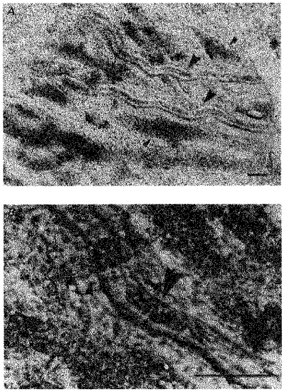

Figures

Figure 1
Figure 1. Micrographs of guinea-pig choroid
A shows the location of arterioles and veins in the choroid at low magnification. The larger arrowheads indicate arterioles (right side is proximal) and the smaller arrowheads indicate veins. Arterioles had smaller diameter and thicker walls than did veins. B shows a typical recording site at higher magnification. The large arrowhead indicates a section of arteriole which contains red blood corpuscles. The smaller arrowhead indicates an area where a vein overlaps the arteriole. Scale bars, 100 μm in both A and B.
Figure 4
Figure 4. The effects of phentolamine, guanethidine, α,β-methylene ATP and tetrodotoxin on the rapid EJPs recorded from choroidal arterioles
The rapid EJPs evoked by trains of stimuli (supramaximal voltage, 50 μs, 0.5 Hz, 10 s; Aa) in choroidal arterioles persisted in the presence of phentolamine (1 μM; Ab) but were abolished by tetrodotoxin (TTX, 1 μM; Ac). EJPs were abolished by guanethidine (10 μM; Bb) and by α,β-methylene-ATP (10 μM; Cb). A, B and C were recorded from 3 different cells. Aa, Ba and Ca are respective control responses. In each trace, the small downward deflection at the beginning of each EJP indicates the artefact of nerve stimulation. The scale bars on the right refer to all traces. Vm refers to resting membrane potential.
Figure 2
Figure 2. Different types of responses recorded from choroidal vessels in response to train of transmural nerve stimuli
Aa, a train of stimuli (supramaximal voltage, 50 μs, 50 Hz, 1 s) initiated a short-latency depolarization, followed by a transient hyperpolarization, a transient depolarization and a prolonged slow hyperpolarization. Ab, each component was abolished by 1 μM TTX, indicating that each resulted from the stimulation of the perivascular nerves. B, in a vessel, tentatively identified as being a vein, a train of stimuli (supramaximal voltage, 50 μs, 10 Hz, 1 s) failed to evoke the same complex sequence of membrane potential changes. C, in the third type of cell, which could not be identified, trains of stimuli (supramaximal voltage, 50 μs, 50 Hz, 1 s) failed to evoke any membrane potential changes. Da and Ea show other examples of recordings obtained from arterioles stimulated with trains of low frequency (supramaximal voltage, 50 μs, 10 Hz, 1 s); Db and Eb, again the membrane changes were abolished by 1 μM TTX. A, D and E were each recorded from 3 different cells. The scale bars on the right refer to all traces. Vm refers to resting membrane potential.
Figure 3
Figure 3. Relationship between [K+]o and the membrane potential of choroidal arterioles
In control solutions (○) the membrane potential was about −37 mV; when [K+]o was increased to either 10 or 15 mM, the membrane hyperpolarized. At higher values of [K+]o the steady-state value of membrane potential was more positive than that detected in control solutions. Prior addition of 50 μM Ba2+ (•) prevented the hyperpolarizations produced by [K+]o (10 and 15 mM). The vertical bars represent ± s.d. (n = 4-25) and the dashed horizontal line indicates the membrane potential recorded in [K+]o = 5.9 mM.
Figure 5
Figure 5. The effects of phentolamine and guanethidine on slow depolarizations recorded from choroidal arterioles
The slow depolarizations evoked by trains of stimuli in two different preparations (supramaximal voltage, 50 μs, 10 Hz, 1 s; Aa and Ba) were abolished by phentolamine (Ab and Bb). In both preparations, the initial purinergic responses persisted. In one of the preparations a slow hyperpolarization was revealed (Ab); in the other a rapid IJP was detected (Bb). In the third preparation both the purinergic and slow depolarization evoked by a train of stimuli (supramaximal voltage, 50 μs, 50 Hz, 1 s, Ca) were abolished by guanethidine to reveal a slow hyperpolarization and an augmented IJP (Cb). A, B and C were recorded from 3 different cells. The scale bars on the right refer to all traces. Vm refers to resting membrane potential.
Figure 6
Figure 6. The effects of atropine and guanethidine on rapid IJPs recorded from choroidal arterioles
Trains of field stimulation (supramaximal voltage, 50 μs, 10 Hz, 1 s), in the presence of 1 μM phentolamine produced rapid IJPs which were abolished by atropine (1 μM; Ab). In another preparation, guanethidine (10 μM; Bb) applied before atropine (1 μM, Bc) abolished the purinergic response but did not affect the initial IJP. Bc, subsequently atropine abolished the initial IJP. Although guanethidine reduced the late slow hyperpolarization (Bb), atropine was without further effect on this component (Bc). A and B were recorded from 2 different cells. Aa and Ba are respective control responses. The scale bars on the right refer to all traces. Vm refers to resting membrane potential.
Figure 7
Figure 7. The effects of L-nitroarginine and L-arginine on the slow hyperpolarization recorded from choroidal arterioles
L-Nitroarginine (LNA, 10 μM; Ab) selectively abolished the slow hyperpolarization compared with controls (Aa) in response to a high-frequency train of stimuli (supramaximal voltage, 50 μs, 50 Hz, 1 s). In another preparation where recordings were made in the presence of 10 μM guanethidine (Ba), again 10 μM LNA selectively abolished the slow hyperpolarization (Bb) and the effect of LNA was partially reversed by L-arginine (1 mM; Bc). The scale bars on the right refer to all traces. Vm refers to resting membrane potential.
Figure 8
Figure 8. The individual effects of propranolol, guanethidine and capsaicin on slow hyperpolarizations recorded from choroidal arterioles
A series of membrane potential changes, initiated in the presence of phentolamine (1 μM) by trains of high-frequency nerve stimuli (supramaximal voltage, 50 μs, 50 Hz, 1 s) are shown. The slow hyperpolarization was reduced in amplitude by each of propranolol (1 μM; Ab) and guanethidine (10 μM; Bb) and abolished by capsaicin (10 μM; Cb). The traces shown in D were recorded in the presence of guanethidine (10 μM); Db, the guanethidine-resistant component was virtually abolished by capsaicin. A, B, C and D were recorded from 4 different cells. Aa, Ba, Ca and Da are respective control responses. The scale bars on the right refer to all traces. Vm refers to resting membrane potential.
Figure 9
Figure 9. The effects of apamin, charybdotoxin and glibenclamide on two distinct hyperpolarizations recorded from choroidal arterioles
A comparison of the effects of apamin (0.1 μM) alone (Ab) and with charybdotoxin (ChTX, 50 nM; Ac) each in the presence of guanethidine (10 μM) and of glibenclamide (Glib, 10 μM) in the presence of phentolamine (1 μM) on the responses of guinea-pig choroidal arterioles to trains of field stimulation (supramaximal voltage, 50 μs, 50 Hz, 1 s). A and B were recorded from 2 different cells. Respective controls Aa and Ba were in the presence of guanethidine and phentolamine. Ab, cholinergic IJP but not slow hyperpolarization was inhibited by apamin. Ac, subsequent addition of ChTX abolished the residual IJP and enhanced the amplitude of the slow hyperpolarization. Bb, glibenclamide almost completely inhibited the slow hyperpolarization without affecting IJP. The scale bar on the right refers to all traces. Vm refers to resting membrane potential.
Figure 10
Figure 10. Immunoreactivity of nerve fibres innervating choroidal arterioles in the guinea-pig to αPGP9.5, αTH, αVIP, αNOS and αSP
A, a preparation treated with αPGP9.5, a marker used to identify all neuronal elements. Numerous, dense nerve fibres immunoreactive to αPGP9.5 were identified surrounding guinea-pig choroidal arterioles (arrows). B and C, same preparation double labelled with αTH (B) and αVIP (C). Choroidal arterioles were densely innervated with αTH immunoreactive fibres, which often formed a plexus around the arterioles (B). Choroidal arterioles were also innervated by nerve fibres immunoreactive to αVIP (C). In preparations double labelled with both αTH and αVIP, nerve fibres immunoreactive to αTH formed a separate population from those fibres which expressed immunoreactivity to αVIP (longer arrows in B indicate fibres immunoreactive to αTH but not to αVIP and longer arrows in C indicate those fibres expressing immunoreactivity to αVIP only). However, even though nerve fibres immunoreactive to αTH were separate from those immunoreactive to αVIP, they were often found running alongside one another, possibly in the same nerve bundle (arrowed in B and C). Choroidal arterioles were also innervated by numerous αNOS-containing nerve fibres, which appeared to be varicose (arrow, D). In separate preparations, varicose nerve fibres expressing immunoreactivity to αSP were also identified (arrow, E), although innervation by αSP immunoreactive fibres were sparse. Scale bars: A, 100 μm; B-E, 50 μm.

Similar articles

Cited by

References

    1. Albert A. Ocular circulation. In: Hart WM Jr, editor. Adler's Physiology of the Eye. 9. St Louis, MO, USA: Mosby-Year Book, Inc.; 1992. pp. 198–227.
    1. Bolton TB, Lang RJ, Takewaki T. Mechanisms of action of noradrenaline and carbachol on smooth muscle of guinea- pig anterior mesenteric artery. The Journal of Physiology. 1984;351:549–572. - PMC - PubMed
    1. Bolton TB, Large WA. Are junction potentials essential? Dual mechanism of smooth muscle cell activation by transmitter released from autonomic nerves. Quarterly Journal of Experimental Physiology. 1986;89:163–171. - PubMed
    1. Brayden JE, Large WA. Electrophysiological analysis of neurogenic vasodilatation in the isolated lingual artery of the rabbit. British Journal of Pharmacology. 1986;89:163–171. - PMC - PubMed
    1. Clarke A, Edwards FR, Hirst GDS, Silverberg GD. Developmental changes in the resting membrane potential of rat cerebral arteries. In: Mulvany MJ, Aalkjaer C, Heagerty AM, Nyborg NCB, Strandgaard S, editors. Resistance Arteries, Structure and Function. Amsterdam: Excerpta Medica; 1991. pp. 147–151.

LinkOut - more resources