. 1998 Jul 1;510 ( Pt 1)(Pt 1):309-20.
doi: 10.1111/j.1469-7793.1998.309bz.x.

Control of motility patterns in the human colonic circular muscle layer by pacemaker activity

Affiliations

Control of motility patterns in the human colonic circular muscle layer by pacemaker activity

M G Rae et al. J Physiol. .

Abstract

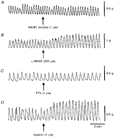
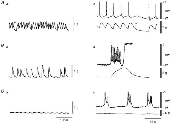
1. This study characterized the electrical and mechanical activities of human colonic muscle strips obtained from either the ascending, descending or sigmoid colon of patient volunteers during elective colon resections. 2. Rhythmic contractile activity was observed in colonic circular muscle strips in the absence of external stimuli. This activity persisted in the presence of atropine, phentolamine, propranolol, tetrodotoxin and Nomega-nitro-L-arginine but was abolished by nifedipine. 3. The activity of whole circular muscle (WCM) was compared with that of the myenteric half (MCM), the submucosal half (SCM) and the interior (ICM) of the circular muscle layer. WCM exhibited a prominent 2-4 contractions min-1 contractile pattern which was also present in strips of SCM. In contrast, MCM and ICM exhibited slow (0.3-0.6 contractions min-1), long duration contractions with superimposed higher frequency contractions (17-18 contractions min-1). 4. Resting membrane potential (Vm), recorded at various positions through the thickness of WCM strips did not differ and averaged -50 mV. 5. Slow waves were observed in 83 % of muscles. They averaged 12 mV in amplitude, 9.4 s in duration and had a frequency of 2-4 contractions min-1. Slow waves were greatest in amplitude near the submucosal edge and decreased with distance away from this edge. Each slow wave was associated with a transient contraction. 6. Near the myenteric edge, rapid fluctuations of Vm with a mean frequency of 18 contractions min-1 were recorded in 67 % of muscles. Spiking activity was common and was superimposed upon slow waves and rapid Vm fluctuations. 7. In summary, slow waves were identified in the human colonic circular muscle layer which arise at or near the submucosal edge. These electrical events give rise to a 2-4 contractions min-1 contractile rhythm which is characteristic of the intact muscle layer. Thus, the nature and spatial organization of pacemaker activity in the human colon bears significant resemblance to other animal models, such as the dog and pig.


Figures

Figure 1
Figure 1. Schematic representation of the cross-sectional preparation of human colon
Dashed lines indicate where cuts were made across the circular muscle layer to create the strips used for contractile measurements. Whole circular muscle (WCM) included a thin intra-taenial strip of longitudinal muscle at one edge and some submucosal connective tissue at the other edge. Myenteric circular muscle (MCM) strips also included the intra-taenial longitudinal muscle as well as half of the circular musle layer. Submucosal circular muscle (SCM) strips included half of the circular muscle layer as well as some submucosal connective tissue. Interior circular muscle (ICM) strips were devoid of both myenteric and submucosal edges and contained approximately one-third of the muscle layer.
Figure 2
Figure 2. Effect of various antagonists on the contractile activity of human colonic muscles
Shown in this figure are rhythmic contractions recorded from strips of whole circular muscle (WCM) obtained from four different patients. In each case a 2-4 contractions min−1 rhythm predominates. A, the contractile activity before and after addition of a ‘NANC’ solution containing atropine, phentolamine and propranolol (all 1 μM). The NANC solution did not change contractile amplitude or pattern in this muscle or in nine others tested. B, the nitric oxide synthase inhibitor Nω-nitro-L-arginine methyl ester (L-NAME; 200 μM) was added in the presence of NANC solution. L-NAME increased contractile amplitude in this and in six other muscles tested (mean increase 54 ± 12 %, n = 7) but the 2-4 contractions min−1 contractile rhythm persisted. C, the fast sodium channel blocker tetrodotoxin (TTX, 1 μM) was applied in the presence of NANC solution. In this case TTX had no effect on spontaneous contractions. Overall, in six muscles there was a small increase in contractile amplitude (i.e. 11 ± 4.6 %, n = 6) but the 2-4 contractions min−1 rhythm persisted. D, the small conductance calcium-activated K+ channel antagonist apamin (1 μM) was applied in the presence of NANC solution plus TTX. Apamin produced an increase in contractile amplitude in this and nine other muscles (i.e. 132 ± 36 %, n = 10) but the 2-4 contractions min−1 contractile rhythm persisted.
Figure 3
Figure 3. Examples of the most common contractile patterns observed in strips of human whole circular muscle (WCM), myenteric circular muscle (MCM), interior circular muscle (ICM) and submucosal circular muscle (SCM)
WCM (A) and SCM (D) both displayed a 2-4 contractions min−1 contractile rhythm. In contrast, both MCM (B) and ICM (C) exhibited slower contractions with superimposed rapid frequency contractions. The inset below B shows a 30 s period of rapid frequency contractions of MCM displayed at a more rapid sweep speed. A and D were recorded from muscle strips of one patient while B and C were recorded from muscle strips of another patient. All muscle strips were from sigmoid colon.
Figure 4
Figure 4. Time-dependent changes in contractile and electrical activity of human colonic whole circular muscle
Aa shows the contractile pattern produced by a muscle strip 133 min after placing it in the recording chamber. This consisted of rapid phasic contractions at a frequency of 9 contractions min−1 with a few brief intermittent periods of relaxation (denoted *). In Ba the contractile activity of the same muscle strip as in A is shown at 207 min. At this time the frequency of contraction had declined to 3 contractions min−1 while the amplitude of contraction increased. This pattern continued for the next hour. Subsequent addition of wortmannin (5 μM) led to a marked reduction in contractile amplitude as shown in Ca obtained 36 min after exposure to wortmannin. Ab, Bb and Cb show examples of the electrical activity which accompanied contractions. Each panel includes an intracellular recording made near the submucosal edge (upper traces) and a contractile recording from the entire muscle layer (lower traces). Ab shows the same contractions as in Aa at a faster sweep speed. The asterisks in Aa and Ab represent the same moment in time. Phasic contractions were associated with actions potentials. Following development of the intermediate rhythm shown in Ba it was not possible to maintain impalements in cells. However, in another muscle strip a brief electrical recording was obtained as shown in Bb. This recording (made 226 min after placing a muscle strip in the recording chamber) reveals that contractions of the type shown in Ba were accompanied by slow waves. Due to the strength of contraction the impalement was lost. By including wortmannin in the superfusate it was possible to maintain impalements during contraction as shown in Cb. Cb shows the same contractions as in Ca at a faster sweep speed. This recording reveals that each phasic contraction was accompanied by a slow wave. Thus, time-dependent changes in contractile pattern were associated with the development of slow waves in the tissue. All recordings were made from strips of sigmoid colon.
Figure 5
Figure 5. Electrical activity at the submucosal edge of whole circular muscle strips of the ascending (a), descending (B and D) or sigmoid (C and E) colon
Records obtained from the tissue of five patients. Slow waves were recorded from all three regions examined and these typically had action potentials superimposed (A, B and C). In some preparations slow waves without spikes were observed (D). In a few muscles only spikes were recorded (E). Although all tissues contracted under control conditions, only C and D had discernible contractions in the presence of wortmannin (5 μM).
Figure 6
Figure 6. Effect of distance from the submucosal edge on slow wave amplitude
A, B and C show examples of the electrical activity recorded from cells near the submucosal edge (5 %), middle (50 %) and myenteric edge (95 %) of muscle strips isolated from the ascending (A), descending (B), and sigmoid colon (C) of three patients. D plots the mean amplitude (± s.e.m.) of slow waves recorded at various distances from the submucosal edge in fifteen muscle strips in which slow waves were present at the submucosal edge. N values for 50 % and 95 % were 9 and 8, respectively. All recordings were obtained in the presence of wortmannin (5 μM). *P < 0.05 and ***P < 0.001, significant differences in slow wave amplitude (i.e. 5 vs. 50 % and 50 vs. 95 %, respectively).
Figure 7
Figure 7. Electrical activity observed in cells near the myenteric edge of whole circular muscle
Records obtained from tissues of four patients. A shows a recording from a muscle strip (sigmoid colon) which did not have any oscillating electrical activity at the myenteric edge. Cell impalement was confirmed by electrically evoking an inhibitory junction potential (IJP, denoted by n.s.; single pulse, 0.3 ms, 15 V). Note the rebound excitation following the IJP. B shows a recording from another muscle strip (descending colon) which exhibited ongoing MPOs (21contractions min−1) which were relatively constant in amplitude. C shows a recording from a third muscle strip (descending colon) in which MPOs gave rise to variable amplitude action potentials. D shows a recording from a fourth muscle strip (descending colon) which exhibited slow waves with superimposed action potentials. All recordings obtained in the presence of wortmannin (5 μM).
Figure 8
Figure 8. Spontaneous transient hyperpolarizations within whole circular muscle strips
Records obtained from tissues of two patients. A shows an intracellular recording from a cell near the myenteric edge of a muscle strip (descending colon). Small (≈1 mV) MPOs are apparent in the recording. IJPs were evoked when nerves were electrically stimulated (NS) with a single pulse (first arrow; 0.3 ms, 15 V) or 5 pulses at 5 Hz (second and third arrows; 0.3 ms, 15 V). Following the third stimulus pulse a burst of transient hyperpolarizations was observed which led to a peak hyperpolarization to −80 mV. B shows an intracellular recording from a cell near the submucosal edge of the muscle strip (sigmoid colon). Slow waves (3 contractions min−1) which occasionally gave rise to action potentials are apparent in the recording. An IJP was elicited at the arrow with a single pulse (NS). This was followed by a period of rebound excitation. One minute later a series of transient hyperpolarizations appeared superimposed upon slow waves which led to a peak hyperpolarization to −76 mV. In C IJPs (upper traces) and transient hyperpolarizations (lower traces) observed during the same impalement have been plotted on an expanded time scale to show that these events were similar in time course. Left traces: electrical events recorded near the myenteric edge. Right traces: electrical events recorded near the submucosal edge. All recordings obtained in the presence of wortmannin (5 μM).

Similar articles

Cited by

References

    1. Burke EP, Gerthoffer WT, Sanders KM, Publicover NG. Wortmannin inhibits contraction without altering electrical activity in canine gastric smooth muscle. American Journal of Physiology. 1996;270:C1405–1412. - PubMed
    1. Berezin I, Huizinga JD, Daniel EE. Interstitial cells of Cajal in the canine colon: a special communication network at the inner border of the circular muscle. Journal of Comparative Neurology. 1988;273:42–51. - PubMed
    1. Chow E, Huizinga JD. Myogenic electrical control activity in longitudinal muscle of human and dog colon. The Journal of Physiology. 1987;392:21–34. - PMC - PubMed
    1. Du C, Conklin JL. Origin of slow waves in the isolated proximal colon of the cat. Journal of the Autonomic Nervous System. 1989;28:167–178. 10.1016/0165-1838(89)90089-1. - DOI - PubMed
    1. Faussone-Pellegrini MS, Pantalone D, Cortesini C. Smooth muscle cells, interstitial cells of Cajal and myenteric plexus interrelationships in the human colon. Acta Anatomica. 1990;139:31–44. - PubMed

Publication types

LinkOut - more resources