. 1998 Jun 15;141(6):1301-10.
doi: 10.1083/jcb.141.6.1301.

Interaction of vault particles with estrogen receptor in the MCF-7 breast cancer cell

Affiliations

Interaction of vault particles with estrogen receptor in the MCF-7 breast cancer cell

C Abbondanza et al. J Cell Biol. .

Abstract

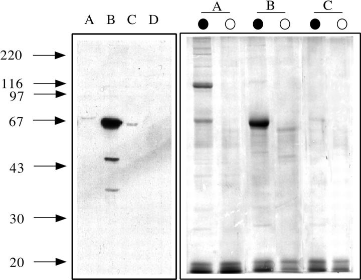
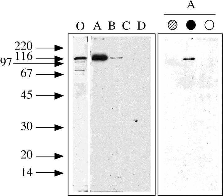
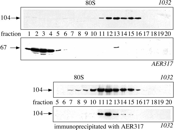
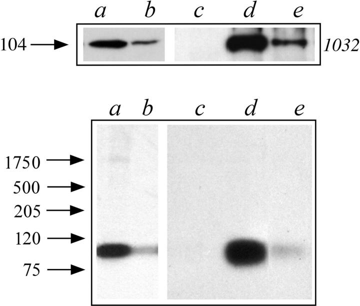
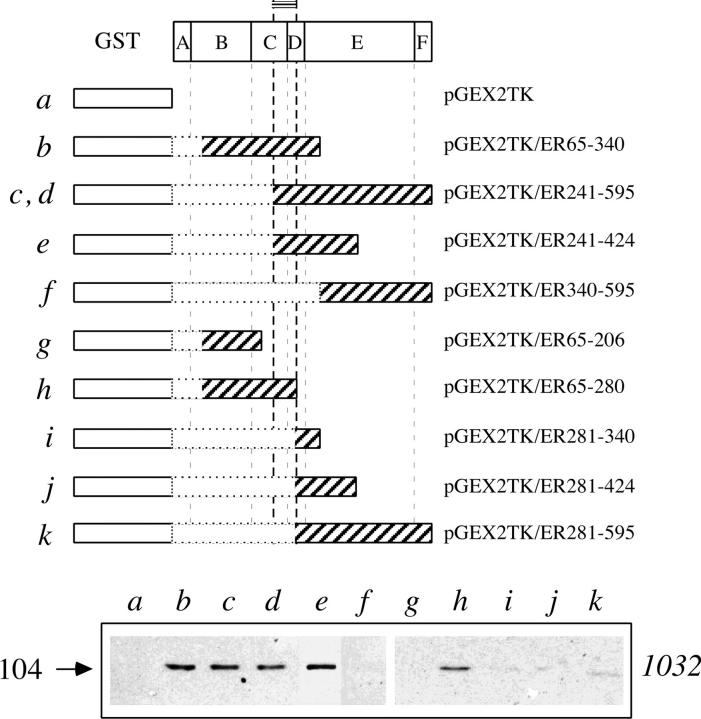
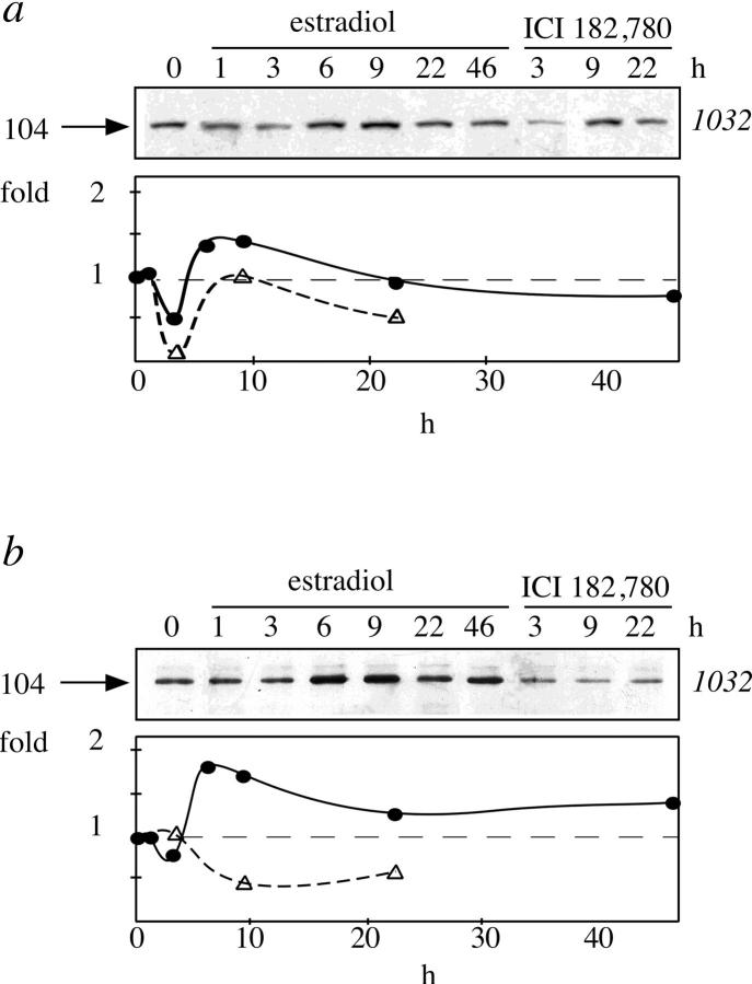
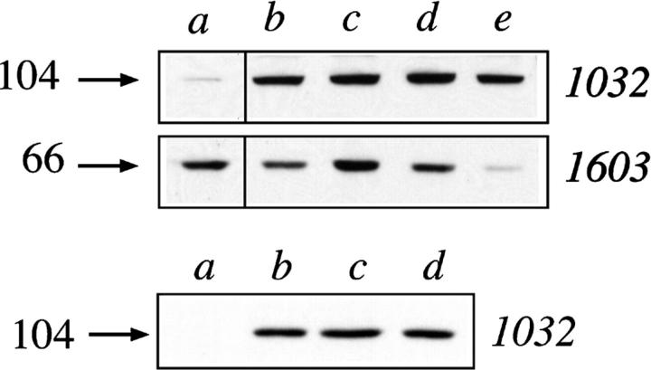
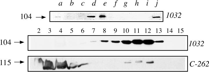
A 104-kD protein was coimmunoprecipitated with the estrogen receptor from the flowtrough of a phosphocellulose chromatography of MCF-7 cell nuclear extract. mAbs to this protein identified several cDNA clones coding for the human 104-kD major vault protein. Vaults are large ribonucleoprotein particles of unknown function present in all eukaryotic cells. They have a complex morphology, including several small molecules of RNA, but a single protein species, the major vault protein, accounts for >70% of their mass. Their shape is reminiscent of the nucleopore central plug, but no proteins of known function have been described to interact with them. Western blot analysis of vaults purified on sucrose gradient showed the presence of estrogen receptor co-migrating with the vault peak. The AER317 antibody to estrogen receptor coimmunoprecipitated the major vault protein and the vault RNA also in the 20,000 g supernatant fraction. Reconstitution experiments of estrogen receptor fragments with the major vault protein mapped the site of the interaction between amino acids 241 and 280 of human estrogen receptor, where the nuclear localization signal sequences are located. Estradiol treatment of cells increased the amount of major vault protein present in the nuclear extract and coimmunoprecipitated with estrogen receptor, whereas the anti-estrogen ICI182,780 had no effect. The hormone-dependent interaction of vaults with estrogen receptor was reproducible in vitro and was prevented by sodium molybdate. Antibodies to progesterone and glucocorticoid receptors were able to coimmunoprecipitate the major vault protein. The association of nuclear receptors with vaults could be related to their intracellular traffic.


Figures

Figure 1
Figure 1
Western blot analysis of estrogen receptor in the nuclear extract separated by phosphocellulose chromatography and identification of proteins coimmunoprecipitated with the estrogen receptor. Arrows on the left side indicate the migration of standard proteins of the indicated molecular weight (kD). (Left) SDS-PAGE followed by Western blot analysis with the mAb to estrogen receptor AER317 of the flowthrough (lane A), 0.3 M (lane B), 0.5 M (lane C), or 0.75 M KCl (lane D) fractions of the phosphocellulose chromatography of MCF-7 cell nuclear extract. (Right) SDS-PAGE analysis followed by Coomassie blue staining of the gel of proteins immunoprecipitated with the mAb to estrogen receptor AER317 (lanes marked by solid circle) or control IgG (lanes marked by open circle) of the flowthrough (lanes A), 0.3 M (lanes B), or 0.5 M (lanes C) fractions of the phosphocellulose chromatography of nuclear extract.
Figure 2
Figure 2
Western blot analysis of the 104-kD protein in the nuclear extract separated by phosphocellulose chromatography and in the proteins coimmunoprecipitated with the estrogen receptor in the flowthrough fraction. Arrows on the left side indicate the migration of standard proteins of the indicated molecular weight (kD). (Left) SDS-PAGE followed by Western blot analysis with mAb 1032 to the major vault protein of MCF-7 cells nuclear extract (lane O) and of the flowthrough (lane A), 0.3 M (lane B), 0.5 M (lane C), or 0.75 M KCl (lane D) fractions of the phosphocellulose chromatography of nuclear extract. (Right) SDS-PAGE followed by Western blot analysis with mAb 1032 to the major vault protein of proteins immunoprecipitated with the mAbs to estrogen receptor AER314 (gray circle) and AER317 (solid circle) or control IgG (open circle) of the flowthrough fraction of the phosphocellulose chromatography of nuclear extract.
Figure 3
Figure 3
Western blot analysis and immunoprecipitation of vaults sedimented through a sucrose gradient. Arrows on the left side indicate the molecular weight (kD) of bands calculated from migration of standard proteins; on the top of the frames is reported the sedimentation of 80 S ribosomal subunit (external marker). (Top) SDS-PAGE followed by Western blot analysis with mAbs 1032 to the major vault protein or AER317 to estrogen receptor, as indicated, of fractions of a 5-ml sucrose gradient separation of the flowthrough of phosphocellulose chromatography of nuclear extract. (Bottom) SDS-PAGE followed by Western blot analysis with mAb 1032 to the major vault protein of proteins immunoprecipitated with the mAb to estrogen receptor AER317 (lower frame) from fractions (upper frame) of a 40-ml sucrose gradient separation of the flowthrough of phosphocellulose chromatography of nuclear extract.
Figure 4
Figure 4
Western blot analysis of the 104-kD protein and Northern blot analysis of vRNA coimmunoprecipitated with estrogen receptor in the 20,000 g supernatant of cell extract. (Top) SDS-PAGE followed by Western blot analysis with mAb 1032 to the major vault protein of 20,000 g supernatant (lane a), or nuclear extract (lane b), or of protein immunoprecipitated from 20,000 g supernatant (lanes c and d), or nuclear extract (lane e) with the mAb to estrogen receptor AER317 (lanes d and e), or control IgG (lane c). The arrow on the left side indicates the molecular weight (kD) of the band calculated from migration of standard proteins. (Bottom) Northern blot analysis with a human vRNA probe (see Materials and Methods) of RNA extracted from aliquots of samples indicated in the top panel with the corresponding letters. Arrows on the left side indicate the migration of standard RNA (nt).
Figure 5
Figure 5
Mapping the estrogen receptor region interacting with the 104-kD protein. SDS-PAGE followed by Western blot analysis with mAb 1032 to the major vault protein of proteins immunopurified on columns containing Sepharose coupled to the indicated fusion proteins. Nuclear extracts were incubated in the absence (lanes a–c and e–k) or in the presence of 10 nM estradiol (lane d). The arrow on the left side indicates the molecular weight (kD) of the band calculated from migration of standard proteins. The domain organization of estrogen receptor is reported in scale. On the top, a double-line segment reports the minimum determined fragment of the estrogen receptor sequence necessary for interaction with the major vault protein.
Figure 6
Figure 6
Time course estrogen effect on the amount of the 104-kD protein present in the nuclear extract and coimmunoprecipitated with the estrogen receptor. SDS-PAGE followed by Western blot analysis with mAb 1032 to the major vault protein of nuclear extracts (a) or of proteins immunoprecipitated from nuclear extracts with the mAb 1603 to estrogen receptor (b) of cells incubated in the presence of 10 nM estradiol or 100 nM ICI182,780 for the indicated time. The arrows on the left side indicate the molecular weight (kD) of the band calculated from migration of standard proteins; the plots on the bottom are the densitometric analysis of the blots (•, estradiol; ▵, ICI182,780).
Figure 7
Figure 7
Western blot analysis of the estrogen receptor associated with the vault- enriched fraction after cell-free activation of the postnuclear supernatant by hormone and temperature. The arrows on the left side indicate the molecular weight (kD) of bands calculated from migration of standard proteins. (Top) SDS-PAGE followed by Western blot analysis with mAb 1032 to the major vault protein or 1603 to estrogen receptor, as indicated, of postnuclear supernatant (lane a), vault-enriched fractions from aliquots of postnuclear supernatant incubated for 30 min at 30°C (lanes b–e), in the presence of 10 nM estradiol (lane c), 100 nM ICI182,780 (lane d), or 5 mM sodium molybdate (lane e). (Bottom) SDS-PAGE followed by Western blot analysis with mAb 1032 to the major vault protein of proteins immunoprecipitated with control antibodies (lane a) or with the mAb 1603 to estrogen receptor (lanes b–d) from the same samples of lanes b–d of the top panel.
Figure 8
Figure 8
Western blot analysis of the 104-kD protein and coimmunoprecipitated by antibodies to other nuclear proteins. The arrows on the left side indicate the molecular weight (kD) of the bands calculated from migration of standard proteins. The arrows on the left side indicate the molecular weight (kD) of the bands calculated from migration of standard proteins. (Top) SDS-PAGE followed by Western blot analysis with mAb 1032 to the major vault protein of nuclear extract (lane j) or of proteins immunoprecipitated from the nuclear extract with antibodies to progesterone receptor (lane a), to glucocorticoid receptor (lane b), to Nurr77 (lane c), to estrogen receptor (lanes d and e, commercial polyclonal and AER317, respectively), to TFIIB (lane f), to SRC-1 (lane g), or with mouse (lane h) and rabbit control antibodies (lane i). (Bottom) SDS-PAGE followed by Western blot analysis with mAb 1032 to the major vault protein or C-262 to progesterone receptor, as indicated, of fractions of a 5-ml sucrose gradient separation of the flowthrough of phosphocellulose chromatography of nuclear extract.

Similar articles

Cited by

References

    1. Abbondanza C, De Falco A, Nigro V, Medici N, Armetta I, Molinari AM, Moncharmont B, Puca GA. Characterization and epitope mapping of a new panel of monoclonal antibodies to estradiol receptor. Steroids. 1993;58:4–12. - PubMed
    1. Ausubel, F.M., R. Breint, R.E. Kingston, D.D. Moore, J.G. Seidman, J.G. Smith, and K. Struhl. 1996. Current Protocols in Molecolar Biology. Vol. 3. Wiley & Sons Inc., New York.
    1. Beato M. Gene regulation by steroid hormones. Cell. 1989;56:335–344. - PubMed
    1. Beato M, Herrlich P, Schütz G. Steroid hormone receptors: many actors in search for a plot. Cell. 1995;83:851–857. - PubMed
    1. Binart N, Chambraud B, Dumas B, Rowlands DA, Bigogne C, Levin JM, Garnier J, Baulieu EE, Catelli MG. The cDNA-derived amino acid sequence of chick heat shock protein Mr90,000 (HSP 90) reveals a “DNA like” structure: potential site of interaction with steroid receptors. Biochem Biophys Res Commun. 1989;159:140–147. - PubMed

Publication types

MeSH terms