. 1998 Jun 15;141(6):1349-56.
doi: 10.1083/jcb.141.6.1349.

A calcium signaling cascade essential for myosin thick filament assembly in Xenopus myocytes

Affiliations

A calcium signaling cascade essential for myosin thick filament assembly in Xenopus myocytes

M B Ferrari et al. J Cell Biol. .

Abstract

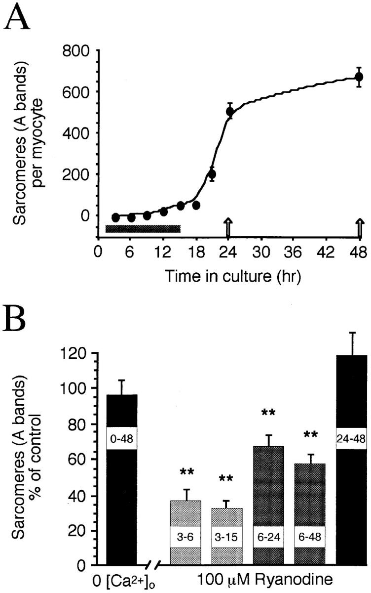
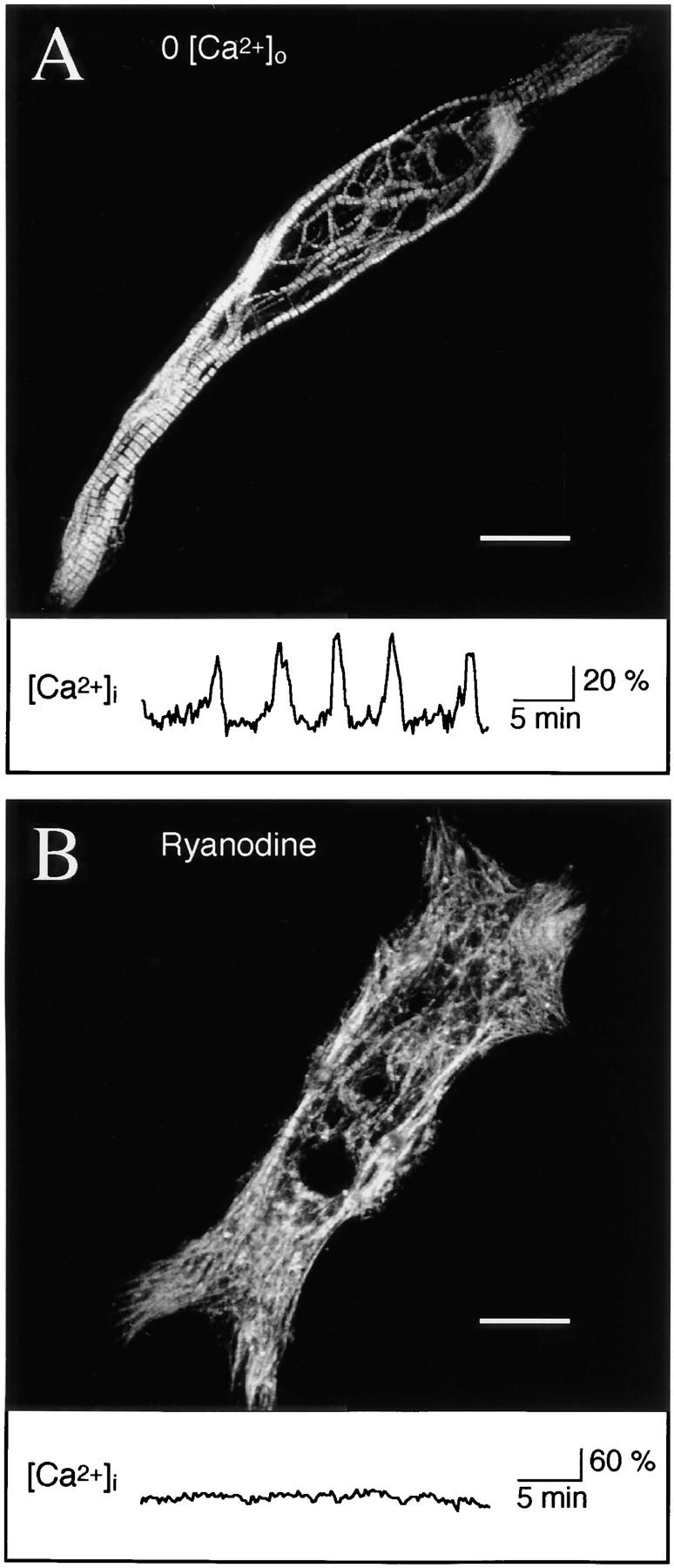
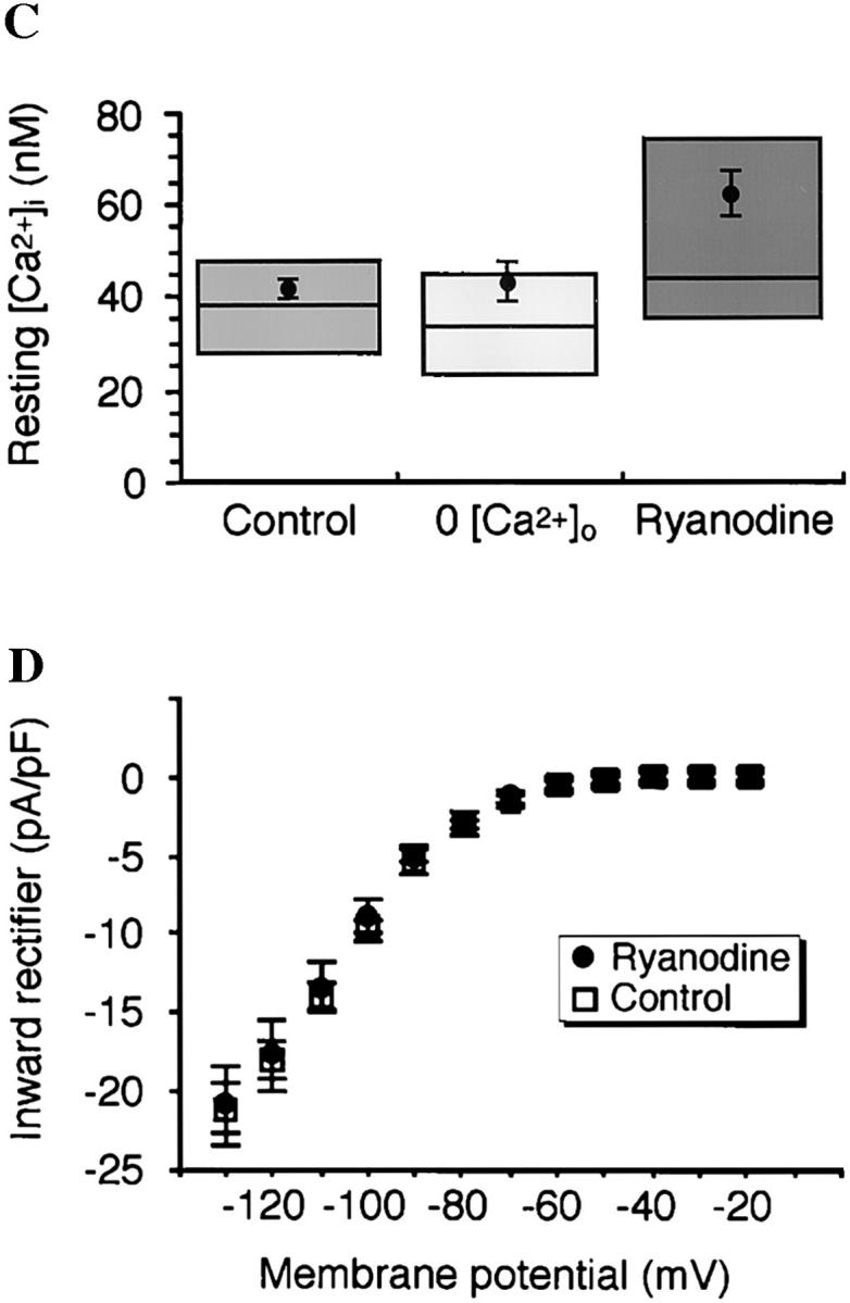
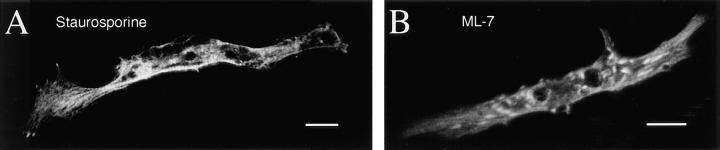
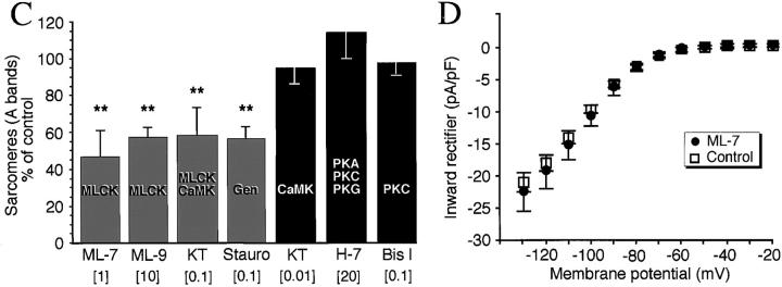
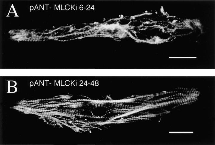
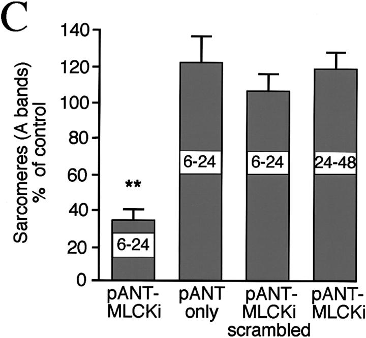
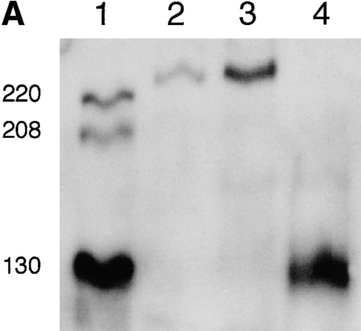
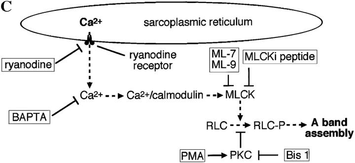
Spontaneous calcium release from intracellular stores occurs during myofibrillogenesis, the process of sarcomeric protein assembly in striated muscle. Preventing these Ca2+ transients disrupts sarcomere formation, but the signal transduction cascade has not been identified. Here we report that specific blockade of Ca2+ release from the ryanodine receptor (RyR) activated Ca2+ store blocks transients and disrupts myosin thick filament (A band) assembly. Inhibition of an embryonic Ca2+/calmodulin-dependent myosin light chain kinase (MLCK) by blocking the ATP-binding site, by allosteric phosphorylation, or by intracellular delivery of a pseudosubstrate peptide, also disrupts sarcomeric organization. The results indicate that both RyRs and MLCK, which have well-described calcium signaling roles in mature muscle contraction, have essential developmental roles during construction of the contractile apparatus.


Figures

Figure 2
Figure 2
Time course and ryanodine sensitivity of A band assembly in Xenopus myocytes. (A) Mean number of sarcomeres per myocyte (n ≥ 30 myocytes per time point) versus time in culture. The period of spontaneous Ca2+ transient production is shown along the x axis (gray bar). After experimental perturbations, sarcomeres were assayed at 24 or 48 h in culture (arrows). (B) Normalized mean sarcomere numbers per myocyte from chronic 0-Ca2+ and ryanodine-treated cultures. 0-Ca2+ cultures were assayed at 48 h; ryanodine was applied at 100 μM in control saline, with 3–6, 3–15, and 6–24 h treatments assayed at 24 h. Note that the early period of ryanodine sensitivity corresponds to the period of Ca2+ transient production shown above. Asterisks, significantly different from controls in this and subsequent figures.
Figure 1
Figure 1
Release of intracellular Ca2+ from RyR stores is necessary for myofibrillogenesis. (A) Myocytes grown 48 h in 0-Ca2+ medium are indistinguishable from controls with respect to bipolar morphology, parallel myofibril alignment, and number and regularity of sarcomeres. Image is a Z-series maximum projection of sections encompassing the full thickness of the cell. In the central region, a loose meshwork of myofibrils surrounds normal cell inclusions including numerous yolk platelets. Trace below shows Ca2+ transients that occur in normal and 0-Ca2+ media up to 15 h in culture (Ferrari et al., 1996). (B) Myocytes grown in the presence of ryanodine (100 μM) at early times show disruption of myofibrils and reduced sarcomere numbers; cell shown was treated with ryanodine from 6 to 48 h in culture. Major phenotypic characteristics are myofibril misalignments, diffuse myosin immunoreactivity throughout the cell, fewer sarcomeres, and localized patches of dense myosin accumulation. Trace below shows Ca2+ transients are blocked by ryanodine (n = 109 cells examined for 30–60 min at 3–9 h in culture; Ferrari et al., 1996). (C) Resting Ca2+ levels are not significantly altered in 0-Ca2+ medium or with the application of 100 μM ryanodine when measured during 3–9 h in culture. Means are from ≥20 cells per condition, boxes enclose 50% of the data, and lines indicate median values. (D) Mean steady-state current–voltage relations of the inward rectifier potassium current at 24–28 h in culture are normal in control and 100 μM ryanodine-treated (6–24 h in culture) cells. Number of myocytes is ≥12 for each condition. Bars, 20 μm.
Figure 1
Figure 1
Release of intracellular Ca2+ from RyR stores is necessary for myofibrillogenesis. (A) Myocytes grown 48 h in 0-Ca2+ medium are indistinguishable from controls with respect to bipolar morphology, parallel myofibril alignment, and number and regularity of sarcomeres. Image is a Z-series maximum projection of sections encompassing the full thickness of the cell. In the central region, a loose meshwork of myofibrils surrounds normal cell inclusions including numerous yolk platelets. Trace below shows Ca2+ transients that occur in normal and 0-Ca2+ media up to 15 h in culture (Ferrari et al., 1996). (B) Myocytes grown in the presence of ryanodine (100 μM) at early times show disruption of myofibrils and reduced sarcomere numbers; cell shown was treated with ryanodine from 6 to 48 h in culture. Major phenotypic characteristics are myofibril misalignments, diffuse myosin immunoreactivity throughout the cell, fewer sarcomeres, and localized patches of dense myosin accumulation. Trace below shows Ca2+ transients are blocked by ryanodine (n = 109 cells examined for 30–60 min at 3–9 h in culture; Ferrari et al., 1996). (C) Resting Ca2+ levels are not significantly altered in 0-Ca2+ medium or with the application of 100 μM ryanodine when measured during 3–9 h in culture. Means are from ≥20 cells per condition, boxes enclose 50% of the data, and lines indicate median values. (D) Mean steady-state current–voltage relations of the inward rectifier potassium current at 24–28 h in culture are normal in control and 100 μM ryanodine-treated (6–24 h in culture) cells. Number of myocytes is ≥12 for each condition. Bars, 20 μm.
Figure 3
Figure 3
Pharmacological inhibition of MLCK disrupts A band formation. All data are from 24 h cultures. (A) Staurosporine (100 nM), a general kinase inhibitor, inhibits thick filament assembly without disrupting bipolar morphology. Myosin is diffusely distributed throughout the cells, with regions of dense accumulation. (B) ML-7 (1 μM), a specific MLCK inhibitor, disrupts myosin incorporation into A bands without affecting bipolar morphology. Myosin is distributed diffusely throughout the cells with localized dense patches. (C) Summary of kinase inhibitor effects on A band assembly. Inhibitors were applied at the concentration indicated (μM) to block the listed kinase(s). Note that KT5926, which inhibits CaMK II with high specificity (10 nM), has no effect, whereas a higher concentration (100 nM) of this agent inhibits MLCK as well, producing the same effects as ML-7 and ML-9. KT, KT 5926; Stauro, staurosporine; Bis I, bisindolylmaleimide 1. (D) Development of the inward rectifier potassium current is unaffected by 6–24 h, 1 μM ML-7 when assayed at 24 h in culture. Bars, 20 μm.
Figure 3
Figure 3
Pharmacological inhibition of MLCK disrupts A band formation. All data are from 24 h cultures. (A) Staurosporine (100 nM), a general kinase inhibitor, inhibits thick filament assembly without disrupting bipolar morphology. Myosin is diffusely distributed throughout the cells, with regions of dense accumulation. (B) ML-7 (1 μM), a specific MLCK inhibitor, disrupts myosin incorporation into A bands without affecting bipolar morphology. Myosin is distributed diffusely throughout the cells with localized dense patches. (C) Summary of kinase inhibitor effects on A band assembly. Inhibitors were applied at the concentration indicated (μM) to block the listed kinase(s). Note that KT5926, which inhibits CaMK II with high specificity (10 nM), has no effect, whereas a higher concentration (100 nM) of this agent inhibits MLCK as well, producing the same effects as ML-7 and ML-9. KT, KT 5926; Stauro, staurosporine; Bis I, bisindolylmaleimide 1. (D) Development of the inward rectifier potassium current is unaffected by 6–24 h, 1 μM ML-7 when assayed at 24 h in culture. Bars, 20 μm.
Figure 4
Figure 4
Activation of PKC disrupts A band assembly. (A) Phorbol 12-myristate, 13-acetate (PMA; 10 nM), a potent activator of PKC, disrupts myofibrillogenesis in the same manner as MLCK inhibitors. Myosin is diffusely distributed throughout these cells, with dense punctate staining located centrally. (B) Co-application of the PKC inhibitor Bis I (100 nM) blocks the action of PMA. Myofibril and sarcomeric structure are normal. (C) PMA application from 6 to 24 h, but not 24 to 48 h, disrupts A band assembly and is rescued by Bis I. Bars, 20 μm.
Figure 4
Figure 4
Activation of PKC disrupts A band assembly. (A) Phorbol 12-myristate, 13-acetate (PMA; 10 nM), a potent activator of PKC, disrupts myofibrillogenesis in the same manner as MLCK inhibitors. Myosin is diffusely distributed throughout these cells, with dense punctate staining located centrally. (B) Co-application of the PKC inhibitor Bis I (100 nM) blocks the action of PMA. Myofibril and sarcomeric structure are normal. (C) PMA application from 6 to 24 h, but not 24 to 48 h, disrupts A band assembly and is rescued by Bis I. Bars, 20 μm.
Figure 5
Figure 5
Molecular inhibition of MLCK blocks A band assembly. (A) Intracellular delivery via antennapedia peptide (pANT) of a pseudosubstrate inhibitory peptide (MLCKi) results in disruption of myofibrillogenesis. (B) Later application of MLCKi at 24 h in culture has no effect on sarcomere assembly. (C) Effects of treatment with synthetic peptides. In addition to late application of MLCKi, pANT alone or pANT-scrambled MLCKi have no significant effect on myofibrillogenesis. Bars, 20 μm.
Figure 5
Figure 5
Molecular inhibition of MLCK blocks A band assembly. (A) Intracellular delivery via antennapedia peptide (pANT) of a pseudosubstrate inhibitory peptide (MLCKi) results in disruption of myofibrillogenesis. (B) Later application of MLCKi at 24 h in culture has no effect on sarcomere assembly. (C) Effects of treatment with synthetic peptides. In addition to late application of MLCKi, pANT alone or pANT-scrambled MLCKi have no significant effect on myofibrillogenesis. Bars, 20 μm.
Figure 6
Figure 6
Detection and developmental regulation of an embryonic MLCK isoform. (A) An embryonic isoform of MLCK is developmentally upregulated during the period of Ca2+ transient production. Western blot of Xenopus embryonic and adult skeletal muscle tissue with R57 antiserum shows that a single isoform running ∼225 kD is upregulated in embryonic myotomal tissue from stage 15 (lane 2) to stage 27 (lane 3), corresponding to 0 and 15 h in culture, respectively. This isoform is absent in adult skeletal muscle (lane 4), where a strong single band at 130 kD is detected. MLCK isoforms at 130, 208, and 220 kD are recognized in the rat A10 cell line (ATCC CRL1476) lysate (lane 1). (B) Striated pattern of labeling with the K36 smooth muscle MLCK mAb is visible in myocyte endfeet at 24 h in culture. The width and spacing of these bands matches the normal A band geometry. (C) Schematic model of Ca2+ transient–dependent MLCK activation and A band assembly in embryonic skeletal muscle. Inhibiting this cascade (dashed lines) at multiple points (boxes) disrupts formation of thick filaments. Bar, 20 μm.
Figure 6
Figure 6
Detection and developmental regulation of an embryonic MLCK isoform. (A) An embryonic isoform of MLCK is developmentally upregulated during the period of Ca2+ transient production. Western blot of Xenopus embryonic and adult skeletal muscle tissue with R57 antiserum shows that a single isoform running ∼225 kD is upregulated in embryonic myotomal tissue from stage 15 (lane 2) to stage 27 (lane 3), corresponding to 0 and 15 h in culture, respectively. This isoform is absent in adult skeletal muscle (lane 4), where a strong single band at 130 kD is detected. MLCK isoforms at 130, 208, and 220 kD are recognized in the rat A10 cell line (ATCC CRL1476) lysate (lane 1). (B) Striated pattern of labeling with the K36 smooth muscle MLCK mAb is visible in myocyte endfeet at 24 h in culture. The width and spacing of these bands matches the normal A band geometry. (C) Schematic model of Ca2+ transient–dependent MLCK activation and A band assembly in embryonic skeletal muscle. Inhibiting this cascade (dashed lines) at multiple points (boxes) disrupts formation of thick filaments. Bar, 20 μm.

Similar articles

Cited by

References

    1. Airey JA, Baring MD, Sutko JL. Ryanodine receptor protein is expressed during differentiation in the muscle cell lines BC3H1 and C2C12. Dev Biol. 1991;148:365–374. - PubMed
    1. Airey JA, Baring MD, Beck CF, Chelliah Y, Deerinck TJ, Ellisman MH, Houenou LJ, McKemy DD, Sutko JL, Talvenheimo J. Failure to make normal α ryanodine receptor is an early event associated with the crooked neck dwarf (cn) mutation in chicken. Dev Dynamics. 1993a;197:169–188. - PubMed
    1. Airey JA, Deerinck TJ, Ellisman MH, Houenou LJ, Ivanenko A, Kenyon JL, McKemy DD, Sutko JL. Crooked neck dwarf (cn) mutant chicken skeletal muscle cells in low density primary cultures fail to express normal alpha ryanodine receptor and exhibit a partial mutant phenotype. Dev Dyn. 1993b;197:189–202. - PubMed
    1. Berridge MJ. The AM and FM of calcium signalling. Nature. 1997;386:759–760. - PubMed
    1. Bouche M, Goldfine SM, Fischman DA. Posttranslational incorporation of contractile proteins into myofibrils in a cell-free system. J Cell Biol. 1988;107:587–596. - PMC - PubMed

Publication types

MeSH terms