. 1998 Jun 15;141(6):1357-70.
doi: 10.1083/jcb.141.6.1357.

The Src homology domain 3 (SH3) of a yeast type I myosin, Myo5p, binds to verprolin and is required for targeting to sites of actin polarization

Affiliations

The Src homology domain 3 (SH3) of a yeast type I myosin, Myo5p, binds to verprolin and is required for targeting to sites of actin polarization

B L Anderson et al. J Cell Biol. .

Abstract

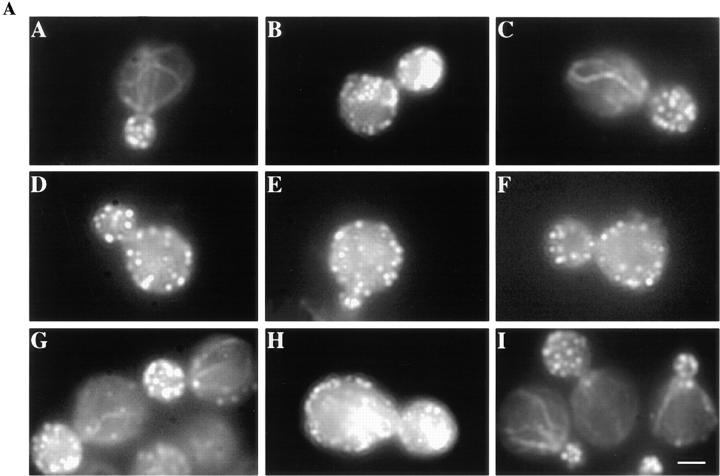
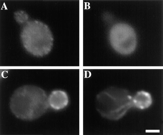
The budding yeast contains two type I myosins, Myo3p and Myo5p, with redundant functions. Deletion of both myosins results in growth defects, loss of actin polarity and polarized cell surface growth, and accumulation of intracellular membranes. Expression of myc-tagged Myo5p in myo3Delta myo5Delta cells fully restores wild-type characteristics. Myo5p is localized as punctate, cortical structures enriched at sites of polarized cell growth. We find that latrunculin-A-induced depolymerization of F-actin results in loss of Myo5p patches. Moreover, incubation of yeast cells at 37 degrees C results in transient depolarization of both Myo5p patches and the actin cytoskeleton. Mutant Myo5 proteins with deletions in nonmotor domains were expressed in myo3Delta myo5Delta cells and the resulting strains were analyzed for Myo5p function. Deletion of the tail homology 2 (TH2) domain, previously implicated in ATP-insensitive actin binding, has no detectable effect on Myo5p function. In contrast, myo3Delta myo5Delta cells expressing mutant Myo5 proteins with deletions of the src homology domain 3 (SH3) or both TH2 and SH3 domains display defects including Myo5p patch depolarization, actin disorganization, and phenotypes associated with actin dysfunction. These findings support a role for the SH3 domain in Myo5p localization and function in budding yeast. The proline-rich protein verprolin (Vrp1p) binds to the SH3 domain of Myo3p or Myo5p in two-hybrid tests, coimmunoprecipitates with Myo5p, and colocalizes with Myo5p. Immunolocalization of the myc-tagged SH3 domain of Myo5p reveals diffuse cytoplasmic staining. Thus, the SH3 domain of Myo5p contributes to but is not sufficient for localization of Myo5p either to patches or to sites of polarized cell growth. Consistent with this, Myo5p patches assemble but do not localize to sites of polarized cell surface growth in a VRP1 deletion mutant. Our studies support a multistep model for Myo5p targeting in yeast. The first step, assembly of Myo5p patches, is dependent upon F-actin, and the second step, polarization of actin patches, requiresVrp1p and the SH3 domain of Myo5p.


Figures

Figure 1
Figure 1
Schematic diagram of Myo5p mutants. pMYO5, plasmid-borne wild-type MYO5 gene bearing three copies of the myc epitope at its carboxy terminus; pTH2Δ, plasmid-borne, myc-tagged MYO5 gene bearing a deletion of the TH2 region; pSH3Δ, plasmid-borne, myc-tagged MYO5 gene bearing a deletion of the SH3 region; pTH2Δ-SH3Δ, plasmid-borne, myc-tagged MYO5 gene bearing deletions of the TH2 and SH3 regions; pSH3, plasmid-borne, myc-tagged SH3 domain of MYO5 gene. With the exception of pSH3, all constructs were expressed in myo3Δ myo5Δ mutant cells. pSH3 was expressed in the myo3Δ strain. All proteins were expressed under control of the MYO5 promoter.
Figure 2
Figure 2
Expression of myc-tagged myo5 mutant proteins. Western blot of whole cell protein extracts (200 μg/ lane) from wild-type (lane A), myo3Δ myo5Δ mutants containing empty vector (lane B), pMYO5 (lane C), pTH2Δ (lane D), pSH3Δ (lane E), and pTH2Δ-SH3Δ (lane F). The blot was probed using 9E10 anti-myc antibody (panel A) and myosin I protein-specific antibody G371 (panel B).
Figure 3
Figure 3
Growth characteristics of myo3Δ,myo5Δ mutants expressing various myo5 constructs. Wild-type (row A), and myo3Δ myo5Δ mutants containing empty vector (row B), pMYO5 (row C), pTH2Δ (row D), pSH3Δ (row E), and pTH2Δ–SH3Δ (row F) were grown on YPD–containing solid media at 30° or 37°C, or in YPD supplemented with 0.75 M KCl at 30°C.
Figure 4
Figure 4
The organization of the yeast actin cytoskeleton in myo3Δ myo5Δ cells rescued with various MYO5 constructs. Wild-type cells (A, panels A, D, and G), myo3Δ myo5Δ cells carrying empty vector (A, panels B, E, and H), pMYO5 (A, panels C, F, and I), pTH2Δ (B, panels A, D, and G), pSH3Δ (B, panels B, E, and H), or pTH2Δ–SH3Δ (B, panels C, F, and I) were fixed before shift to 37°C (A and B, panels A, B, and C), 30 min after a shift to 37°C (A and B, panels D, E, and F), and 90 min after a shift to 37°C (A and B, panels G, H, and I). Cells were converted to spheroplasts, stained with rhodamine– phalloidin, and then viewed as described in Materials and Methods. Bar, 1 μm.
Figure 4
Figure 4
The organization of the yeast actin cytoskeleton in myo3Δ myo5Δ cells rescued with various MYO5 constructs. Wild-type cells (A, panels A, D, and G), myo3Δ myo5Δ cells carrying empty vector (A, panels B, E, and H), pMYO5 (A, panels C, F, and I), pTH2Δ (B, panels A, D, and G), pSH3Δ (B, panels B, E, and H), or pTH2Δ–SH3Δ (B, panels C, F, and I) were fixed before shift to 37°C (A and B, panels A, B, and C), 30 min after a shift to 37°C (A and B, panels D, E, and F), and 90 min after a shift to 37°C (A and B, panels G, H, and I). Cells were converted to spheroplasts, stained with rhodamine– phalloidin, and then viewed as described in Materials and Methods. Bar, 1 μm.
Figure 5
Figure 5
Actin cytoskeletal polarization and repolarization after shift to 37°C are defective in pSH3Δ- or pTH2Δ–SH3Δ-expressing cells. Wild-type cells (A), myo3Δ myo5Δ cells carrying empty vector (B) and pMYO5 (C), pTH2Δ (D), pSH3Δ (E), or pTH2Δ-SH3Δ (F) were removed from liquid culture at the indicated times after shift to 37°C, fixed, converted to spheroplasts, and then stained with rhodamine–phalloidin. Cells were scored as having a depolarized actin cytoskeleton if they contained more than six actin patches within the mother cell. The graphs indicate the percentage of cells that retained polarization of the actin cytoskeleton. These percentages are the average of values from three experiments. n > 100 for each experiment.
Figure 6
Figure 6
Loss of actin patch polarity and Myo5p localization coincide after a shift to 37°C. myo3Δ myo5Δ cells expressing pMYO5 were grown to mid-log phase at 30°C (A and B) and shifted to 37°C. After 30 (C and D) or 90 min (E and F) of incubation, aliquots were removed, fixed, and then converted to spheroplasts. In this double-label experiment, actin (A, C, and E) and myc-tagged Myo5p (B, D, and F) were visualized as described above. Asymmetric Myo5p localization and actin patch polarization are lost after incubation at 37°C for 30 min (C and D) and restored after 90 min of incubation at 37°C (E and F). Bar, 1 μm.
Figure 7
Figure 7
Localization of Myo5p in LAT-A treated cells. myo3Δ myo5Δ cells expressing pMYO5 were grown to mid-log phase at 30°C. Aliquots were then removed and the cells were treated with 0.15 mM LAT-A dissolved in DMSO (C and D) or with equal volume of DMSO (A and B). After 15 min of incubation at 30°C cells were fixed, converted to spheroplasts, and then double labeled with anti-myc antibody (A and C) and rhodamine–phalloidin (B and D). Asymmetric Myo5p localization is lost in LAT-A– treated cells. Bar, 1 μm.
Figure 8
Figure 8
Localization of wild-type and mutant MYO5 gene products. Mid-log phase myo3Δ myo5Δ cells expressing pMYO5 (A and B), pTH2Δ (C and D), pSH3Δ (E and F), or pTH2Δ– SH3Δ (G and H) were fixed, converted to spheroplasts, and then stained for actin (A, C, E, and G) and myc (B, D, F, and H) as described above. The asymmetric localization of wild-type Myo5p patches in the bud is lost after deletion of the SH3 or TH2 and SH3 domains, but not after deletion of the TH2 domain alone. Arrows point to examples of colocalization of Myo5p patches with actin patches. Bar, 1 μm.
Figure 9
Figure 9
Localization of the SH3 domain of Myo5p. Mid-log phase myo3Δ cells expressing pMYO5 (A and B) or pSH3, the myc-tagged SH3 domain of Myo5p, (C and D) were fixed, converted to spheroplasts, and then stained for actin (A and C) and myc (B and D) as described above. The SH3 domain of Myo5p does not show the same localization as full-length Myo5p. Bar, 1 μm.
Figure 10
Figure 10
Myo5p colocalizes with Vrp1p and mislocalizes in a vrp1 deletion mutant. myo3Δ myo5Δ cells expressing HA-Vrp1p and Myc-Myo5p (A–D), were grown to mid-log phase, fixed, converted to spheroplasts, and then stained for HA-tagged Vrp1p (A and C), actin (B), and myc-tagged Myo5p (D). Arrows point to examples of Myo5p andVrp1p colocalization. To evaluate the effect of VRP1 deletion on Myo5p localization, a vrp1Δ mutant (G and H) and the corresponding wild-type strain (E and F), which express myc-Myo5p, were grown to mid-log phase, fixed, and stained for myc-Myo5p (E and G) and actin (F and H). Bar, 1 μm.
Figure 11
Figure 11
Vrp1p coimmunoprecipitates with Myo5p. Yeast strains that express no tagged constructs (HA10-1b), myc-tagged Myo5p (HA31-9c bearing pMYO5), or myc-tagged Myo5p and HA-tagged Vrp1p (VHA-1 bearing pMYO5) were grown to mid-log phase, disrupted, and then subjected to immunoprecipitation in the presence and absence of anti-myc antibody. Recovery of myc-tagged Myo5p was confirmed by Western blot analysis using anti-myc antibodies (left). Coimmunoprecipitation of HA-tagged Vrp1p with myc-tagged Myo5p was evaluated by decorating the same blot with anti-HA antibodies (right). The first lanes of each blot are whole cell lysates from VHA-1 bearing pMYO5 that illustrate the presence and electrophoretic mobility of tagged proteins.

Similar articles

Cited by

References

    1. Adams RJ, Pollard TD. Binding of myosin I to membrane lipids. Nature. 1989;340:565–568. - PubMed
    1. Adams AE, Botstein D, Drubin DG. Requirement of yeast fimbrin for actin organization and morphogenesis in vivo. . Nature. 1991;354:404–408. - PubMed
    1. Altschul SF, Gish W, Miller W, Myers EW, Lipman DJ. Basic local alignment search tool. J Mol Biol. 1990;215:403–410. - PubMed
    1. Amatruda JF, Gattermeir DJ, Karpova TS, Cooper JA. Effects of null mutations and overexpression of capping protein on morphogenesis, actin distribution, and polarized secretion in yeast. J Cell Biol. 1992;119:1151–1162. - PMC - PubMed
    1. Ayscough KR, Stryker J, Pokala N, Sanders M, Crews P, Drubin DG. High rates of actin filament turnover in budding yeast and roles for actin in establishment and maintenance of cell polarity revealed using the actin inhibitor latrunculin-a. J Cell Biol. 1997;137:399–416. - PMC - PubMed

Publication types