. 1998 Jun 15;141(6):1383-91.
doi: 10.1083/jcb.141.6.1383.

Spindle assembly and mitosis without centrosomes in parthenogenetic Sciara embryos

Affiliations

Spindle assembly and mitosis without centrosomes in parthenogenetic Sciara embryos

B de Saint Phalle et al. J Cell Biol. .

Abstract

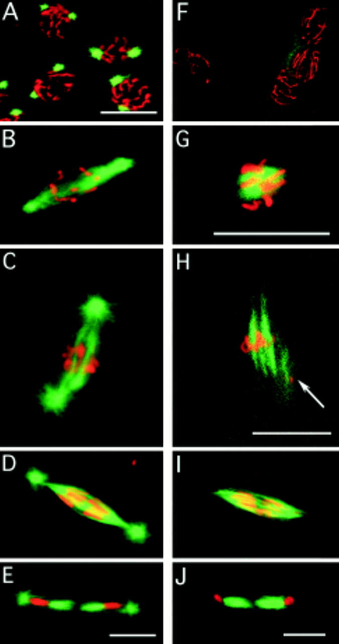
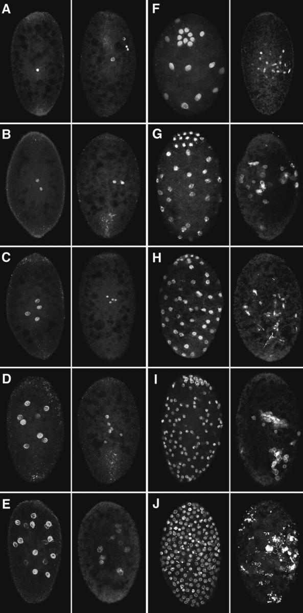
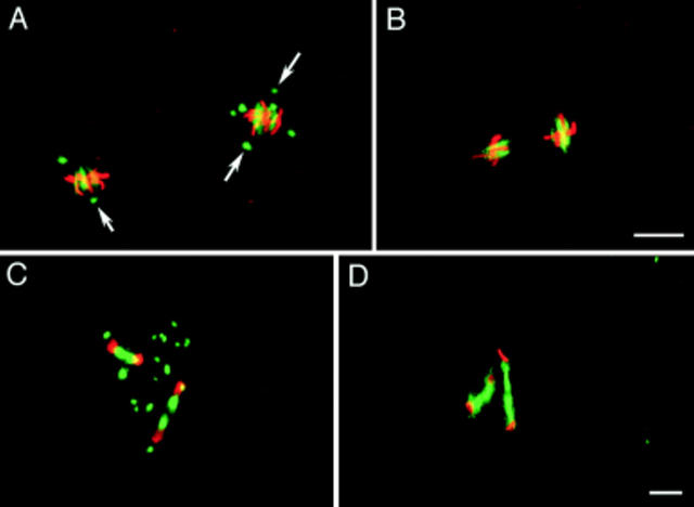
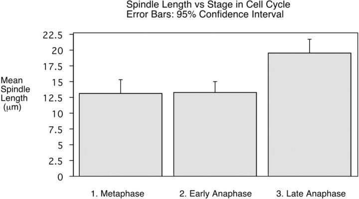
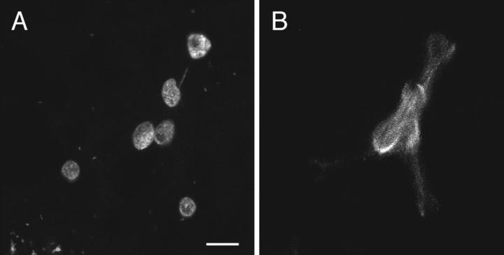
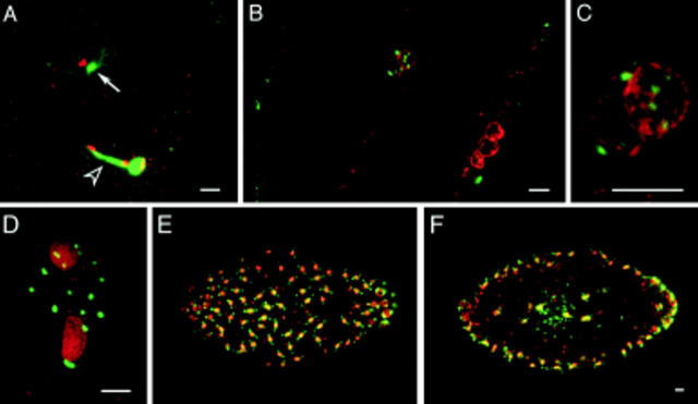
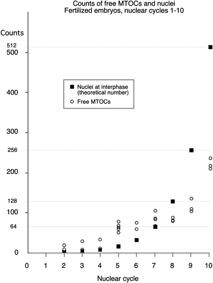
In Sciara, unfertilized embryos initiate parthenogenetic development without centrosomes. By comparing these embryos with normal fertilized embryos, spindle assembly and other microtubule-based events can be examined in the presence and absence of centrosomes. In both cases, functional mitotic spindles are formed that successfully proceed through anaphase and telophase, forming two daughter nuclei separated by a midbody. The spindles assembled without centrosomes are anastral, and it is likely that their microtubules are nucleated at or near the chromosomes. These spindles undergo anaphase B and successfully segregate sister chromosomes. However, without centrosomes the distance between the daughter nuclei in the next interphase is greatly reduced. This suggests that centrosomes are required to maintain nuclear spacing during the telophase to interphase transition. As in Drosophila, the initial embryonic divisions of Sciara are synchronous and syncytial. The nuclei in fertilized centrosome-bearing embryos maintain an even distribution as they divide and migrate to the cortex. In contrast, as division proceeds in embryos lacking centrosomes, nuclei collide and form large irregularly shaped nuclear clusters. These nuclei are not evenly distributed and never successfully migrate to the cortex. This phenotype is probably a direct result of a failure to form astral microtubules in parthenogenetic embryos lacking centrosomes. These results indicate that the primary function of centrosomes is to provide astral microtubules for proper nuclear spacing and migration during the syncytial divisions. Fertilized Sciara embryos produce a large population of centrosomes not associated with nuclei. These free centrosomes do not form spindles or migrate to the cortex and replicate at a significantly reduced rate. This suggests that the centrosome must maintain a proper association with the nucleus for migration and normal replication to occur.


Figures

Figure 3
Figure 3
Spindle assembly in the absence of centrosomes. Embryos are double stained for their DNA (red) and microtubules (green). Fertilized embryos: (A) In prophase, the chromosomes are condensed on the nuclear membrane, and the centrosomes have migrated to approximately a 180 degree position. (B) After nuclear membrane breakdown, the centrosomes of fertilized embryos nucleate microtubules that are preferentially directed toward the chromosomes. (C) Metaphase fertilized embryos have a spindle between the two centrosomes. (D) In early anaphase, the spindle is sharply pointed, and the chromosomes are starting to segregate. (E) In telophase, the midbody (central spindle) has formed between the two daughter nuclei of the division. Unfertilized embryos: (F) In prophase, the chromosomes are condensed on the nuclear membrane, but there are no MTOCs. (G) Tubulin is apparent on and near the chromosomes as the spindle starts to assemble. (H) Arrays of microtubules arise from the chromatin. The arrow marks a second metaphase nucleus partly in the plane of focus with a spindle also assembling. The close apposition of nuclei is typical of unfertilized embryos. (I) The appearance of the anaphase spindle is very similar to the anaphase spindle of fertilized embryos, except that centrosomes with astral microtubules are conspicuously absent. (J) In telophase, the midbody (central spindle) has formed between the two daughter nuclei of the division, even though there are no centrosomes. Bars, 10 μm.
Figure 1
Figure 1
Unfertilized embryos undergo many rounds of nuclear division but exhibit an abnormal nuclear distribution. Syncytial development in fertilized (left-hand panels) and unfertilized (right-hand panels) embryos stained for their DNA. Cycles 1–10 are depicted in A–J. Because of extensive nuclear clumping, the cycles after nuclear cycle 5 in the unfertilized embryos are only estimates. Approximate width of the embryos is 150 μm, length 200 μm.
Figure 2
Figure 2
Unfertilized embryos do not possess MTOCs. Nuclear cycle 2 embryos are stained for their DNA (red) and microtubules (green). (A) In a fertilized embryo, two of the MTOCs are associated with spindle poles while the others surround the metaphase plate. (B) In the equivalently staged unfertilized embryo, no MTOCs are present, and by counting the chromosome arms it can be seen that the nuclei are haploid. This may be why unfertilized nuclei seem smaller (see also Fig. 1). (C and D) Telophase of nuclear cycle 2 in fertilized and unfertilized embryos, respectively. While both exhibit robust midbodies, no MTOCs are present in the unfertilized embryo. Bars, 10 μm.
Figure 4
Figure 4
Anaphase B elongation of the spindle in unfertilized embryos. Spindle length was measured in fixed embryos. The average length of the spindle at metaphase is 13.15 μm (n = 8, SD 2.65 μm). In early anaphase, when the centromeres have separated but there is no gap between the segregating chromosomes, the average spindle length is 13.32 μm (n = 12, SD 2.58 μm). In late anaphase, when a gap has appeared between the segregating chromosomes, the average spindle length is 19.55 μm (n = 17, SD 4.20 μm). The difference between the length of early anaphase and late anaphase spindles was statistically highly significant (P = 0.0001 by an unpaired t test). The mean length of the spindle in late anaphase is likely to be an underestimate because for the majority of the spindles measured, the chromosomes had not yet reached the poles.
Figure 5
Figure 5
In unfertilized embryos, the spindles are improperly oriented and the products of neighboring nuclear divisions collide with one another. An unfertilized embryo in telophase of nuclear cycle 3, stained for both DNA (A) and tubulin (B). The spindles are in a single plane and are oriented so that the nuclear products from three separate divisions will collide with one another. This is a magnified view of the embryo depicted in Fig. 1 D. Bar, 10 μm.
Figure 6
Figure 6
Free MTOCs are present in fertilized Sciara embryos. These embryos are double stained for their DNA (red) and microtubules (green). (A) During telophase of female meiosis II, the centrosome associated with the decondensing male pronucleus is nucleating a large aster (arrow), and a large midbody separates the female pronucleus and the second polar body. (B) The asters of eight MTOCs are in the plane of focus surrounding the fusing pronuclei. Three polar bodies reside at the cortex. (C) Magnified view of pronuclei. (D) During interphase of nuclear cycle 2, the two nuclei are encompassed by 16 MTOCs. E and F depict surface and medial views of a nuclear cycle 8 embryo and demonstrate that the free MTOCs do not migrate to the cortex. Bars, 10 μm.
Figure 7
Figure 7
Free MTOC duplication during nuclear cycles 1–10. The number of free MTOCs, on the y axis, plotted against the nuclear cycle of the embryo on the x axis. The theoretical number of nuclei (2n −1, where n is the nuclear cycle) is also shown for each cycle. MTOCs not associated with nuclei (free MTOCs) were counted in a series of optical sections taken at 5-μm increments in mitotic embryos. The number of free MTOCs does not increase exponentially, but it does increase from ∼20 in cycle 1 to ∼200 in cycle 10.

References

    1. Aist JR, Bayles CJ, Tao W, Berns MW. Direct evidence for the existence, structural basis and function of astral forces during anaphase B in vivo. J Cell Sci. 1991;100:279–288. - PubMed
    1. Baker J, Theurkauf WE, Schubiger G. Dynamic changes in microtubule configuration correlate with nuclear migration in the preblastoderm Drosophilaembryo. J Cell Biol. 1993;122:113–121. - PMC - PubMed
    1. Balczon R. The centrosome in animal cells and its functional homologs in plant and yeast cells. Int Rev Cytol. 1996;169:25–82. - PubMed
    1. Boleti H, Karsenti E, Vernos I. Xklp2, a novel Xenopuscentrosomal kinesin-like protein required for centrosome separation during mitosis. Cell. 1996;84:49–59. - PubMed
    1. Cassimeris L, Rieder CL, Salmon ED. Microtubule assembly and kinetochore directional instability in vertebrate monopolar spindles: implications for the mechanism of chromosome congression. J Cell Sci. 1994;107:285–297. - PubMed

Publication types