. 1998 Jun 15;141(6):1393-406.
doi: 10.1083/jcb.141.6.1393.

Mammalian p55CDC mediates association of the spindle checkpoint protein Mad2 with the cyclosome/anaphase-promoting complex, and is involved in regulating anaphase onset and late mitotic events

Affiliations

Mammalian p55CDC mediates association of the spindle checkpoint protein Mad2 with the cyclosome/anaphase-promoting complex, and is involved in regulating anaphase onset and late mitotic events

M Kallio et al. J Cell Biol. .

Abstract

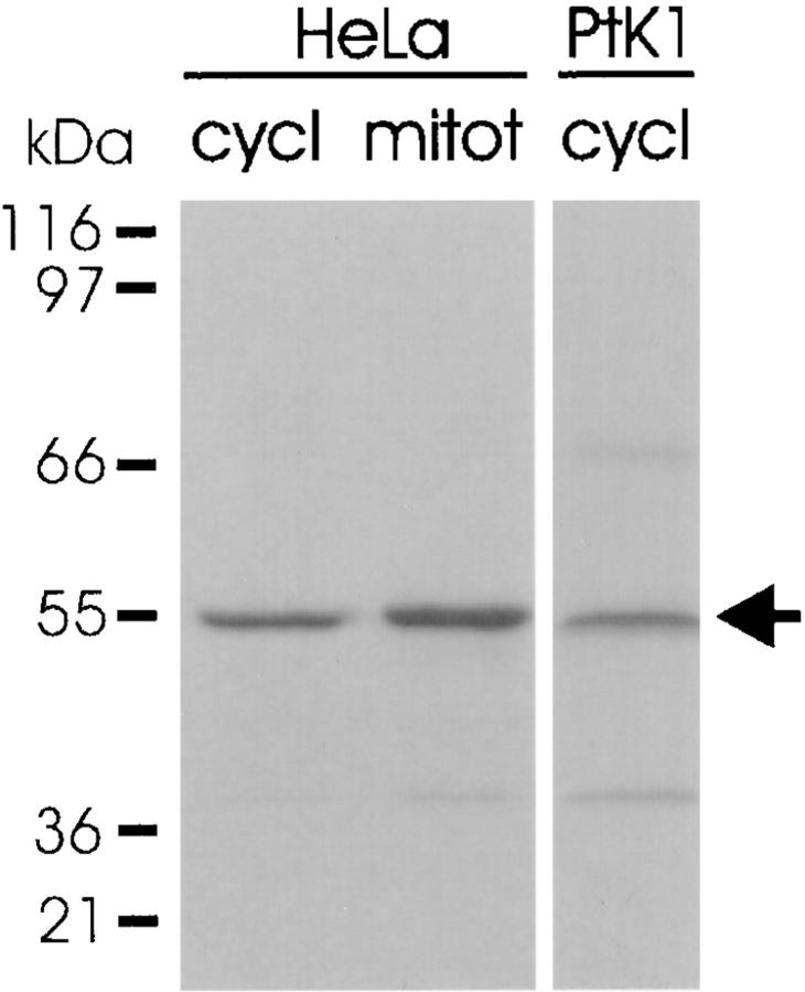
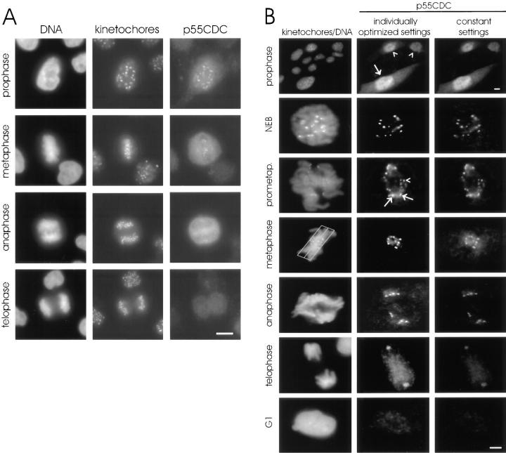
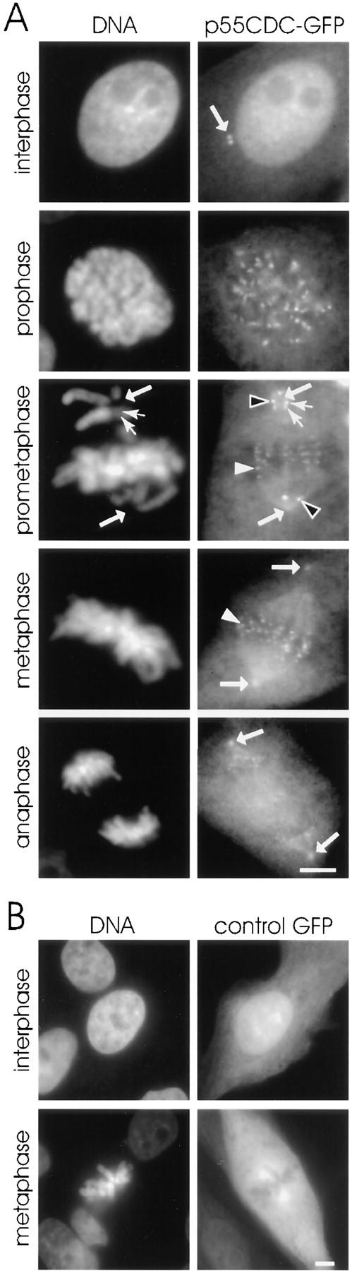
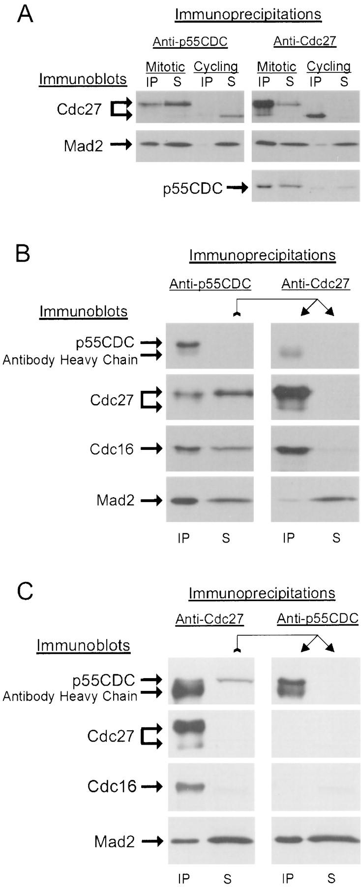
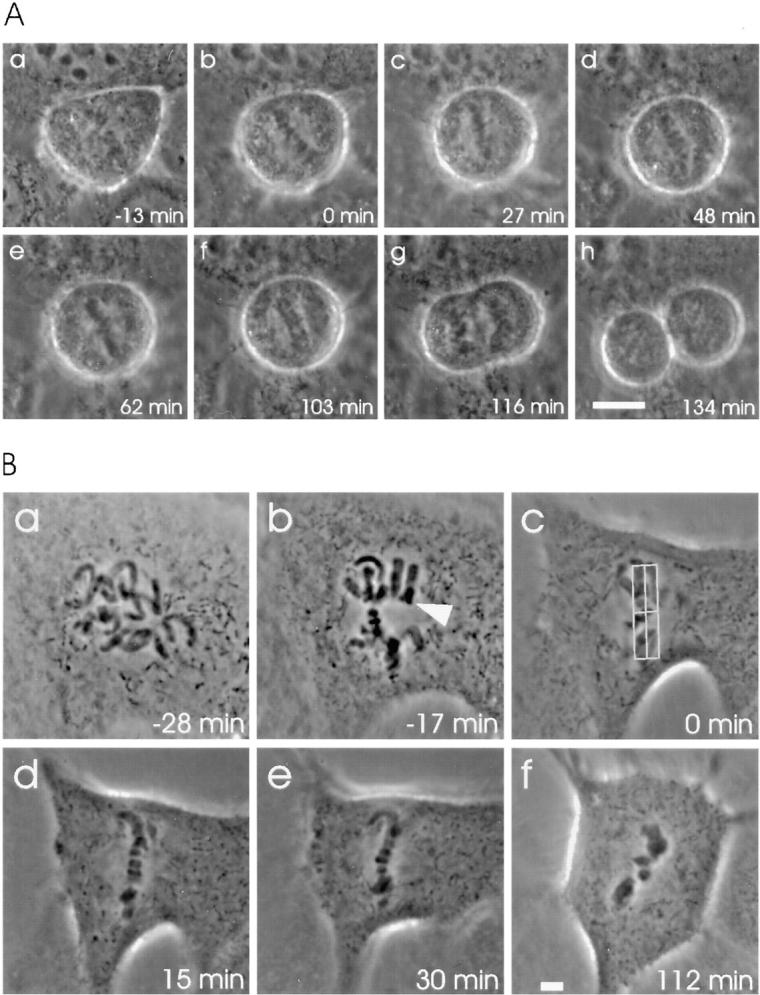
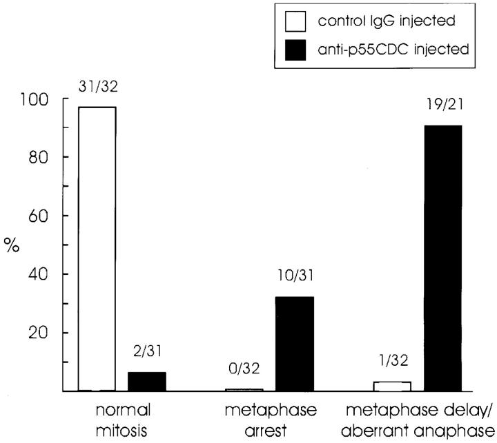
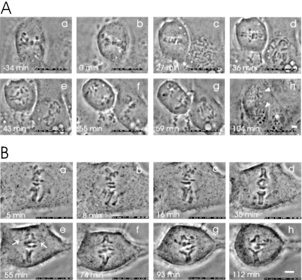
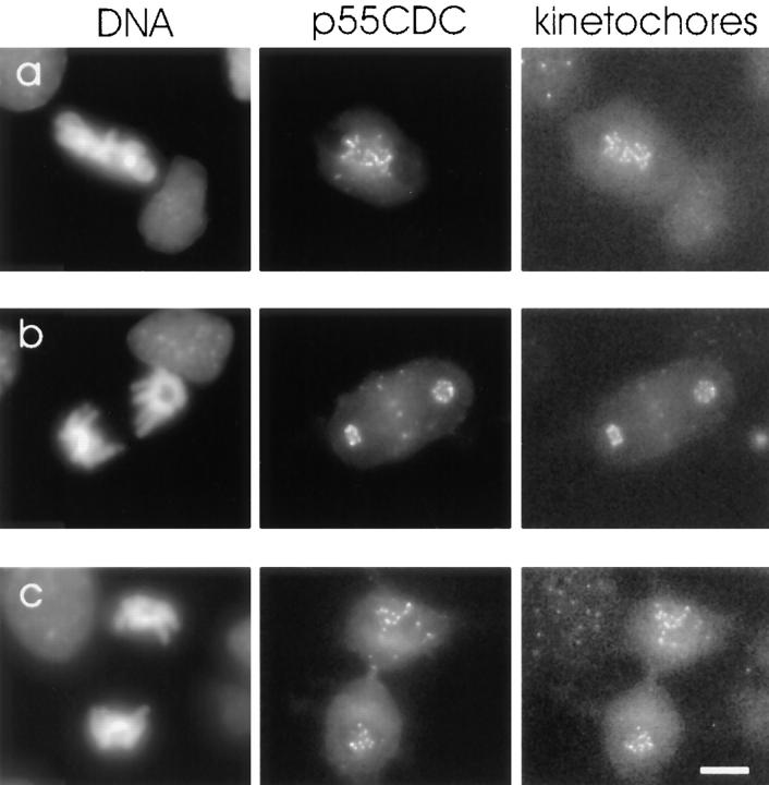
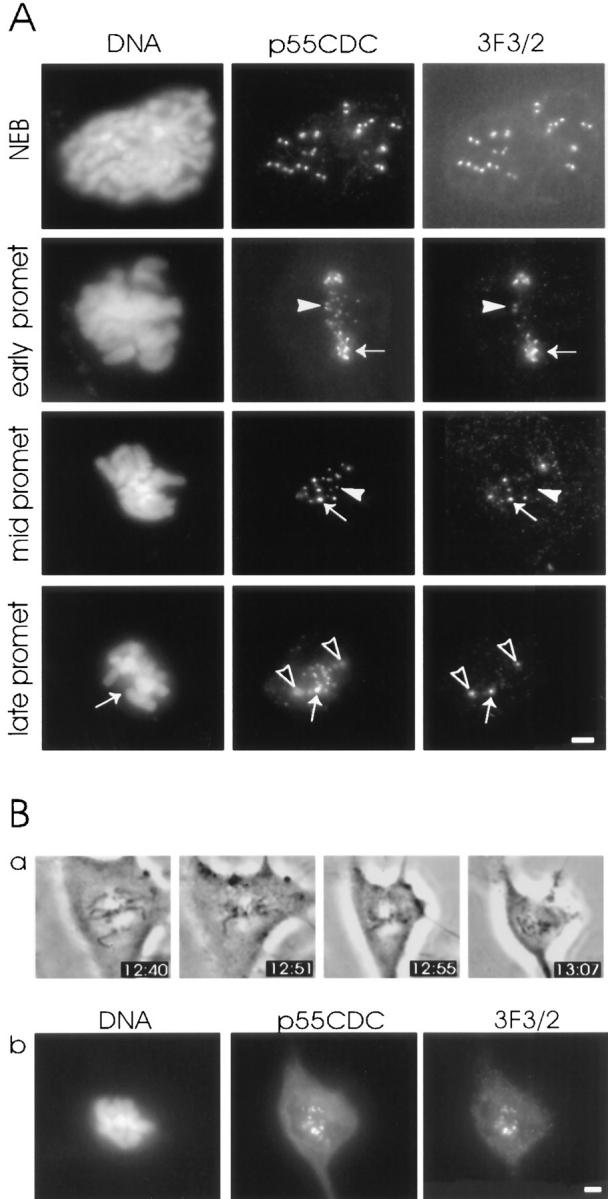
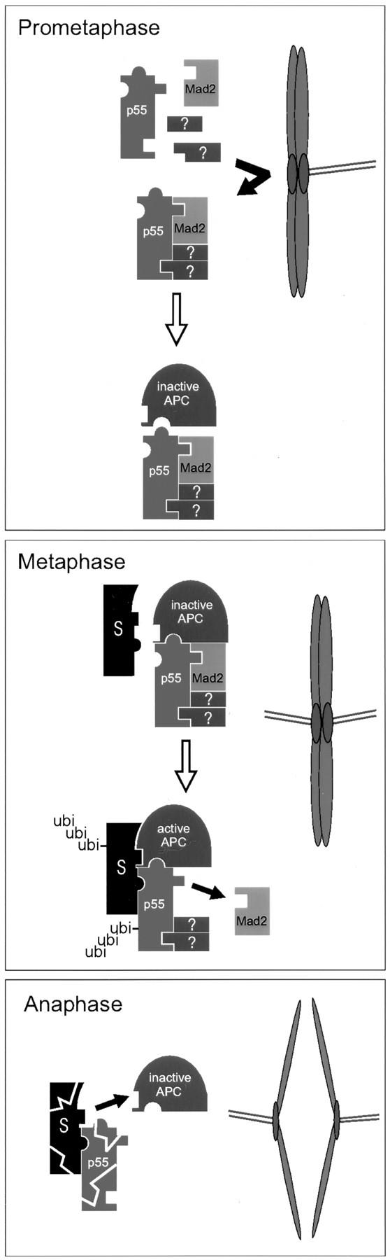
We have investigated the function of p55CDC, a mammalian protein related to Cdc20 and Hct1/Cdh1 in Saccharomyces cerevisiae, and Fizzy and Fizzy-related in Drosophila. Immunofluorescence studies and expression of a p55CDC-GFP chimera demonstrate that p55CDC is concentrated at the kinetochores in M phase cells from late prophase to telophase. Some p55CDC is also associated with the spindle microtubules and spindle poles, and some is diffuse in the cytoplasm. At anaphase, the concentration of p55CDC at the kinetochores gradually diminishes, and is gone by late telophase. In extracts prepared from M phase, but not from interphase HeLa cells, p55CDC coimmunoprecipitates with three important elements of the M phase checkpoint machinery: Cdc27, Cdc16, and Mad2. p55CDC is required for binding Mad2 with the Cdc27 and Cdc16. Thus, it is likely that p55CDC mediates the association of Mad2 with the cyclosome/anaphase-promoting complex. Microinjection of anti-p55CDC antibody into mitotic mammalian cells induces arrest or delay at metaphase, and impairs progression of late mitotic events. These studies suggest that mammalian p55CDC may be part of a regulatory and targeting complex for the anaphase-promoting complex.


Figures

Figure 1
Figure 1
Expression of p55CDC in proliferating mammalian cells. The antibody recognizes a 55-kD band (arrow) in whole HeLa cell extracts from both cycling cultures (cycl) and after mitotic arrest (mitot). In whole cycling PtK1 cell extracts, the anti-p55CDC antibody recognizes primarily a 55-kD band, although slight reactivity is seen with two other bands at ∼40 and ∼70 kD.
Figure 2
Figure 2
Immunofluorescent localization of p55CDC in HeLa and PtK1 cells. (A) HeLa cells. The left column shows DNA (DAPI staining), the middle column shows kinetochores (human autoimmune antibody), and the right column shows p55CDC labeling. At prophase, diffuse labeling is seen throughout the cells with a concentration at the kinetochores. The kinetochore localization is apparent from late prophase to telophase. During anaphase, kinetochore labeling diminishes and is lost by the end of telophase. The diffuse cytoplasmic labeling persists until the M-to-G1 transition. (B) PtK1 cells. The left column shows DNA (Yo-Pro staining) and kinetochores (human autoimmune antibody). The middle column demonstrates p55CDC staining at different stages of mitosis where intensity and contrast settings have been optimized for each individual panel. This technique permits visualization of the staining at anaphase and telophase. The right column shows p55CDC labeling in which intensity and contrast settings were kept constant for all stages, allowing comparison of the relative intensity of labeling at different stages of mitosis. The top set of panels, taken at low magnification, shows a field of cells with some in G2 (arrowheads) and one in prophase (arrow). p55CDC relocalizes from the diffuse cytoplasmic/nuclear pool to the kinetochores of chromosomes just before NEB. Kinetochores show different intensities of labeling during prometaphase. At prometaphase, the kinetochores near the spindle poles often possess brighter signals (arrows) compared with ones that are closer to the spindle equator (arrowhead). The box (3 μm on either side of the spindle equator) used to define metaphase is shown. During anaphase, the anti-p55CDC signal gradually diminishes at the kinetochores, and ultimately disappears by late telophase. Bars, 5 μm.
Figure 3
Figure 3
(A) Fixed LLC-PK1 cells expressing chimeric GFP-p55CDC. In interphase cells, a pair of dots at the centrosome (arrow) is brightly labeled throughout the cell cycle. In mitotic cells, the p55CDC-GFP concentrates at the kinetochores from late prophase to late anaphase. A pool of the protein also remains diffusely distributed. In addition, the spindle poles (arrows) and the kinetochore microtubules show concentrations of the p55CDC-GFP. During prometaphase, the intensity of the kinetochore p55CDC-GFP signal is more intense at the kinetochores of unaligned chromosomes located near the spindle poles (black arrowheads) compared with the kinetochores of chromosomes aligned at metaphase plate (white arrowheads). No apparent difference is seen in intensity of the p55CDC-GFP signal between the leading and the trailing kinetochore of individual unaligned chromosome (small arrows in prometaphase row). In anaphase cells, the spindle poles (big arrows) remain p55CDC-GFP-positive while kinetochore labeling diminishes. (B) An interphase and a mitotic LLC-PK1 cell expressing the GFP alone show no specific fluorescent kinetochore, spindle microtubule, or spindle pole signals. Bars, 5 μm.
Figure 4
Figure 4
p55CDC binds to Mad2 protein and to the APC. (A) Cdc27 and Mad2 proteins are detected in anti-p55CDC immunoprecipitates from mitotic, but not interphase HeLa cell extracts (Cdc27 protein is indicated in two locations since it undergoes a phosphorylation shift in M phase). Mad2 and p55CDC proteins are detected in anti-Cdc27 immunoprecipitates from mitotic, but not interphase HeLa cell extracts. (B) A sequential immunoprecipitation experiment showing that p55CDC is necessary for Mad2 binding to the APC. A mitotic extract of HeLa cells was first immunodepleted of p55CDC protein (left). Some of the APC remains in the supernatant. The p55CDC-depleted extract was then used in a second immunoprecipitation with anti-Cdc27 antibody. In the absence of p55CDC, almost none of the remaining Mad2 coprecipitates with the Cdc27 protein. (C) Sequential immunoprecipitation experiment demonstrating that p55CDC and Mad2 form complexes independent of the APC. The mitotic HeLa cell extract was first immunodepleted of APC components by immunoprecipitation with anti-Cdc27 protein. The APC-depleted extract was then used for a second immunoprecipitation with anti-p55CDC. In the absence of APC components, a substantial portion of the Mad2 protein is complexed with p55CDC.
Figure 5
Figure 5
Microinjection with antibodies against p55CDC hampers the metaphase-to-anaphase transition. (A) Anti-p55CDC microinjection induces a transient metaphase arrest in HeLa cells. The antibody (2.0 mg/ml in the micropipette) was injected at prometaphase 18 min before the metaphase plate formed (time 0 = metaphase). (a) The first image was taken 5 min after injection. (b–f) During the metaphase arrest, the chromosomes underwent occasional stretching, but were rapidly pulled back to form a tight metaphase plate. (f) The onset of anaphase occurred 103 min after the tight metaphase plate formed for the first time. (f–g) Separation and movement of the sister chromatids required 13 min, which is slightly longer than the average of control cells. (g–h) Cytokinesis and exit from mitosis appeared normal. (B) Anti-p55CDC microinjection induces metaphase arrest in PtK1 cells. The cell was injected with anti-p55CDC (2.0 mg/ml) at late prophase a few minutes before breakdown of the nuclear envelope. (a) The first image was taken 5 min later, when the cell was at early prometaphase. (b) The last chromosome (arrowhead) became bioriented 17 min before the metaphase plate was formed. Progression through prometaphase appeared normal. (c) The last chromosome aligned at the spindle equator, and a metaphase plate was formed. The box (3 μm on either side of the spindle equator) used to define the start of metaphase is shown. (c–f) The cell did not initiate anaphase during the time it was monitored (total time = 168 min after the beginning of metaphase). Bars, 5 μm.
Figure 6
Figure 6
Cell cycle progression in PtK1 cells injected with nonspecific rabbit IgG (8.0 mg/ml) or anti-p55CDC (1.0–2.5 mg/ml). The numbers above the bars denote the proportion of cells in each category. Control IgG or anti-p55CDC injected cells were considered to exhibit a delay at metaphase if the duration of metaphase was more than double the mean duration of metaphase in the control cells. Cells were considered to exhibit an aberrant anaphase if they arrested during anaphase or if the duration of anaphase was more than double the mean duration of anaphase in control cells.
Figure 7
Figure 7
Microinjection of mitotic PtK1 cells with anti-p55CDC antibodies impairs normal progression of late mitotic events. (A) Slow separation of sister chromatids and impeded exit from mitosis by anti-p55CDC microinjection. The cell was microinjected with anti-p55CDC antibodies (1.5 mg/ ml in the micropipette) at early prometaphase (6 min after NEB). (a) The first image was taken 13 min later when the cell was at late prometaphase. (b) The chromosomes reached the metaphase plate. (c) Anaphase initiated after a short delay of ∼26 min. (c–g) Separation of sister chromatids was very slow, requiring over 50 min before the sister chromatids reached the spindle poles. The cell failed to undergo cytokinesis, and exited mitosis as a binucleate cell (arrowheads in h). To the right of the microinjected cell, an untreated cell progressed through a normal mitosis. The asterisk in h denotes one of the progeny of this normal division. (B) Inhibition of sister chromatid separation by anti-p55CDC microinjection. The cell was injected at late prometaphase with anti-p55CDC antibodies (1.5 mg/ml). (a) The first evidence of chromatid segregation was seen 5 min after the start of metaphase (time point = 0 min). (b–h) Despite pulling forces evident from stretching of the kinetochore regions (e, arrows), full separation of sister chromatid arms was never achieved. Bars, 5 μm.
Figure 8
Figure 8
Tracking of anti-p55CDC antibody injected into living cells. Cells were microinjected with anti-p55CDC, observed for a time, and then fixed for immunofluorescence to determine the localization of the injected antibody. Cells were colabeled with human autoimmune antibodies to localize kinetochores. (a) PtK1 cell arrested at metaphase after microinjection with anti-p55CDC at early prometaphase. (b) An anti-p55CDC–microinjected PtK1 cell at late anaphase. The cell was microinjected at late prometaphase. (c) A anti-p55CDC–microinjected PtK1 cell at late telophase. The antibodies were introduced at late prometaphase. Note that the microinjected anti-p55CDC antibody remained bound to kinetochores even at late telophase. Bar, 10 μm.
Figure 9
Figure 9
Expression of 3F3/2 phosphoepitope at the kinetochores of anti-p55CDC microinjected PtK1 cells. (A) Similar to p55CDC signal, the intensity of the fluorescent label of 3F3/2 epitope varies among chromosomes with different positions along the spindle. The kinetochores of chromosomes closer to spindle poles possess brighter 3F3/2 and p55CDC signals (arrows in early prometaphase and mid prometaphase rows) compared with kinetochores of aligned chromosomes (arrowheads in early prometaphase and mid prometaphase rows). At late prometaphase and metaphase the 3F3/2 signal disappears from the kinetochores of aligned chromosomes, while p55CDC is still present at all kinetochores. One misaligned chromosome having a bright p55CDC signal also shows strong labeling with the 3F3/2 antibody (arrows in late prometaphase row). The spindle poles that are labeled by both p55CDC and 3F3/2 are denoted with open arrowheads. (B) The 3F3/2 phosphoepitope expression reappears at the kinetochores of anti-p55CDC injected cells at metaphase if the spindle is destroyed with nocodazole. (a) At 12:35 the early prometaphase cell was injected with antibodies against p55CDC (1.0 mg/ml). At 12:55, all the chromosomes were aligned at the spindle equator. Nocodazole (5 μg/ml) was added at 12:57. (b) The cell was fixed 15 min later, and was immunolabeled to detect injected anti-p55CDC and the 3F3/2 phosphoepitope. Bars, 5 μm.
Figure 10
Figure 10
Model for p55CDC function. p55CDC and Mad2 proteins associate with unattached kinetochores in prometaphase cells (Figs. 2 and 3; Chen et al., 1996) where p55CDC and Mad2 complexes are formed (Fig. 5 A). p55CDC/Mad2 complex formation does not require the presence of APC (Fig. 5 C). The complex may also contain other yet uncharacterized elements (question marks). p55CDC/Mad2 complex production ceases when all chromosomes are properly oriented in the spindle. The p55CDC/ Mad2 complex then binds the APC (Fig. 5 B). The APC activity is inhibited by Mad2 (Li et al., 1997). p55CDC participates in targeting the APC to proteolytic substrates (S). Substrate binding may occur before or after activation of the APC (binding to inactive APC/p55CDC/Mad2 complex shown). The APC is activated upon loss of Mad2. This activation leads to ubiquitination of mitotic target proteins and p55CDC by the APC. At anaphase onset, the polyubiquitinated substrate proteins and p55CDC are proteolysed by the proteosome. During late mitotic events, APC may associate with other p55CDC-like protein/substrate complexes and be reused for their destruction.

References

    1. Campbell MS, Gorbsky GJ. Microinjection of mitotic cells with 3F3/2 anti-phosphoepitope antibody delays the onset of anaphase. J Cell Biol. 1995;129:1195–1204. - PMC - PubMed
    1. Chen R-H, Waters JC, Salmon ED, Murray AW. Association of spindle assembly checkpoint component XMAD2 with unattached kinetochores. Science. 1996;274:242–246. - PubMed
    1. Cohen-Fix O, Peters J-M, Kirschner MW, Koshland D. Anaphase initiation in Saccharomyces cerevisiae is controlled by the APC-dependent degradation of the anaphase inhibitor Pds1p. Genes Dev. 1996;10:3081–3093. - PubMed
    1. Cyert MS, Scherson T, Kirschner MW. Monoclonal antibodies specific for thiophosphorylated proteins recognize XenopusMPF. Dev Biol. 1988;129:209–216. - PubMed
    1. Dawson IA, Roth S, Akam M, Artavanis-Tsakonas S. Mutations of the fizzy locus cause metaphase arrest in Drosophila melanogasterembryos. Development. 1993;117:359–376. - PubMed

Publication types

MeSH terms