. 1998 Jun 15;141(6):1449-65.
doi: 10.1083/jcb.141.6.1449.

Tyrosine phosphorylation and src family kinases control keratinocyte cell-cell adhesion

Affiliations

Tyrosine phosphorylation and src family kinases control keratinocyte cell-cell adhesion

E Calautti et al. J Cell Biol. .

Abstract

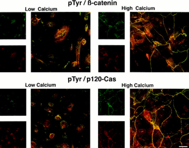
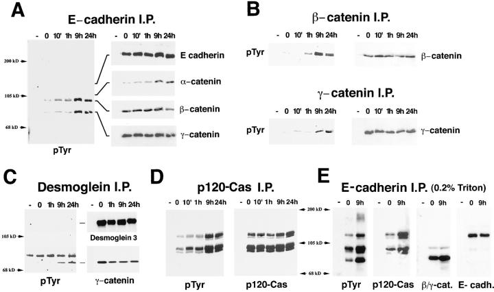
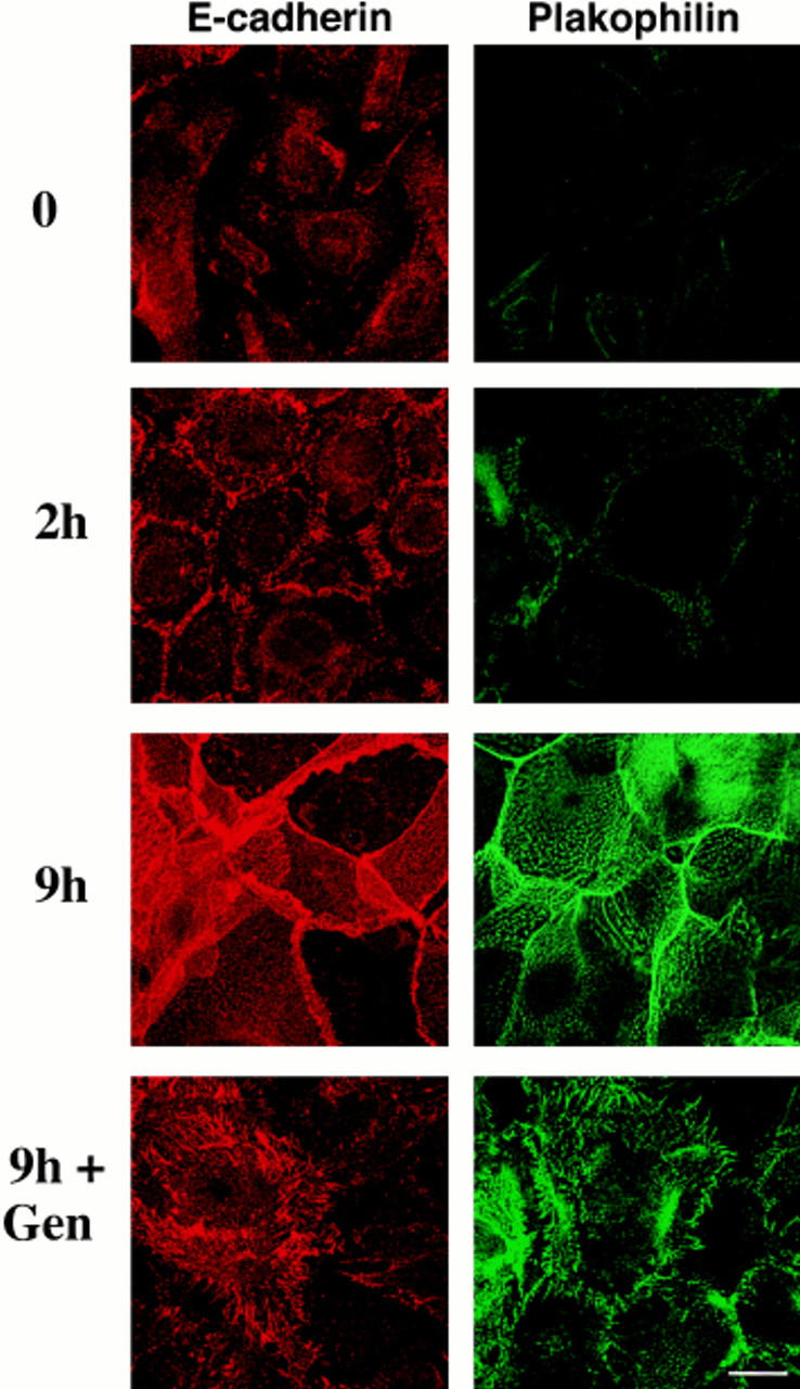
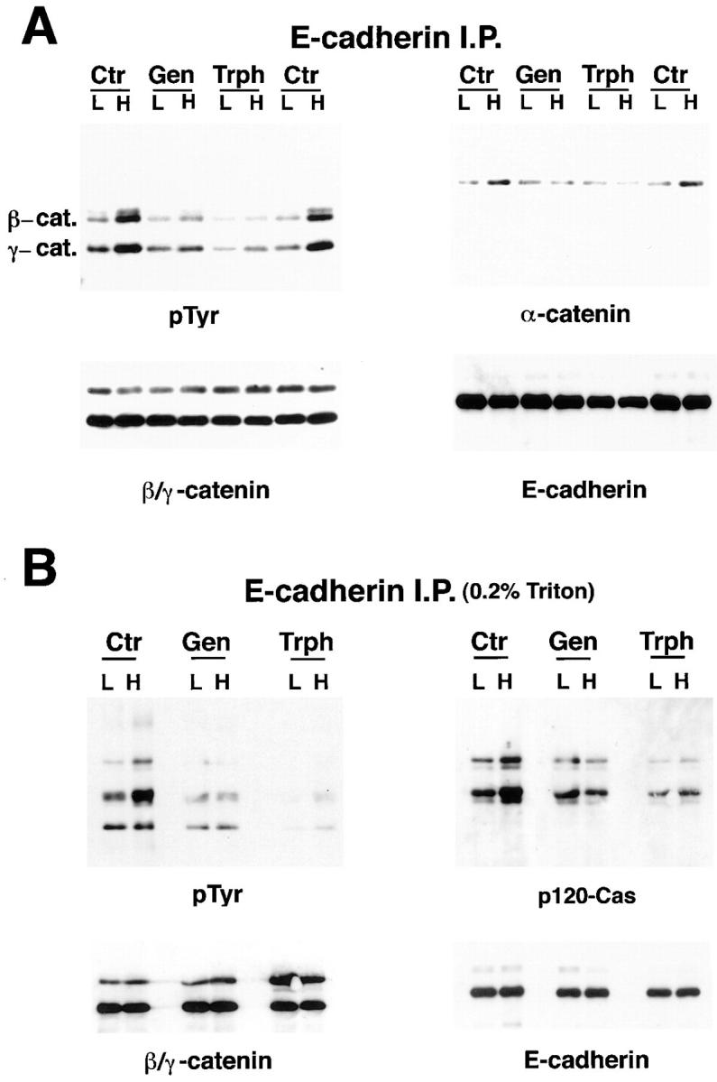
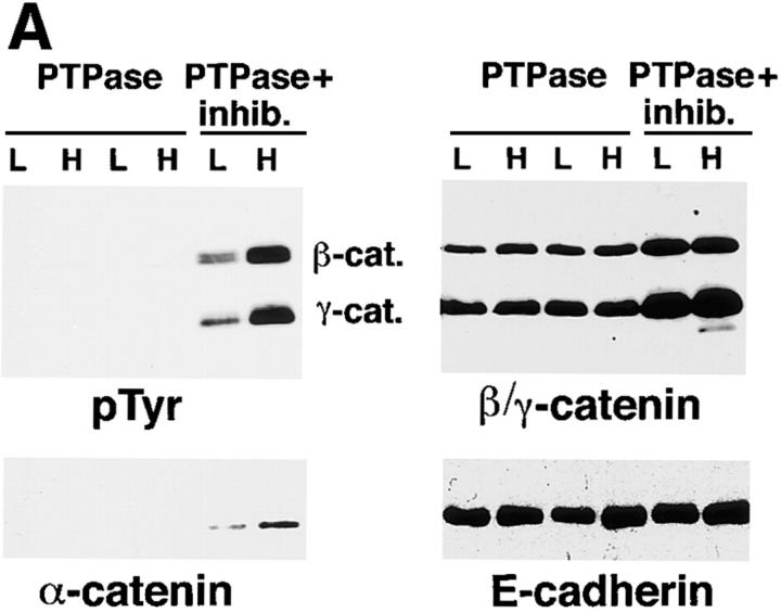
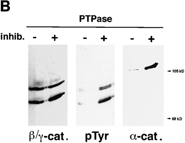
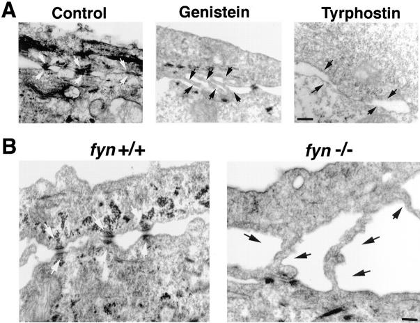
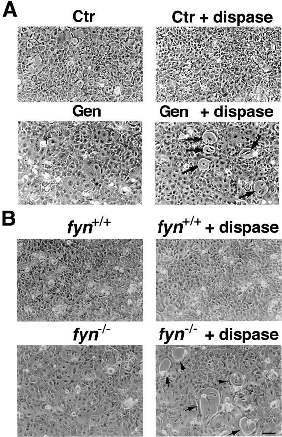
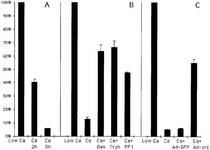
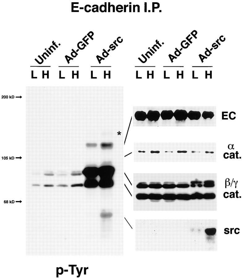
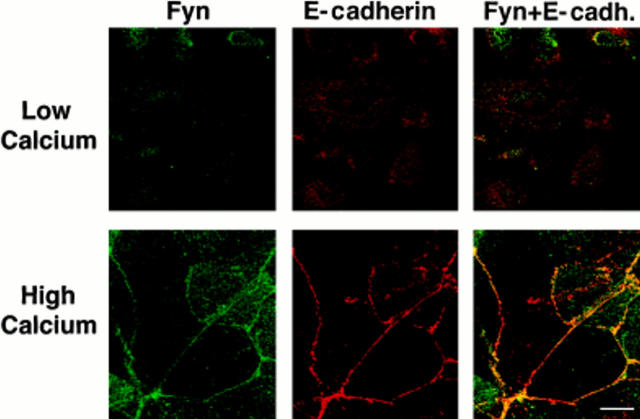
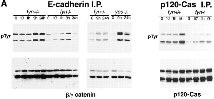
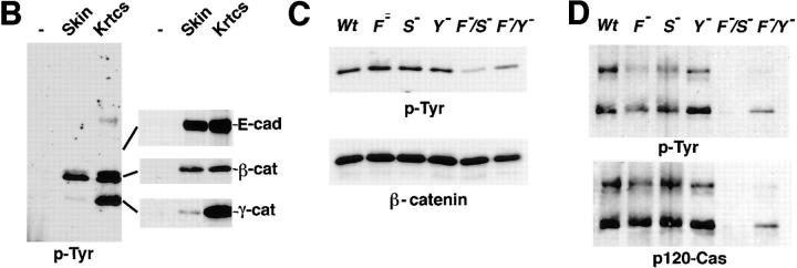
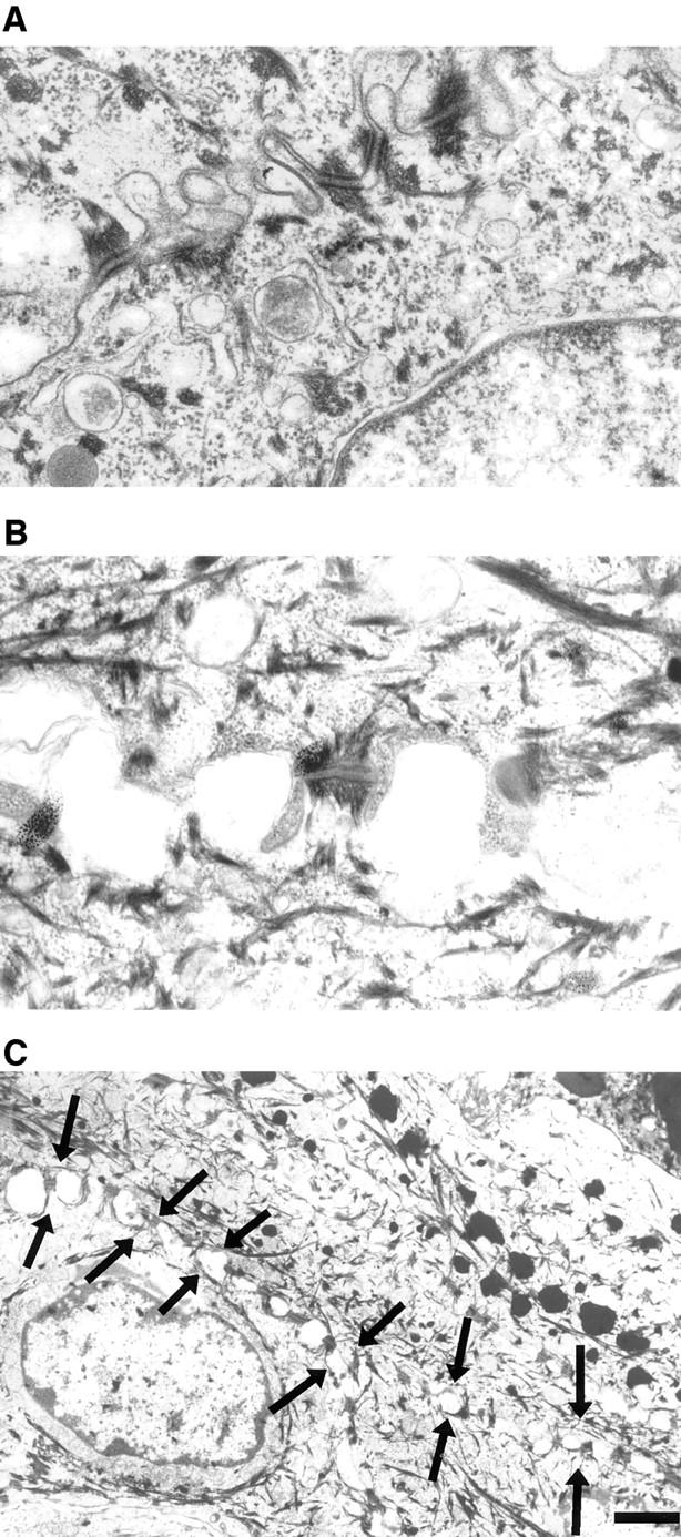
In their progression from the basal to upper differentiated layers of the epidermis, keratinocytes undergo significant structural changes, including establishment of close intercellular contacts. An important but so far unexplored question is how these early structural events are related to the biochemical pathways that trigger differentiation. We show here that beta-catenin, gamma-catenin/plakoglobin, and p120-Cas are all significantly tyrosine phosphorylated in primary mouse keratinocytes induced to differentiate by calcium, with a time course similar to that of cell junction formation. Together with these changes, there is an increased association of alpha-catenin and p120-Cas with E-cadherin, which is prevented by tyrosine kinase inhibition. Treatment of E-cadherin complexes with tyrosine-specific phosphatase reveals that the strength of alpha-catenin association is directly dependent on tyrosine phosphorylation. In parallel with the biochemical effects, tyrosine kinase inhibition suppresses formation of cell adhesive structures, and causes a significant reduction in adhesive strength of differentiating keratinocytes. The Fyn tyrosine kinase colocalizes with E-cadherin at the cell membrane in calcium-treated keratinocytes. Consistent with an involvement of this kinase, fyn-deficient keratinocytes have strongly decreased tyrosine phosphorylation levels of beta- and gamma-catenins and p120-Cas, and structural and functional abnormalities in cell adhesion similar to those caused by tyrosine kinase inhibitors. Whereas skin of fyn-/- mice appears normal, skin of mice with a disruption in both the fyn and src genes shows intrinsically reduced tyrosine phosphorylation of beta-catenin, strongly decreased p120-Cas levels, and important structural changes consistent with impaired keratinocyte cell adhesion. Thus, unlike what has been proposed for oncogene-transformed or mitogenically stimulated cells, in differentiating keratinocytes tyrosine phosphorylation plays a positive role in control of cell adhesion, and this regulatory function appears to be important both in vitro and in vivo.


Figures

Figure 1
Figure 1
Colocalization of phosphotyrosine with β-catenin and p120-Cas in the cortical cytoskeleton of differentiating keratinocytes. Mouse primary keratinocytes in low calcium medium or at 9 h of calcium exposure were pre-extracted in 0.2% Triton X-100 buffer before paraformaldehyde fixation as described in Materials and Methods. Cells were double stained with anti-phosphotyrosine antibodies and FITC-conjugated secondaries (green) and antibodies against either β-catenin (top panels) or p120-Cas (bottom panels), and Texas red–conjugated secondaries (red). Samples were analyzed by confocal microscopy and green and red images (small panels) were superimposed (large panels), so that sites of staining overlap are visualized as yellow. Bars: (large panels) 15 μm; (small panels) 33 μm.
Figure 2
Figure 2
Tyrosine phosphorylation and cadherin association of α-, β-, and γ-catenins and p120-Cas in growing versus differentiating keratinocytes. (A–D) Keratinocytes in low calcium medium (0), and at various times after calcium addition were lysed in 0.5% NP-40 lysis buffer and immunoprecipitated with antibodies against E-cadherin (A), β-catenin, or γ-catenin/ plakoglobin (B), desmoglein 3 (C), or p120-Cas (D). In all cases, control immunoprecipitations with unrelated antibodies were included (−). Immune complexes were analyzed by SDS-PAGE and anti-phosphotyrosine immunoblotting (left panels). The same blots were subsequently reprobed with antibodies against specific proteins as indicated (right panels). Positions of these molecules in the anti-phosphotyrosine immunoblots are indicated. In C, the band recognized by anti-phosphotyrosine antibodies that migrates above γ-catenin is nonspecific, since it is also detected in the nonimmune control. (E) Association of p120-Cas with E-cadherin, as detected by immunoprecipitation under milder stringency conditions than in the previous experiments. Keratinocytes in low calcium medium (0), and at 9 h after calcium addition (2 mM) were lysed in a 0.2% Triton X-100 lysis buffer and immunoprecipitated with E-cadherin–specific monoclonals or with unrelated control monoclonals (−). Immune complexes were analyzed by SDS-PAGE and sequential immunoblotting with antibodies against phosphotyrosine (p-Tyr), p120-Cas (p120-Cas), a mixture of antibodies against β- and γ-catenins (β/γ-cat.) and E-cadherin (E-cadh.).
Figure 3
Figure 3
Time course of cell adhesion formation in mouse primary keratinocytes after calcium treatment, and as a function of tyrosine phosphorylation. Mouse primary keratinocytes in low calcium medium or at 2 and 9 h of calcium exposure were pre-extracted in 0.2% Triton X-100 buffer before paraformaldehyde fixation as described in Materials and Methods. Parallel experiments were performed with cells pretreated for 2 h with 100 μM Genistein (Gen) and incubated for additional 9 h under high calcium conditions. Cells were stained with antibodies against E-cadherin (left panels, red) or plakophilin (right panels, green). Samples were analyzed by confocal microscopy. Each image results from the projection of eight different focal plans of the same field, and is representative of what observed in the individual focal plans. Bar, 15 μm.
Figure 4
Figure 4
E-cadherin association of α-, β-, and γ-catenins and p120-Cas in growing versus differentiating keratinocytes as affected by tyrosine kinase inhibition. Growing keratinocytes were pretreated for 2 h with either 100 μM Genistein (Gen), Tyrphostin 23 (Trph), or with DMSO solvent alone (Ctr). Incubation was then continued for additional 9 h under either low (L) or high (H) calcium conditions. Cells were lysed in either 0.5% NP-40 lysis buffer (A) or 0.2% Triton X-100 lysis buffer (B), and immunoprecipitated with anti–E-cadherin antibodies. Immune complexes were separated by SDS-PAGE and immunoblotted sequentially, as indicated, with antibodies against phosphotyrosine, α-catenin, a mixture of anti–β- and anti–γ-catenin antibodies, antibodies against p120-Cas, and against E-cadherin. In the experiment of A, two independent sets of control samples were included, to illustrate the reproducibility of the calcium-induced increase in α-catenin association with E-cadherin under normal conditions.
Figure 5
Figure 5
Strength of α-catenin association with E-cadherin complexes as a direct function of tyrosine phosphorylation. (A) Keratinocytes in low calcium medium (L) and at 9 h after high calcium addition (H) were lysed in 0.5% NP-40 lysis buffer, and immunoprecipitated with mAbs against E-cadherin. Immunocomplexes were treated with tyrosine-specific phosphatase (PTPase) ± phosphatase-specific inhibitors, followed by washing under high stringency conditions, as described in Materials and Methods. Samples were analyzed by SDS-PAGE and anti-phosphotyrosine immunoblotting (top panel). The same blots were subsequently reprobed with antibodies against α-, β-, and γ-catenins, and E-cadherin, as indicated. (B) A second independent experiment was performed, similar to the previous one, except that E-cadherin immunoprecipitated samples from keratinocytes at 9 h of calcium treatment were normalized for amounts of β- and γ-catenin levels (by a preliminary immunoblotting experiment), before the immunoblot analysis shown here. Densitometric quantitation of the film indicated that the relative ratio of α-catenin in the immunoprecipitates treated with phosphatase plus versus minus inhibitor was 6.2, while that of β- and γ-catenin was 1.2 and 0.8, respectively.
Figure 5
Figure 5
Strength of α-catenin association with E-cadherin complexes as a direct function of tyrosine phosphorylation. (A) Keratinocytes in low calcium medium (L) and at 9 h after high calcium addition (H) were lysed in 0.5% NP-40 lysis buffer, and immunoprecipitated with mAbs against E-cadherin. Immunocomplexes were treated with tyrosine-specific phosphatase (PTPase) ± phosphatase-specific inhibitors, followed by washing under high stringency conditions, as described in Materials and Methods. Samples were analyzed by SDS-PAGE and anti-phosphotyrosine immunoblotting (top panel). The same blots were subsequently reprobed with antibodies against α-, β-, and γ-catenins, and E-cadherin, as indicated. (B) A second independent experiment was performed, similar to the previous one, except that E-cadherin immunoprecipitated samples from keratinocytes at 9 h of calcium treatment were normalized for amounts of β- and γ-catenin levels (by a preliminary immunoblotting experiment), before the immunoblot analysis shown here. Densitometric quantitation of the film indicated that the relative ratio of α-catenin in the immunoprecipitates treated with phosphatase plus versus minus inhibitor was 6.2, while that of β- and γ-catenin was 1.2 and 0.8, respectively.
Figure 6
Figure 6
Inefficient cell–cell junction formation in calcium-treated keratinocytes as a consequence of tyrosine kinase inhibition or lack of the Fyn kinase. (A) Confluent keratinocyte cultures under growing conditions were either kept as untreated controls (Control) or pretreated for 2 h with 100 μM Genistein (Gen) or Tyrphostin 23 (Trph). Incubation was then continued for additional 9 h under high calcium conditions. Cells were fixed and processed for electron microscopy as described in Materials and Methods. The protrusions from neighboring cells in the Genistein-treated cultures were consistently observed. (B) Primary keratinocytes derived from fyn−/− mice and wild-type littermates were exposed to high calcium concentrations (2 mM) for 9 h. Cells were fixed and processed for electron microscopy. Note that in the fyn−/− cultures, cell borders were far apart and connected by protrusions of the cell membrane similar to those found with genistein-treated keratinocytes (as shown in A). Note the presence of well formed cell adhesive junctions in control cells and fyn+/+ cells (A and B, white arrows) and the lack of well-formed junctions and of cytoskeleton organization in the tyrosine kinase inhibitor treated and fyn−/− cells (A and B, black arrows). Adherens junctions and desmosomes can be defined as electron dense intercellular adhesive structures connected with the actin and keratin cytoskeleton, respectively. Unlike in vivo, in most of our EM photographs of cultured keratinocytes it is hard to see electron-dense junctional structures in connection with either the actin or keratin cytoskeleton, and therefore to conclusively distinguish between adherens junctions and desmosomes. In all cases, the observed alterations (>70% reduction in mature cell junction formation) were observed throughout the dishes (in each case examining at least 10 different fields), and were confirmed in a minimal of two independent experiments. Bars: (A) 200 nm; (B) 100 nm.
Figure 7
Figure 7
Decreased strength of calcium-induced cell adhesion as a consequence of tyrosine kinase inhibition or lack of the Fyn kinase. (A) Primary keratinocytes under growing conditions were either tested as untreated controls (Ctr) or pretreated for 2 h with Genistein. Incubation was then continued for additional 24 h under high calcium conditions. Cultures were examined as such (left panels) or after dispase treatment for 5 min (right panels). Arrows, the focal areas of cell detachment that occurred in the tyrosine kinase inhibitor-treated cultures. No such areas were evident in control cultures even after prolonged dispase exposure (>30 min). Instead, control cells eventually detached from the dish as a confluent sheet. Similar results were observed with keratinocyte cultures switched to high calcium conditions for only 9 h (not shown). (B) Primary keratinocytes derived from fyn−/− mice and wild-type littermates were exposed to high calcium concentrations (2 mM) for 9 h. Cultures were examined as such (left panels) or after treatment with dispase for 5 min (right panels). Arrows, the focal areas of cell detachment that occurred in the fyn−/− cultures already at this time. There were no cells missing in the monolayer of fyn knockout keratinocytes before dispase treatment. As we previously reported (Calautti et al., 1995), the fyn knockout keratinocytes fail to stratify and are larger than normal, which explains the different morphological appearance of these cultures relative to the wild-type controls even before dispase treatment. Similar results were observed in two other independent experiments. Bar, 60 μm.
Figure 8
Figure 8
Quantitative measurements of keratinocyte cell adhesion as determined by a novel dispase-based assay. (A) Primary keratinocytes under low calcium conditions (Low Ca) or treated with calcium for 2 or 9 h (Ca) were incubated with dispase for 35 min as described in Materials and Methods. Data are expressed as percentage of single cells released by mechanical disruption after dispase treatment versus total number of cells recovered after subsequent treatment of the same samples with trypsin. (B) A similar assay was performed with keratinocytes under low calcium conditions or incubated with calcium for 9 h (Ca) in the absence or presence of Genistein (100 μM) (Gen), Thyrphostin (100 μM) (Trph), or PP1 (1 μg/ml). Cells were preincubated with the inhibitors for 2 h before calcium treatment. (C) Keratinocytes under low calcium conditions were infected with a control adenovirus expressing the green fluorescent protein (Ad-GFP) or a virus expressing a constitutively active form of c-src (Ad-src). 24 h after infection, cells were switched to high calcium medium and incubation was continued for additional 24 h (Ca). Cells were analyzed by the dispase assay as before, together with control uninfected cells kept in either low or high calcium medium for 24 h.
Figure 9
Figure 9
Tyrosine phosphorylation and protein composition of E-cadherin complexes in control versus src-transformed keratinocytes. Keratinocytes under low calcium conditions were infected with either a green fluorescent protein control adenovirus (Ad-GFP) or a virus expressing constitutively active c-src (Ad-src). 24 h after infection cells were incubated in either low (L) or high (H) calcium medium for additional 9 h, in parallel with control uninfected cells (Uninf.). Keratinocytes were lysed in 0.5% NP-40 lysis buffer, and immunoprecipitated with mAbs against E-cadherin. Samples were analyzed by SDS-PAGE and anti-phosphotyrosine immunoblotting (p-Tyr, left). The same blot was subsequently reprobed with antibodies against α-catenin (α-cat.), β-catenin, and γ-catenin (β/γ-cat.), E-cadherin (EC), and Src as indicated (right panels). The asterisk on the anti-phosphotyrosine immunoblot indicates one of the additional tyrosine phosphorylated proteins that are present in the E-cadherin immunoprecipitates from Src-expressing cells, and which become more evident after prolonged exposures.
Figure 10
Figure 10
Colocalization of Fyn and E-cadherin at sites of cell–cell adhesion in calcium-treated keratinocytes. Mouse primary keratinocytes in low calcium medium (top row) or at 9 h of calcium exposure (bottom row) were pre-extracted in 0.2% Triton X-100 buffer before paraformaldehyde fixation as described in Materials and Methods. Cells were double stained with affinity-purified anti-Fyn polyclonal antibodies and FITC-conjugated secondaries (green) and mAbs against E-cadherin and Texas red–conjugated secondaries (red). Samples were analyzed by confocal microscopy and green and red images were superimposed (right panels), so that sites of staining overlap are visualized as yellow. Images correspond to single focal plans. Bar, 15 μm.
Figure 11
Figure 11
Tyrosine phosphorylation of β- and γ-catenins and p120-Cas in wild-type versus fyn−/− keratinocytes, and in the skin of mice with single versus double knockout mutations of fyn- related genes. (A) Primary keratinocytes derived from fyn−/− mice and genetically matched wild-type controls were either kept in low calcium medium (0) or exposed to high calcium concentrations (mm) for the indicated amounts of time. Cells were lysed in 0.5% NP-40 lysis buffer, and immunoprecipitated with mAbs against either E-cadherin (A) or p120-Cas (B). In A, a second independent experiment is also shown, with E-cadherin immunoprecipitates from keratinocytes with a disruption of the fyn versus yes kinase gene. Immune complexes were analyzed by SDS-PAGE and immunoblotting with anti-phosphotyrosine antibodies (top panels) and with a mixture of antibodies against β- and γ-catenins or anti–p120-Cas (bottom panels). (B) Pattern of tyrosine phosphorylation and of β- and γ-catenin association in E-cadherin complexes derived from newborn wild-type mouse skin (Skin) versus cultured keratinocytes (Krcts). Total extracts of newborn mouse skin along with extracts from cultured keratinocytes at 9 h after calcium exposure were immunoprecipitated with anti–E-cadherin antibodies or with unrelated control antibodies (−). Immunoprecipitates were analyzed by SDS-PAGE and sequential immunoblotting with antibodies against phosphotyrosine (p-Tyr), β- and γ-catenins and E-cadherin, as indicated. (C) tyrosine phosphorylation of E-cadherin associated proteins (β-catenin) in the skin of newborn mice with single versus double knockout mutations of fyn-related genes. Newborn mice with single knockout mutations of the fyn (F ), src (S ), and yes (Y ) genes, and with double fyn/src (F /S ) and fyn/yes (F /Y ) mutations and wild-type littermates, were killed immediately after birth. Total skin extracts were normalized for E-cadherin amounts by preliminary SDS-PAGE and immunoblotting with E-cadherin antibodies. Normalized extracts were immunoprecipitated with anti–E-cadherin antibodies followed by SDS-PAGE and immunoblotting with anti-phosphotyrosine antibodies (top panel). The same blot was reprobed with antibodies against β-catenin (bottom panel). (D) Tyrosine phosphorylation and total protein levels of p120-Cas in the skin of newborn mice with single versus double knockout mutations of fyn-related genes. Same amounts of the extracts used above were immunoprecipitated with anti–p120-Cas antibodies followed by SDS-PAGE and immunoblotting with anti-phosphotyrosine antibodies (top panel). The same blot was reprobed with antibodies against p120-Cas (bottom panel).
Figure 11
Figure 11
Tyrosine phosphorylation of β- and γ-catenins and p120-Cas in wild-type versus fyn−/− keratinocytes, and in the skin of mice with single versus double knockout mutations of fyn- related genes. (A) Primary keratinocytes derived from fyn−/− mice and genetically matched wild-type controls were either kept in low calcium medium (0) or exposed to high calcium concentrations (mm) for the indicated amounts of time. Cells were lysed in 0.5% NP-40 lysis buffer, and immunoprecipitated with mAbs against either E-cadherin (A) or p120-Cas (B). In A, a second independent experiment is also shown, with E-cadherin immunoprecipitates from keratinocytes with a disruption of the fyn versus yes kinase gene. Immune complexes were analyzed by SDS-PAGE and immunoblotting with anti-phosphotyrosine antibodies (top panels) and with a mixture of antibodies against β- and γ-catenins or anti–p120-Cas (bottom panels). (B) Pattern of tyrosine phosphorylation and of β- and γ-catenin association in E-cadherin complexes derived from newborn wild-type mouse skin (Skin) versus cultured keratinocytes (Krcts). Total extracts of newborn mouse skin along with extracts from cultured keratinocytes at 9 h after calcium exposure were immunoprecipitated with anti–E-cadherin antibodies or with unrelated control antibodies (−). Immunoprecipitates were analyzed by SDS-PAGE and sequential immunoblotting with antibodies against phosphotyrosine (p-Tyr), β- and γ-catenins and E-cadherin, as indicated. (C) tyrosine phosphorylation of E-cadherin associated proteins (β-catenin) in the skin of newborn mice with single versus double knockout mutations of fyn-related genes. Newborn mice with single knockout mutations of the fyn (F ), src (S ), and yes (Y ) genes, and with double fyn/src (F /S ) and fyn/yes (F /Y ) mutations and wild-type littermates, were killed immediately after birth. Total skin extracts were normalized for E-cadherin amounts by preliminary SDS-PAGE and immunoblotting with E-cadherin antibodies. Normalized extracts were immunoprecipitated with anti–E-cadherin antibodies followed by SDS-PAGE and immunoblotting with anti-phosphotyrosine antibodies (top panel). The same blot was reprobed with antibodies against β-catenin (bottom panel). (D) Tyrosine phosphorylation and total protein levels of p120-Cas in the skin of newborn mice with single versus double knockout mutations of fyn-related genes. Same amounts of the extracts used above were immunoprecipitated with anti–p120-Cas antibodies followed by SDS-PAGE and immunoblotting with anti-phosphotyrosine antibodies (top panel). The same blot was reprobed with antibodies against p120-Cas (bottom panel).
Figure 12
Figure 12
Altered cell–cell adhesion in the skin of double fyn/src knockouts as directly assessed by ultra structural analysis. Newborn mice with single versus double knockout mutations of the fyn (F ), src (S ), and yes (Y ) genes, and wild-type littermates, were killed immediately after birth and skin samples were processed for electron microscopy. (A) Ultrastructural analysis of skin from a wild-type newborn animal. (B and C) Ultrastructural analysis of the skin from a double fyn/src knockout animal at high (B) and low (C) magnification. Similar alterations were found in the skin of three double fyn/src knockouts, derived from two independent litters. Arrows point to the interdesmosomal areas of cell detachment found in the fyn/src mutant skin. The skin of all other mutant animals was found to have desmosomal structures and interdesmosomal spaces similar to the wild-type controls. Bars: (A and B) 300 nm; (C) 1.4 μm.

Similar articles

Cited by

References

    1. Aberle H, Schwartz H, Hoschuetzky H, Kemler R. Single amino acid substitutions in proteins of the armadillo gene family abolish their binding to α-catenin. J Biol Chem. 1996;271:1520–1526. - PubMed
    1. Amagai M, Fujimori T, Masunaga T, Shimizu H, Nishikawa T, Shimizu N, Takeichi M, Hashimoto T. Delayed assembly of desmosomes in keratinocytes with disrupted classic- cadherin-mediated cell adhesion by a dominant negative mutant. J Invest Dermatol. 1995;104:27–32. - PubMed
    1. Angres B, Barth A, Nelson WJ. Mechanism for transition from initial to stable cell–cell adhesion: Kinetic analysis of E-cadherin–mediated adhesion using a quantitative adhesion assay. J Cell Biol. 1996;134:549–557. - PMC - PubMed
    1. Behrens J, Vakaet L, Friis R, Winterhager E, Van Roy F, Mareel MM, Birchmeier W. Loss of epithelial differentiation and gain of invasiveness correlates with tyrosine phosphorylation of the E-cadherin/β-catenin complex in cells transformed with a temperature-sensitive v-SRC gene. J Cell Biol. 1993;120:757–766. - PMC - PubMed
    1. Behrens J, von Kries JP, Kuhl M, Bruhn L, Wedlich D, Grosschedl R, Birchmeier W. Functional interaction of β-catenin with the transcription factor LEF-1. Nature. 1996;382:638–642. - PubMed

Publication types

MeSH terms

Substances