. 1998 Jul;66(7):3029-34.
doi: 10.1128/IAI.66.7.3029-3034.1998.

Expression of Legionella pneumophila virulence traits in response to growth conditions

Affiliations

Expression of Legionella pneumophila virulence traits in response to growth conditions

B Byrne et al. Infect Immun. 1998 Jul.

Abstract

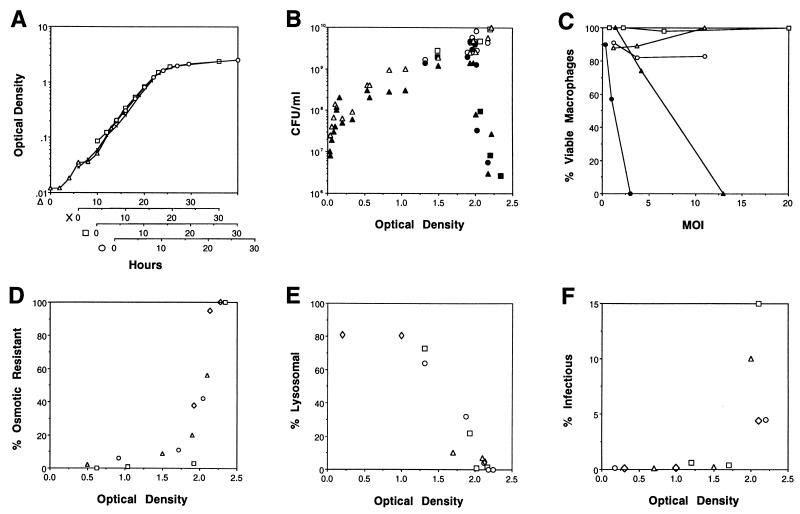
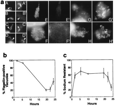
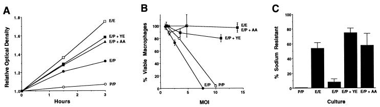
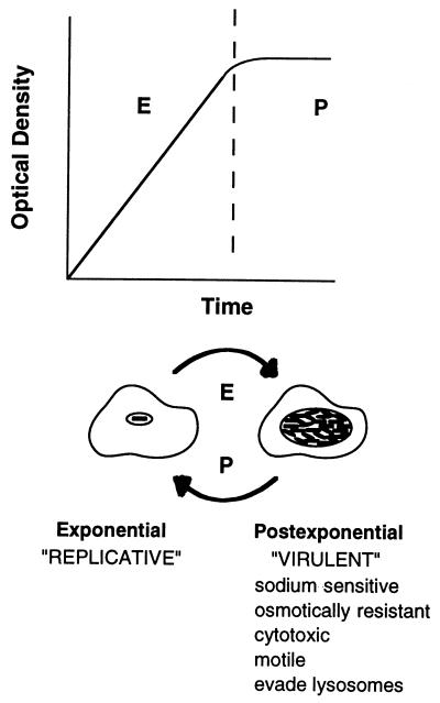
In nature, Legionella pneumophila replicates exclusively as an intracellular parasite of amoebae, but it also persists in the environment as a free-living microbe. Studies of how this opportunistic pathogen recognizes and responds to distinct extracellular and intracellular environments identified a link between the growth phase and expression of traits previously correlated with virulence. When cultured in broth, only post-exponential-phase L. pneumophila was sodium sensitive, cytotoxic, osmotically resistant, competent to evade macrophage lysosomes, infectious, and motile. Likewise, the L. pneumophila phenotype changed during growth in macrophages. During the intracellular replication period, this bacterium was sodium resistant and lacked flagella; concomitant with macrophage lysis, L. pneumophila became sodium sensitive and flagellated. Expression of the virulent phenotype was a response to starvation, since exponential-phase L. pneumophila became cytotoxic, sodium sensitive, and motile after incubation in broth from stationary-phase cultures, except when it was supplemented with amino acids. Together, these data indicate that while nutrients are plentiful, intracellular L. pneumophila organisms are dedicated to replication; when amino acids become limiting, the progeny express virulence factors to escape the spent host, to disperse and survive in the aquatic environment, and to reestablish a protected intracellular niche favorable for growth.


Figures

FIG. 1
FIG. 1
Characteristics of L. pneumophila exiting the exponential phase of growth. (A) Correspondence between the OD600 and the growth phase of L. pneumophila AYET broth cultures. An exponential-phase culture was diluted 1:350 (▵), 1:120 (×), 1:35 (□), or 1:16 (○) and incubated at 37°C for 15 h; then the OD600 of each subculture was determined at the times shown. Results from four representative cultures were superimposed such that their growth curves overlapped; similar results were obtained in several other experiments. (B) L. pneumophila sodium sensitivity was determined by plating cultures with the OD600s shown on CYET medium without (open symbols) and with (closed symbols) 100 mM NaCl. Three experiments performed in duplicate are represented by different symbols (triangles, circles, and squares). (C) L. pneumophila cytotoxicity was judged by the capacity of viable macrophages to reduce the colorimetric dye Alamar blue after a 1-h incubation with bacteria obtained from AYET cultures grown to OD600s of 0.374 (○), 1.519 (▵), 1.882 (□), 2.151 (•), and 2.231 (▴). The means of values for triplicate samples are shown; standard errors ranged from 1.5 to 8%. The multiplicity of infection (MOI) was calculated by plating the respective broth culture on CYET. The means of values for triplicate samples are shown; standard errors ranged from 2.5 to 14%. (D) Osmotic resistance of L. pneumophila cultures grown to the OD600s indicated was determined by incubation for 1 h in AYET that did or did not contain 0.3 M KCl, dilution into water, and then plating of duplicate samples on CYET to quantify the CFU. Results from each of four experiments are represented by different symbols (triangles, squares, circles, and diamonds). (E) The ability of L. pneumophila grown to the indicated OD600s to evade macrophage lysosomes was quantified by fluorescence microscopy, using the endocytic probe Texas red-ovalbumin. Data were obtained from four experiments, in which the number of phagosomes scored was 35 (◊), 75 (□), 50 (○), or 100 (▵). As a positive control, bacteria grown to an OD600 of >2 and then killed with formalin were analyzed; a mean of 67% (standard error = 13) of such particles colocalized with Texas red-ovalbumin. (F) The capacity of L. pneumophila grown to the OD600s indicated to enter and survive in macrophages was defined as protection from gentamicin added 2 h after infection. The mean percentage of infectious L. pneumophila was determined for duplicate or triplicate samples in four experiments, each represented by a different symbol.
FIG. 2
FIG. 2
Characteristics of L. pneumophila cells growing in macrophages. (a) Representative images of macrophages at the initial (A and B), replicative (C to F), and necrotic stages (G and H) of L. pneumophila infection. After infection for 2 h (A), 6 h (C and D), 19.5 h (G), 22 h (F and H), and 24 h (E), bacteria were stained with DAPI (A to H) and flagella were labeled with a flagellum-specific mouse monoclonal antibody and a Texas red-conjugated anti-mouse immunoglobulin G secondary antibody (A′ to H′). Because initiation of intracellular replication was not synchronous, by 19.5 h postinfection, samples included both the replicative and necrotic stages of infection. Nonmotile bacteria obtained from cultures grown to an OD600 of <2 and incubated with macrophages for 2 h served as a negative control (B). Arrowheads indicate junctions between cells and flagella and serve as reference points. (b) Flagellum production by intracellular L. pneumophila was scored by immunofluorescence microscopic analysis of macrophages infected with motile and sodium-sensitive bacteria obtained from post-exponential-phase broth cultures. Because L. pneumophila vacuoles which contained one or more flagellum were scored as positive, the percentage of positive vacuoles reflects both flagellum production and stability. Because necrotic macrophages often detached and were washed from the coverslips during the immunostaining procedure, flagellin-positive vacuoles in the samples analyzed 22 h postinfection were likely underestimated. Shown are the means and standard errors determined by scoring at least 30 phagosomes at each time point in each of three experiments. (c) L. pneumophila sodium resistance was determined by plating dilutions of the infection medium (0 h) or pooled macrophage lysates and culture supernatants (2 to 24 h) on CYET medium which did or did not contain 100 mM NaCl. Shown are the means and standard errors of values from four to six experiments performed in triplicate.
FIG. 3
FIG. 3
Growth, cytotoxicity, and sodium resistance of exponential-phase L. pneumophila cultured in post-exponential-phase supernatants. Cells collected from an exponential-phase culture with an OD600 of 0.62 were incubated for the periods of time shown in supernatant prepared from the identical exponential-phase culture (E/E; OD600 = 0.57 at 0 h) (open squares) or from a post-exponential-phase culture with an OD600 of 2.09 supplemented with H2O (E/P; OD600 = 0.65 at 0 h) (closed circles), with yeast extract (E/P + YE; OD600 = 0.65 at 0 h) (closed squares), or with a cocktail of five amino acids (E/P + AA; OD600 = 0.62 at 0 h) (closed triangles). As a positive control for the virulent phenotype, cells collected from a post-exponential-phase culture with an OD600 of 2.09 were incubated for the periods of times shown in supernatant prepared from the identical post-exponential-phase culture (P/P; OD600 = 1.94 at 0 h) (open circles). (A) The relative level of bacterial growth under each set of conditions is represented as the ratio of the OD600 at the indicated time to the OD600 at 0 h. (B) The cytotoxicity of L. pneumophila cultured for 3 h as described above was determined as described in the legend to Fig. 1C. Shown are the averages of values for duplicate samples; error bars indicate the ranges of the values. The cytoxicity observed was cell dependent, since post-exponential-phase culture supernatants alone were not cytopathic (data not shown). Panels A and B depict results from a single experiment; similar results were obtained in four other experiments. (C) The sodium resistance of the L. pneumophila cultures defined above was determined as described in the legends of Fig. 1 and 2. Shown are the means and standard errors for data from five experiments performed in duplicate.
FIG. 4
FIG. 4
Model for L. pneumophila phenotypic regulation in response to growth conditions as described in the text. E, exponential growth phase; P, post-exponential phase of growth.

References

    1. Abu Kwaik Y, Eisenstein B I, Engleberg N C. Phenotypic modulation by Legionella pneumophila upon infection of macrophages. Infect Immun. 1993;61:1320–1329. - PMC - PubMed
    1. Berger K H, Isberg R R. Two distinct defects in intracellular growth complemented by a single genetic locus in Legionella pneumophila. Mol Microbiol. 1993;7:7–19. - PubMed
    1. Bermudez L E, Petrofsky M, Goodman J. Exposure to low oxygen tension and increased osmolarity enhance the ability of Mycobacterium avium to enter intestinal epithelial (HT-29) cells. Infect Immun. 1997;65:3768–3773. - PMC - PubMed
    1. Bermudez L E, Parker A, Goodman J R. Growth within macrophages increases the efficiency of Mycobacterium avium in invading other macrophages by a complement receptor-independent pathway. Infect Immun. 1997;65:1916–1925. - PMC - PubMed
    1. Catrenich C E, Johnson W. Characterization of the selective inhibition of growth of virulent Legionella pneumophila by supplemented Mueller-Hinton medium. Infect Immun. 1989;57:1862–1864. - PMC - PubMed

Publication types

LinkOut - more resources