. 1998 Jul;66(7):3113-9.
doi: 10.1128/IAI.66.7.3113-3119.1998.

Phenotypic effect of isogenic uspA1 and uspA2 mutations on Moraxella catarrhalis 035E

Affiliations

Phenotypic effect of isogenic uspA1 and uspA2 mutations on Moraxella catarrhalis 035E

C Aebi et al. Infect Immun. 1998 Jul.

Abstract

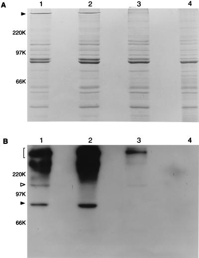
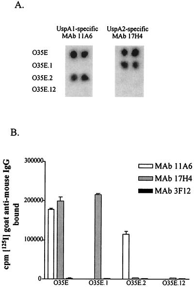
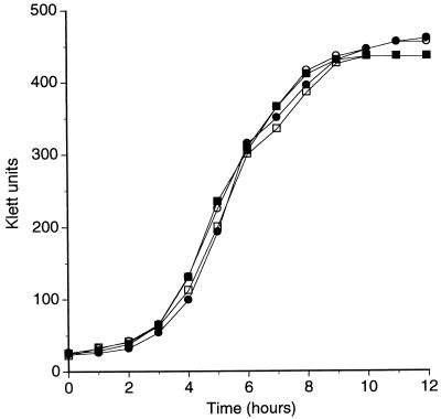
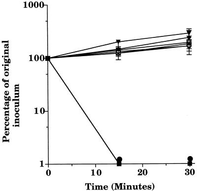
The UspA surface antigen of Moraxella catarrhalis was recently shown to be comprised of two different proteins (UspA1 and UspA2) which share an internal region containing 140 amino acids with 93% identity (C. Aebi, I. Maciver, J. L. Latimer, L. D. Cope, M. K. Stevens, S. E. Thomas, G. H. McCracken, Jr., and E. J. Hansen, Infect. Immun. 65:4367-4377, 1997). Isogenic uspA1, uspA2, and uspA1 uspA2 mutants were tested in a number of in vitro systems to determine what effect these mutations, either individually or together, might exert on the phenotype of M. catarrhalis 035E. Monoclonal antibodies specific for UspA1 or UspA2 were used in an indirect antibody accessibility assay to prove that both of these proteins were expressed on the surface of M. catarrhalis. All three mutants grew in vitro at the same rate and did not exhibit autoagglutination or hemagglutination properties that were detectably different from those of the wild-type parent strain. When tested for the ability to adhere to human epithelial cells, the wild-type parent strain and the uspA2 mutant readily attached to Chang conjunctival cells. In contrast, the uspA1 mutant and the uspA1 uspA2 double mutant both attached to these epithelial cells at a level nearly 2 orders of magnitude lower than that obtained with the wild-type parent strain, a result which suggested that expression of UspA1 by M. catarrhalis is essential for attachment to these epithelial cells. Both the wild-type parent strain and the uspA1 mutant were resistant to the bactericidal activity of normal human serum, whereas the uspA2 mutant and the uspA1 uspA2 double mutant were readily killed by this serum. This latter result indicated that the presence of UspA2 is essential for expression of serum resistance by M. catarrhalis.


Figures

FIG. 1
FIG. 1
Detection of the UspA1 and UspA2 proteins in wild-type and mutant strains of M. catarrhalis 035E. Proteins (10 μg) present in EDTA-extracted outer membrane vesicles from the wild-type strain (lane 1), uspA1 mutant 035E.1 (lane 2), uspA2 mutant 035E.2 (lane 3), and isogenic uspA1 uspA2 double mutant 035E.12 (lane 4) were resolved by SDS-PAGE and either stained with Coomassie blue (A) or transferred to nitrocellulose and probed with MAb 17C7 followed by radioiodinated goat anti-mouse immunoglobulin in Western blot analysis (B). In panel A, the closed arrowhead indicates the very high molecular weight form of the UspA antigen, in which UspA2 likely predominates. In panel B, the bracket on the left indicates the region of the autoradiograph containing the very high molecular weight forms of the UspA1 and UspA2 proteins that bind MAb 17C7. The open arrowhead indicates the 120-kDa, putative monomeric form of UspA1. The closed arrowhead indicates the 85-kDa, putative monomeric form of UspA2. This autoradiograph was overexposed to allow detection of the relatively minor, putative monomeric form of UspA1 in lanes 1 and 3; this overexposure resulted in detection of a diffuse region of MAb 17C7 reactivity in the region of the autoradiograph near and immediately above the 220-kDa position marker in lanes 1 and 2. This material is present only in those strains which express UspA2 (lanes 1 and 2) and likely represents different size aggregates of UspA2. Molecular weight position markers (in kilodaltons) are present on the left.
FIG. 2
FIG. 2
Binding of MAbs to wild-type and mutant strains of M. catarrhalis. Colony paste of wild-type strain 035E, uspA1 mutant 035E.1, uspA2 mutant 035E.2, and uspA1 uspA2 double mutant 035E.12 spotted in duplicate on filter paper was probed with MAbs 11A6 and 17H4 in the colony blot radioimmunoassay (A) to prove the specificity of these MAbs for UspA1 and UspA2, respectively. These two MAbs were then tested for the ability to bind to whole cells of these same strains in the indirect antibody accessibility assay (B). Binding of the UspA1- and UspA2-specific MAbs to whole cells of these four strains is reflected by the amount (in counts per minute) of radioiodinated goat anti-mouse IgG bound to MAbs attached to the surface of the bacterial cells. MAb 3F12, a murine IgG MAb specific for the major outer membrane protein of H. ducreyi (34), was used as a negative control.
FIG. 3
FIG. 3
Comparison of the growth of the wild-type and mutant strains of M. catarrhalis in vitro. Wild-type strain 035E (closed squares), uspA1 mutant 035E.1 (open squares), uspA2 mutant 035E.2 (closed circles), and uspA1 uspA2 double mutant 035E.12 (open circles) from overnight broth cultures were diluted to a density of 35 Klett units in BHI broth and subsequently allowed to grow at 37°C with shaking. Growth was followed by means of turbidity measurements.
FIG. 4
FIG. 4
Susceptibility of wild-type and mutant strains of M. catarrhalis to killing by normal human serum. Cells of wild-type strain 035E (diamonds), uspA1 mutant 035E.1 (triangles), uspA2 mutant 035E.2 (circles), and uspA1 uspA2 double mutant 035E.12 (squares) from logarithmic-phase BHI broth cultures were incubated in the presence of 10% (vol/vol) normal human serum (closed symbols) or heat-inactivated normal human serum (open symbols). Data are presented as the percentage of the original inoculum remaining at each time point; error bars are included.

References

    1. Aebi C, Maciver I, Latimer J L, Cope L D, Stevens M K, Thomas S E, McCracken G H, Jr, Hansen E J. A protective epitope of Moraxella catarrhalis is encoded by two different genes. Infect Immun. 1997;65:4367–4377. - PMC - PubMed
    1. Aebi C, Stone B, Beucher M, Cope L D, Maciver I, Thomas S E, McCracken G H, Jr, Sparling P F, Hansen E J. Expression of the CopB outer membrane protein by Moraxella catarrhalis is regulated by iron and affects iron acquisition from transferrin and lactoferrin. Infect Immun. 1996;64:2024–2030. - PMC - PubMed
    1. Ahmed K, Matsumoto K, Rikitomi N, Nagatake T. Attachment of Moraxella catarrhalis to pharyngeal epithelial cells is mediated by a glycosphingolipid receptor. FEMS Microbiol Lett. 1996;135:305–309. - PubMed
    1. Barenkamp S J, St. Geme J W., III Identification of a second family of high-molecular-weight adhesion proteins expressed by non-typable Haemophilus influenzae. Mol Microbiol. 1996;19:1215–1223. - PubMed
    1. Bartos L C, Murphy T F. Comparison of the outer membrane proteins of 50 strains of Branhamella catarrhalis. J Infect Dis. 1988;158:761–765. - PubMed

Publication types

Substances

LinkOut - more resources