. 1998 Jul;18(7):3647-58.
doi: 10.1128/MCB.18.7.3647.

RB and c-Myc activate expression of the E-cadherin gene in epithelial cells through interaction with transcription factor AP-2

Affiliations

RB and c-Myc activate expression of the E-cadherin gene in epithelial cells through interaction with transcription factor AP-2

E Batsché et al. Mol Cell Biol. 1998 Jul.

Abstract

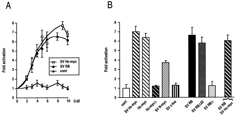
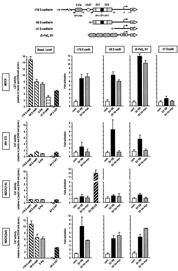
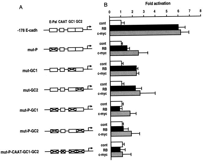
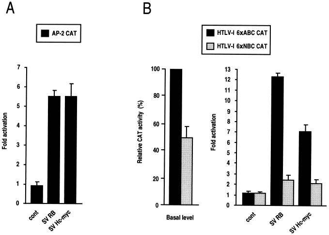
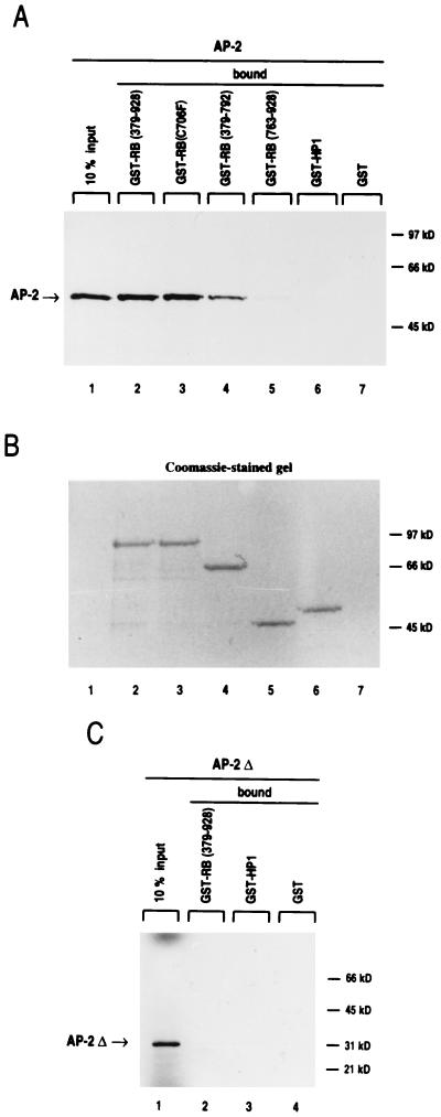
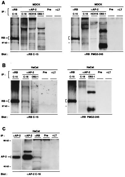
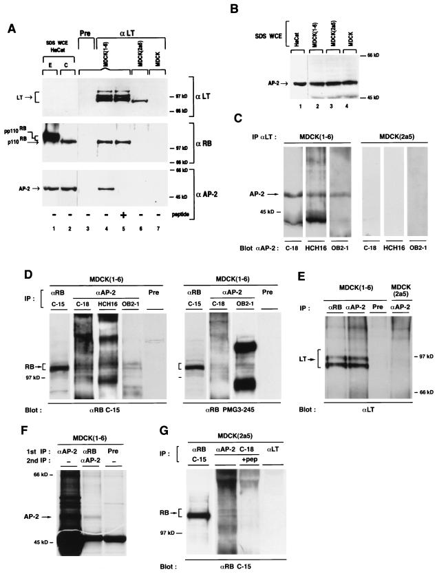
E-cadherin plays a pivotal role in the biogenesis of the first epithelium during development, and its down-regulation is associated with metastasis of carcinomas. We recently reported that inactivation of RB family proteins by simian virus 40 large T antigen (LT) in MDCK epithelial cells results in a mesenchymal conversion associated with invasiveness and a down-regulation of c-Myc. Reexpression of RB or c-Myc in such cells allows the reexpression of epithelial markers including E-cadherin. Here we show that both RB and c-Myc specifically activate transcription of the E-cadherin promoter in epithelial cells but not in NIH 3T3 mesenchymal cells. This transcriptional activity is mediated in both cases by the transcription factor AP-2. In vitro AP-2 and RB interaction involves the N-terminal domain of AP-2 and the oncoprotein binding domain and C-terminal domain of RB. In vivo physical interaction between RB and AP-2 was demonstrated in MDCK and HaCat cells. In LT-transformed MDCK cells, LT, RB, and AP-2 were all coimmunoprecipitated by each of the corresponding antibodies, and a mutation of the RB binding domain of the oncoprotein inhibited its binding to both RB and AP-2. Taken together, our results suggest that there is a tripartite complex between LT, RB, and AP-2 and that the physical and functional interactions between LT and AP-2 are mediated by RB. Moreover, they define RB and c-Myc as coactivators of AP-2 in epithelial cells and shed new light on the significance of the LT-RB complex, linking it to the dedifferentiation processes occurring during tumor progression. These data confirm the important role for RB and c-Myc in the maintenance of the epithelial phenotype and reveal a novel mechanism of gene activation by c-Myc.


Figures

FIG. 1
FIG. 1
Activation of the murine E-cadherin promoter by RB and Myc in MDCK epithelial cells. (A) Dose-dependent effects of SV RB and SV Hc-myc on transcriptional activity of the −178 E-cadherin promoter. Three micrograms of −178 E-cadherin CAT was cotransfected with pUC DNA (baseline value) or with increasing amounts of SV RB and SV Hc-myc expression vectors. Total DNA was always kept constant by adding pUC DNA. As a control, −178 E-cadherin CAT was cotransfected with an expression plasmid without insert, pSV2Δ (cont). The values indicated are averages expressed as fold activation of CAT activity relative to the baseline value obtained by cotransfecting −178 E-cadherin promoter CAT with pUC DNA. Each value is expressed after normalization for transfection efficiency, using pHβALacZ as an internal control. The results are from at least three experiments performed in duplicate, and standard deviation bars are shown. (B) Activation of E-cadherin transcriptional activity by different nuclear regulators. Three micrograms of −178 E-cadherin CAT was cotransfected into MDCK cells with 7 μg of the indicated vectors.
FIG. 2
FIG. 2
Cell-type-specific activation of E-cadherin promoter by RB and Myc. The reporters used in this study are depicted at the top. For graphs of basal level, the −178, −58, and −21 E-cadherin CAT, (E-Pal)4 SV CAT, and AP-2 CAT constructs (3 μg of each) were transfected into MDCK, NIH 3T3, MDCK(1-6), and MDCK(2a5) cells. Basal CAT activities are expressed after normalization with the β-actin promoter and SV40 promoter/enhancer activities and are presented, except for AP-2 CAT, relative to the −178 E-cadherin promoter in MDCK(1-6) cells, which is set at 1. AP-2 CAT activities are presented relative to its activity in MDCK(1-6) cells, which is set at 1. For graphs related to cell type specificity, 2 μg [1 μg for (E-Pal)4 SV CAT] of each E-cadherin promoter construct was cotransfected with 7 μg [or 10 μg for (E-Pal)4 SV CAT] of RB, RBΔ22, and Myc expression vectors or the control pSV2Δ (cont) in the indicated cell lines. The values indicated are averages expressed as fold activation of CAT activity relative to the baseline value obtained by cotransfecting each E-cadherin CAT construct with pUC DNA.
FIG. 3
FIG. 3
Specific point mutations of the AP-2 binding sites of the E-cadherin promoter result in the loss of RB- and Myc-mediated activation. (A) Diagrams of mutant constructs of the −178 E-cadherin promoter. Point mutations abolishing AP-2-like and AP-2 binding within the E-Pal and GC boxes are indicated by crosses (21). (B) The E-cadherin constructs (2 μg of each) were cotransfected with 7 μg of RB and Myc expression vectors (SV RB and SV Hc-myc, respectively) or the control pSV2Δ (cont) in MDCK epithelial cells. The values are averages expressed as fold activation of CAT activities relative to the baseline obtained by cotransfecting each E-cadherin CAT construct with 7 μg of pUC DNA.
FIG. 4
FIG. 4
RB and Myc activate expression of the HTLV-1 LTR through AP-2. (A) MDCK cells were cotransfected with 3 μg of AP-2 cona CAT (AP-2 CAT) reporter containing three metallothionein AP-2 binding sites linked to the conaβ2 promoter and either 5 μg of RB and Myc expression vectors or pSV2Δ (cont). Controls with only the conaβ2 promoter were also performed (not shown). (B) MDCK cells were cotransfected with 3 μg of either HTLV-1 6×ABC or HTLV-1 6×NBC (see Materials and Methods) and with 5 μg of RB and Myc expression vectors or pSV2Δ (cont). The panel on the left shows the basal level of the wild-type and mutated HTLV-1 reporters. All values indicated are averages expressed as fold activation of CAT activity relative to the baseline value obtained by cotransfecting each E-cadherin CAT construct with pUC DNA.
FIG. 5
FIG. 5
AP-2 and RB proteins interact in vitro. (A and B) Both the RB small pocket and the C-terminal domain are required for interaction with AP-2. GST, GST-RB(379-928), GST-RB(379-928; 706C-to-F mutation) [GST(C706F)], GST-RB(379-792), GST-RB(763-928), and GST-HP1 fusion proteins immobilized on glutathione-agarose beads were used to bind radiolabeled AP-2 protein. Lane 1 shows 10% of the input of in vitro-translated [35S]AP-2. In lanes 2 to 7, the same amounts of [35S]AP-2 were incubated with glutathione-agarose beads containing similar amounts of the various GST fusion proteins. GST-HP1, a chromatin protein unrelated to RB, was used as negative control. Nine to 10% of AP-2 bound to GST-RB(379-928) and to GST-RB(C706F), 4.5 to 5% bound to GST-RB(379-792), and 1 to 2% bound to GST-RB(763-928). (B) Before drying and autoradiography, the gel was stained with Coomassie blue to ensure that all fusion proteins were correctly recovered. (C) N-terminally deleted AP-2 protein does not bind to RB. A 35S-labeled N-terminal deletion AP-2 protein (AP-2Δ) was incubated with various GST fusion proteins (lanes 2 to 4). Similar amounts of radiolabeled AP-2 and AP-2Δ were used in panels A and C, and gels were exposed for the same length of time.
FIG. 6
FIG. 6
RB and AP-2 interact in vivo. (A) RB–AP-2 complexes in MDCK cells. Lysates of MDCK cells were immunoprecipitated (IP) with the three αAP-2, C-18, HCH16, and OB2-1, αRB (C-15) as a positive control, or rabbit preimmune serum (Pre) and αLT as negative controls. Immune complexes were separated on a 7.5% polyacrylamide gel and transferred to a PVDF membrane, which was then probed with αRB (C-15 and PMG3-245). (B) Stable RB–AP-2 complexes in HaCat cells. Coimmunoprecipitations similar to those described for panel A were performed with HaCat cells with αAP-2 (C-18 and OB2-1). (C) HaCat lysates were also subjected to the reverse immunoprecipitation with αRB (PMG3-245) or αAP-2 (C-18) as a positive control. As negative controls, mouse preimmune serum (Pre) and αLT were used. After transfer, the PVDF membrane was probed with αAP-2 (C-18). A short exposure of the positive control is also shown.
FIG. 7
FIG. 7
In vivo interaction between LT, RB, and AP-2. (A) αLT coimmunoprecipitates RB and AP-2 in LT-transformed MDCK cells when the RB oncoprotein binding domain is not mutated. Total lysates of MDCK(1-6), MDCK(2a5), and MDCK cells were immunoprecipitated (IP) with mouse preimmune serum (Pre) or the mouse monoclonal 419 αLT coupled to protein A/G-agarose beads. Immune complexes were separated on an SDS–7.5% polyacrylamide gel, and proteins were transferred onto a PVDF membrane. After transfer, the membrane was cut into two pieces. The upper part was successively probed with the monoclonal PMG3-245 αRB and then αLT. In lanes 1 and 2, 30-μg aliquots of 1% SDS whole-cell HaCat extract (SDS WCE) of exponential (E) and confluent (C) cells were loaded on the gel as positive controls for hyper- and hypophosphorylated RB. Lane 5 was isolated from the lower part; then lanes 1 to 4, 6, and 7 were probed with the rabbit polyclonal αAP-2 (C-18) raised against the C-terminal peptide of AP-2 (Santa Cruz). Lane 5 alone was incubated with αAP-2 and a 10-fold excess of the cognate peptide. (B) The 52-kDa AP-2 protein is expressed in all cells tested. Aliquots (30 μg) of 1% SDS whole-cell extracts (SDS WCE) of HaCat, MDCK(1-6), MDCK(2a5), and MDCK cells were separated on a 7.5% polyacrylamide gel, transferred to a PVDF membrane, and probed with the polyclonal αAP-2 (C-18). (C) The LT-immunoprecipitated 52-kDa protein is recognized by three different αAP-2. Total lysates of MDCK(1-6) and MDCK(2a5) cells were immunoprecipitated with αLT as described for panel A, and the different Western blots were probed with the rabbit polyclonal C-18, the rabbit polyclonal antiserum HCH16 against the N-terminal peptide of AP-2 (9), and the mouse polyclonal OB2-1 antiserum directed against the whole human AP-2 protein (9). (D) RB is coimmunoprecipitated by several distinct αAP-2 in MDCK(1-6) cells. Total lysates of MDCK(1-6) cells were immunoprecipitated with αAP-2 (C-18, HCH16, and OB2-1), αRB (C-15) as positive control, or rabbit preimmune serum (Pre) as a negative control. After separation on a 7.5% polyacrylamide gel and transfer, the PVDF membrane was probed with αRB (C-15 and PMG3-245). (E) LT is coimmunoprecipitated with αAP-2 only in MDCK(1-6) and not in MDCK(2a5) cells. Total lysates of MDCK(1-6) and MDCK(2a5) cells were subjected to precipitation with rabbit preimmune serum (Pre), αRB (C-15), or αAP-2 (C-18) as indicated. After separation on 7.5% polyacrylamide gels and transfer, the PVDF membrane was probed with αLT. (F) AP-2 is coimmunoprecipitated by αRB in MDCK(1-6) cells. To allow easier detection of AP-2, cell lysates of MDCK(1-6) cells (7 × 10⁷) labeled with [35S]methionine were subjected to precipitation with rabbit preimmune serum (Pre), αRB (C-15), or αAP-2 (C-18) as a positive control. The washed RB immunoprecipitate was then dissociated with 1% SDS and reprecipitated with αAP-2 (C-18). Immune complexes were separated on a 10% polyacrylamide gel. The gel was fluorographed and dried, and proteins were detected by autoradiography. (G) RB coprecipitates with αAP-2 in MDCK(2a5) cells. Lysates of MDCK(2a5) cells were immunoprecipitated with αAP-2 (C-18) in the absence or in the presence of the cognate peptide (pep) in threefold excess, αRB (C-15) as a positive control, or αLT as a negative control. Immunoprecipitates were analyzed on a 7.5% polyacrylamide gel; proteins were transferred onto a PVDF membrane and probed with αRB (C-15).

Similar articles

Cited by

References

    1. Adnane J P, Robbins D. The retinoblastoma susceptibility gene product regulates Myc-mediated transcription. Oncogene. 1995;10:381–387. - PubMed
    1. Batsché E, Lipp M, Crémisi C. Transcriptional repression and activation in the same cell type of the human c-myc promoter by the retinoblastoma gene protein: antagonisation of both effects by SV40 T antigen. Oncogene. 1994;9:2235–2243. - PubMed
    1. Bauer R, Imhof A, Pscherer A, Kopp H, Moser M, Seegers S, Kerscher M, Tainsky M, Hofstaedter F, Buettner R. The genomic structure of the human AP-2 transcription factor. Nucleic Acids Res. 1994;22:1413–1420. - PMC - PubMed
    1. Behrens J, Löwrick O, Klein-Hitpass L, Birchmeier W. E-cadherin promoter: functional analysis of a G-C-rich region and an epithelial cell-specific palindromic regulatory element. Proc Natl Acad Sci USA. 1991;88:11495–11499. - PMC - PubMed
    1. Behrens J, Mareel M M, Van Roy F F, Birchmeier W. Dissecting tumor cell invasion: epithelial cells acquire invasive properties after the loss of uvomorulin-mediated cell-cell adhesion. J Cell Biol. 1989;108:2435–2447. - PMC - PubMed

Publication types

MeSH terms