. 1998 Jul;18(7):3727-34.
doi: 10.1128/MCB.18.7.3727.

SBA1 encodes a yeast hsp90 cochaperone that is homologous to vertebrate p23 proteins

Affiliations

SBA1 encodes a yeast hsp90 cochaperone that is homologous to vertebrate p23 proteins

Y Fang et al. Mol Cell Biol. 1998 Jul.

Abstract

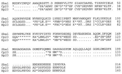
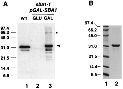
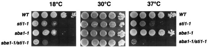
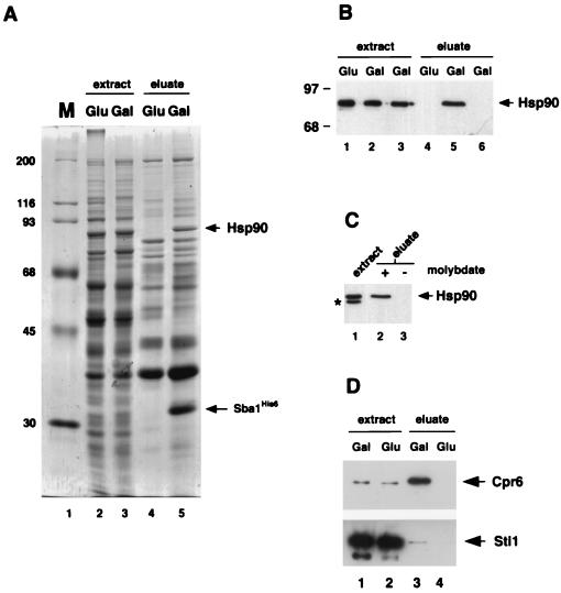
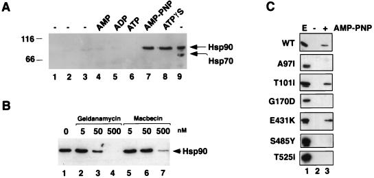
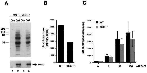
The Saccharomyces cerevisiae SBA1 gene was cloned by PCR amplification from yeast genomic DNA following its identification as encoding an ortholog of human p23, an Hsp90 cochaperone. The SBA1 gene product is constitutively expressed and nonessential, although a disruption mutant grew more slowly than the wild type at both 18 and 37 degreesC. A double deletion of SBA1 and STI1, encoding an Hsp90 cochaperone, displayed synthetic growth defects. Affinity isolation of histidine-tagged Sba1p (Sba1(His6)) after expression in yeast led to coisolation of Hsp90 and the cyclophilin homolog Cpr6. Using an in vitro assembly assay, purified Sba1(His6) bound to Hsp90 only in the presence of adenosine 5'-O-(3-thiotriphosphate) or adenyl-imidodiphosphate. Furthermore, interaction between purified Sba1(His6) and Hsp90 in yeast extracts was inhibited by the benzoquinoid ansamycins geldanamycin and macbecin. The in vitro assay was also used to identify residues in Hsp90 that are important for complex formation with Sba1(His6), and residues in both the N-terminal nucleotide binding domain and C-terminal half were characterized. In vivo analysis of known Hsp90 substrate proteins revealed that Sba1 loss of function had only a mild effect on the activity of the tyrosine kinase v-Src and steroid hormone receptors.


Figures

FIG. 1
FIG. 1
Sequence comparison of Sba1 with chicken (Cp23) and human (Hp23) p23 proteins. Identical matches are denoted by asterisks; gaps in the sequence are denoted by periods.
FIG. 2
FIG. 2
Characterization of Sba1 by gel electrophoresis. (A) Western blot analysis of Sba1 in yeast whole-cell extracts using polyclonal anti-Sba1. Lane 1, 50 μg of whole-cell extract from wild-type (WT) cells; lane 2, 50 μg of whole-cell extract from sba1-1 cells containing pGAL-SBA1 (YF256) grown in glucose-containing medium; lane 3, 5 μg of whole-cell extract from sba1-1 cells containing pGAL-SBA1 grown in galactose-containing medium. The arrowhead shows positions of Sba1 (lane 1) and Sba1His6 (lane 3). The asterisk shows the band at 60 kDa that reacts with anti-Sba1 in cells overexpressing Sba1His6. (B) Coomassie blue-stained gel of 2 μg of Sba1His6 (lane 2) after overexpression in E. coli and protein purification. Molecular size markers (in kilodaltons) shown in lane 1.
FIG. 3
FIG. 3
Growth phenotype of sba1-1 cells. Each panel shows a serial dilution of yeast cells spotted (3 μl of culture starting at 2 × 10⁸ cells/ml) from left to right of each plate, using wild-type (WT), sti1-1, sba1-1, and double-mutant (sba1-1/sti1-1) strains. Each plate was incubated at the indicated temperature for either 4 days (30 and 37°C) or 7 days (18°C).
FIG. 4
FIG. 4
Sba1His6 binding to Hsp90. (A) Coomassie blue-stained gel of whole-cell extracts from strain YF256 (sba1-1 pGAL-SBA1) (lanes 2 and 3) and eluates after affinity chromatography with Ni-NTA resin (lanes 4 and 5). Extracts were prepared from cells grown in glucose-containing medium (lanes 2 and 4) or galactose-containing medium (lanes 3 and 5). Molecular size markers (M, in kilodaltons) are shown in lane 1. Bands corresponding to Sba1His6 and Hsp90 are indicated by arrows. (B) Detection of Hsp90 by Western blot analysis in whole-cell extracts (lanes 1 to 3) and eluates after Ni-NTA resin affinity chromatography (lanes 4 to 6). Extracts and eluates were prepared from strain YF256 grown in glucose (lanes 1 and 4)- or galactose (lanes 2 and 5)-containing medium and wild-type strain W3031b grown in galactose-containing medium (lanes 3 and 6). (C). Western blot analysis of Hsp90 after isolation of Sba1His6 protein complexes on Ni-NTA resin and washing in buffers plus (lane 2) or minus (lane 3) sodium molybdate. A Western blot of the whole-cell extract is shown in lane 1; the band labeled with an asterisk is an Hsp90 degradation product. Data are from nonconsecutive lanes of the same gel and Western blot. (D) Western blot analysis of Cpr6 (upper panel) and Sti1 (lower panel) after isolation of Sba1His6 protein complexes on Ni-NTA resin. Lanes 1 and 2, whole-cell extracts of cells grown in galactose or glucose; lanes 3 and 4, eluates after affinity chromatography on Ni-NTA resin.
FIG. 5
FIG. 5
In vitro association of Sba1His6 with Hsp90. (A) Purified Sba1His6 was prebound to Ni-NTA resin and added to desalted whole-cell extracts from wild-type yeast cells and incubated at 30°C for 30 min. Hsp90 binding to Sba1His6 (lanes 1 to 8; lane 9 is whole-cell extract loaded directly onto the gel) was determined after reisolation of Sba1His6 and Western blotting using anti-Hsp90. Lane 1, Ni-NTA-Sba1His6 incubated in buffer; lane 2, Ni-NTA resin without prebound Sba1His6 incubated with extract; lane 3; Ni-NTA-Sba1His6 incubated with extract but without further addition; lane 4, Ni-NTA-Sba1His6 incubated with extract plus 5 mM AMP; lane 5, Ni-NTA-Sba1His6 incubated with extract plus 5 mM ADP; lane 6, Ni-NTA-Sba1His6 incubated with extract plus 5 mM ATP; lane 7, Ni-NTA-Sba1His6 incubated with extract plus 5 mM AMP-PNP; lane 8, Ni-NTA-Sba1His6 incubated with extract plus 5 mM ATPγS; lane 9, 2.5 μg of desalted whole-cell extract. The blot was first probed with anti-Hsp90 and subsequently reprobed with anti-Hsp70. Molecular size markers are shown in kilodaltons at left. (B) Binding of Hsp90 to Sba1His6 is competed by benzoquinoid ansamycins. Ni-NTA-Sba1His6 resin was incubated with desalted extracts as described above plus 5 mM AMP-PNP in the presence of solvent (dimethyl sulfoxide; lane 1), geldanamycin (lanes 2 to 4), or macbecin (lanes 5 to 7) at the concentrations indicated. After 30 min at 30°C, the resin was reisolated and washed, and bound proteins were resolved by SDS-PAGE. Hsp90 was detected by Western blotting. (C) Binding of wild-type (wt) and different mutant forms of Hsp90 to Ni-NTA-Sba1His6 resin. Lane 1, whole-cell extract (E); lane 2, incubation of Ni-NTA-Sba1His6 resin with extracts in the absence of AMP-PNP; lane 3, incubation of Ni-NTA-Sba1His6 resin with extracts in the presence of AMP-PNP. The Western blots were probed with anti-Hsp90.
FIG. 6
FIG. 6
v-Src and AR activities in wild-type and sba1-1 mutant yeast cells. (A) Western blot analysis of phosphotyrosine activity (upper panel) and the level of v-Src protein (lower panel) in wild-type (WT; strain AFY200; lanes 1 and 2) or sba1-1 (strain AFY202; lanes 3 and 4) cells after growth in glucose (lanes 1 and 3) or galactose (lanes 2 and 4) for 6 h. Molecular size markers are shown in kilodaltons at the left. (B) Quantitation of the level of phosphotyrosine in wild-type and sba1-1 cells from the Western blot shown in panel A. (C) lacZ gene expression in wild-type (strain YF267) and sba1-1 cells (strain YF268) after addition of dihydrotestosterone (DHT) as indicated. Cells were assayed for β-galactosidase 2 h after addition of hormone. Data are means of three independent assays.

Similar articles

Cited by

References

    1. Blagosklonny M V, Toretsky J, Bohen S, Neckers L. Mutant conformation of p53 translated in vitro or in vivo requires functional HSP90. Proc Natl Acad Sci USA. 1996;93:8379–8383. - PMC - PubMed
    1. Boeke J D, Trueheart J, Natsoulis G, Fink G R. 5-Fluoroorotic acid as a selective agent in yeast molecular genetics. Methods Enzymol. 1987;154:164–175. - PubMed
    1. Bohen S P, Yamamoto K R. Isolation of Hsp90 mutants by screening for decreased steroid receptor function. Proc Natl Acad Sci USA. 1993;90:11424–11428. - PMC - PubMed
    1. Borkovich K A, Farrelly F W, Finkelstein D B, Taulien J, Lindquist S. Hsp82 is an essential protein that is required in higher concentrations for growth of cells at higher temperatures. Mol Cell Biol. 1989;9:3919–3930. - PMC - PubMed
    1. Bose S, Weikl T, Bügl H, Buchner J. Chaperone function of Hsp90-associated proteins. Science. 1996;274:1715–1717. - PubMed

Publication types

MeSH terms