. 1998 Jul;18(7):3838-50.
doi: 10.1128/MCB.18.7.3838.

Identification of major binding proteins and substrates for the SH2-containing protein tyrosine phosphatase SHP-1 in macrophages

Affiliations

Identification of major binding proteins and substrates for the SH2-containing protein tyrosine phosphatase SHP-1 in macrophages

J F Timms et al. Mol Cell Biol. 1998 Jul.

Abstract

The protein tyrosine phosphatase SHP-1 is a critical regulator of macrophage biology, but its detailed mechanism of action remains largely undefined. SHP-1 associates with a 130-kDa tyrosyl-phosphorylated species (P130) in macrophages, suggesting that P130 might be an SHP-1 regulator and/or substrate. Here we show that P130 consists of two transmembrane glycoproteins, which we identify as PIR-B/p91A and the signal-regulatory protein (SIRP) family member BIT. These proteins also form separate complexes with SHP-2. BIT, but not PIR-B, is in a complex with the colony-stimulating factor 1 receptor (CSF-1R), suggesting that BIT may direct SHP-1 to the CSF-1R. BIT and PIR-B bind preferentially to substrate-trapping mutants of SHP-1 and are hyperphosphorylated in macrophages from motheaten viable mice, which express catalytically impaired forms of SHP-1, indicating that these proteins are SHP-1 substrates. However, BIT and PIR-B are hypophosphorylated in motheaten macrophages, which completely lack SHP-1 expression. These data suggest a model in which SHP-1 dephosphorylates specific sites on BIT and PIR-B while protecting other sites from dephosphorylation via its SH2 domains. Finally, BIT and PIR-B associate with two tyrosyl phosphoproteins and a tyrosine kinase activity. Tyrosyl phosphorylation of these proteins and the level of the associated kinase activity are increased in the absence of SHP-1. Our data suggest that BIT and PIR-B recruit multiple signaling molecules to receptor complexes, where they are regulated by SHP-1 and/or SHP-2.


Figures

FIG. 1
FIG. 1
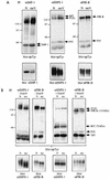
P130 is a membrane-localized glycoprotein and associates with both SHP-1 and SHP-2. (A) SHP-1–P130 complex localizes to the membrane compartment. BAC1.2F5 cells were starved of CSF-1 for 20 h and then stimulated with 2,000 U of CSF-1 per ml for 1 min (+) or left unstimulated (−). Cells were fractionated as described in Materials and Methods, and aliquots of the P1, P100, and S100 fractions were immunoprecipitated (IP) with anti-SHP-1 (αSHP-1) or anti-CSF-1R antibodies. Immune complexes were analyzed by SDS-PAGE (10% gel) and anti-pTyr immunoblotting. The anti-SHP-1 antibodies used for this panel are weakly cross-reactive with SHP-2, accounting for the 70-kDa phosphotyrosyl species; SHP-1-specific reagents are used in all subsequent experiments. Whole-cell lysates (WCL) and each fraction also were analyzed. Blots were reprobed with an anti-SHP-1 MAb to test for protein levels, and blots of each fraction were reprobed with anti-MAPK to monitor contamination of the P1 and P100 fractions with soluble cytoplasmic proteins. The positions of migration of Gibco BRL protein molecular size standards are shown in kilodaltons at the left. (B) P130 is a glycoprotein. Anti-SHP-1 immunoprecipitates from randomly growing BAC1.2F5 cells were treated with endo F as described in Materials and Methods. Deglycosylated (+) and untreated (−) samples were analyzed by SDS-PAGE (8% gel) and anti-pTyr immunoblotting (left panel). Lectin binding assays (right panel) were carried out as described in Materials and Methods, using A. hypogaea lectin (AHL), L. culinaris lectin (LCL), concanavalin A (ConA), or wheat germ agglutinin (WGA). Bound proteins were analyzed by SDS-PAGE (8% gel) and anti-pTyr immunoblotting. An anti-SHP-1 immunoprecipitate was loaded as a control (Ctrl IP). (C) P130 can coimmunoprecipitate with SHP-1 and SHP-2. Specific antibodies to SHP-1 and SHP-2 were used in consecutive immunoprecipitations from the lysates of a randomly growing normal macrophage cell line (N). Bound proteins were analyzed by SDS-PAGE (8% gel) and anti-pTyr immunoblotting. The migration of molecular size standards is indicated in kilodaltons at the left.
FIG. 2
FIG. 2
P130 consists of two glycoproteins, one a SIRP family member and the other PIR-B/p91A. (A) P130 consists of two distinct glycoproteins in primary BMM and cell lines derived from them. SHP-1 immunoprecipitates (IP) from randomly growing normal primary BMM (left panel) or N and meV cell lines (right panel) were treated with endo F (+) or left untreated (−). Deglycosylated proteins were analyzed by SDS-PAGE (8% gel) and anti-pTyr immunoblotting. The migration of molecular size standards is indicated in kilodaltons at the left. (B) Identification of the two glycoproteins as an SHPS-1-related protein and PIR-B/p91A. Preimmune (PI), anti-SHP-1, and anti-SHPS-1 immunoprecipitates from the meV cell line were treated with endo F (+) or left untreated (−) and then immunoblotted with anti-SHPS-1 (left panel) and anti-pTyr (middle panel) antisera. The anti-pTyr blot was stripped and reprobed with anti-PIR-B antisera (right panel).
FIG. 3
FIG. 3
Association of BIT with the CSF-1R. (A) Transient association and phosphorylation of a ∼160-kDa protein in BIT immunoprecipitates (IP). BAC1.2F5 cells were starved of CSF-1 for 22 h and then stimulated with 2,000 U of CSF-1 per ml for the indicated times or left unstimulated (0). BIT was immunoprecipitated from these lysates by using an anti-SHPS-1 antiserum, and bound proteins were analyzed by SDS-PAGE (8% gel) and anti-pTyr immunoblotting. The migration of molecular size standards is indicated in kilodaltons at the left. (B) BIT is associated constitutively with the CSF-1R. The CSF-1R was immunoprecipitated from the same set of lysates and treated with endo F or left untreated. The 1-min lysate was also immunoprecipitated with a nonimmune serum (NI). The presence of BIT in these immunoprecipitates was assessed by immunoblotting with the anti-SHPS-1 antiserum, which is able to recognize murine BIT. Whole-cell lysate (WCL) also was tested as a positive control. The migration of molecular size standards is indicated in kilodaltons at the left.
FIG. 4
FIG. 4
BIT and PIR-B are substrates of SHP-1 in macrophages. (A) Schematic representation of wild-type and mutant forms of SHP-1 used in this study. The properties of the PTP-inactive substrate-trapping and PTP-inactive nontrapping mutants are described in detail in the text. (B) P130 complex binds to substrate trapping mutants of SHP-1. GST fusion protein binding assays from BAC1.2F5 cell lysates were carried out as described in Materials and Methods, using 10 μg of either GST alone, GST–SHP-1(WT), GST–SHP-1(C453S) (C→S) or GST-SHP-1(D419A) (D→A). Bound proteins were eluted with 1 and 20 mM sodium orthovanadate or left untreated (0 mM). The amount of P130 remaining in the complex was analyzed by SDS-PAGE and anti-pTyr immunoblotting. (C) BIT binds preferentially to SHP-1(C453S) in vaccinia virus-infected BAC1.2F5 cells. Starved cells were either left uninfected (Ctrl) or infected with empty vaccinia virus (Empty), recombinant virus encoding wild-type SHP-1 (WT), or epitope-tagged SHP-1(C453S) (C→S) as described in Materials and Methods. Control and infected cells were stimulated with CSF-1 for 1 min or left unstimulated (0). Lysates were prepared and immunoprecipitated with anti-SHP-1 CTM antibodies, and immunoprecipitates were analyzed by SDS-PAGE and anti-pTyr immunoblotting (top panel). SHP-1 protein levels were analyzed in whole-cell lysates by immunoblotting with anti-SHP-1 MAb (lower panel). (D) Comparison of SHP-1-associated BIT phosphorylation in cells infected with phosphatase dead mutants of SHP-1. The experiment in Figure 4D was repeated with recombinant viruses for the expression of SHP-1 mutants D419A, R459M, and ΔP. SHP-1 immunoprecipitates from uninfected and virus infected BAC1.2F5 cells were analyzed by anti-pTyr immunoblotting. Levels of SHP-1 in immune complexes (middle panel) and whole-cell lysates (lower panel) were checked by reprobing and blotting with anti-SHP-1 MAb. (E) BIT phosphorylation in recombinant vaccinia virus-infected cells. BIT was immunoprecipitated from starved, uninfected and virus-infected BAC1.2F5 cells by using the anti-SHPS-1 antiserum, which is able to recognize murine BIT. Relative BIT tyrosine phosphorylation was analyzed by anti-pTyr blotting (top panel) and anti-SHPS-1 blotting (middle panel). The level of expression of each recombinant protein was checked by anti-SHP-1 blotting of whole-cell lysates (lower panel).
FIG. 5
FIG. 5
BIT and PIR-B tyrosine phosphorylation in N, meV, and me/me primary BMM. (A) BIT and PIR-B are hyperphosphorylated in primary BMM from meν/meν mice. The relative levels of tyrosine phosphorylation of BIT and PIR-B in anti-SHP-1, anti-SHPS-1 (which recognizes murine BIT), and anti-PIR-B immunoprecipitates (IP) from N and meV primary BMM were compared by anti-pTyr immunoblotting (upper panels) and by reprobing and immunoblotting for protein levels (lower panels). The migration of molecular size standards is indicated in kilodaltons at the left. (B) BIT and PIR-B are hypophosphorylated in primary BMM from meν/meν mice. As in panel A, relative tyrosine phosphorylation levels in anti-SHPS-1 and anti-PIR-B immunoprecipitates from normal and meν/meν primary BMM were compared by anti-pTyr immunoblotting (upper panels) and immunoblotting for protein levels (lower panels). In addition, immunoprecipitates were endo F-treated to facilitate the visualization of BIT and PIR-B (right-hand panels). The migration of molecular size standards is indicated in kilodaltons at the left.
FIG. 6
FIG. 6
Characterization of BIT- and PIR-B-associated proteins. (A) BIT and PIR-B associate with a PTK activity in primary BMM. BIT and PIR-B from normal and me/me primary BMM were assayed for autokinase activity for the times indicated and analyzed by autoradiography (left panel) and anti-pTyr immunoblotting (right panel). Assay conditions are described in Materials and Methods. The migration of molecular size standards is indicated in kilodaltons at the left. IP, immunoprecipitation. (B) V8 endoprotease mapping of BIT- and PIR-B-associated P55 and P120 proteins. The autophosphorylated 55- and 120-kDa proteins from radiolabeled anti-SHPS-1 and anti-PIR-B immune complexes were excised from gels and subjected to in gel treatment with the indicated amounts of V8 protease as described in Materials and Methods. Labeled digests were analyzed by SDS-PAGE (15% gel) and autoradiography. The migration of molecular size standards is indicated in kilodaltons at the left of each panel.
FIG. 7
FIG. 7
Model of BIT and PIR-B phosphorylation in normal, meν/mev, and me/me BMM. In normal cells, SHP-1 can dephosphorylate certain sites while affording other sites protection through its SH2 domains. In meν/meν cells, SHP-1 still is capable of protecting sites via its SH2 domains but is unable to dephosphorylate substrate sites. Thus, BIT and PIR-B will be hyperphosphorylated in motheaten viable (+++) compared to normal (++) cells. In this way, the PTP-inactive nontrapping mutants of SHP-1 would resemble the meν/meν forms of SHP-1. In me/me cells, there is no SH2 domain protection, and these sites are dephosphorylated by other cellular PTPs. Although substrate sites would be hyperphosphorylated as in meν/meν cells, the SH2 domain-binding sites would be less phosphorylated than normal, resulting in an overall decrease in BIT and/or PIR-B tyrosyl phosphorylation (+). BIT and PIR-B tyrosyl phosphorylation in cells expressing trapping mutants of SHP-1 might be expected to be even higher than in meν/meν BMM (++++), due to an additional PTP domain protection of sites normally targeted by SHP-1.

Similar articles

Cited by

References

    1. Adachi M, Fisher E H, Ihle J, Imai K, Jirik F, Neel B, Pawson T, Shen S-H, Thomas M, Ullrich A, Zhao Z. Mammalian SH2-containing protein tyrosine phosphatases. Cell. 1996;85:15. - PubMed
    1. Barford D, Flint A J, Tonks N K. Crystal structure of human protein tyrosine phosphatase 1B. Science. 1994;263:1397–1404. - PubMed
    1. Bignon J S, Siminovitch K A. Identification of PTP1C mutation as a genetic defect in motheaten and viable motheaten mice: a step toward defining the roles of protein tyrosine phosphatases in the regulation of hemopoietic cell differentiation and function. Clin Immunol Immunopathol. 1994;73:168–179. - PubMed
    1. Brown M, McCormack M, Zinn K G, Farrell M P, Bikel I, Livingston D M. A recombinant murine retrovirus for simian virus 40 large T cDNA transforms mouse fibroblasts to anchorage-independent growth. J Virol. 1986;60:290–293. - PMC - PubMed
    1. Burshtyn, D. N., and E. O. Long. Regulation through inhibitory receptors: lessons from natural killer cells. Trends Cell Biol., in press. - PubMed

Publication types

MeSH terms

Substances