. 1998 Jul;18(7):3862-70.
doi: 10.1128/MCB.18.7.3862.

Inhibition of clathrin-mediated endocytosis selectively attenuates specific insulin receptor signal transduction pathways

Affiliations

Inhibition of clathrin-mediated endocytosis selectively attenuates specific insulin receptor signal transduction pathways

B P Ceresa et al. Mol Cell Biol. 1998 Jul.

Abstract

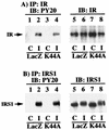
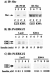
To examine the role of clathrin-dependent insulin receptor internalization in insulin-stimulated signal transduction events, we expressed a dominant-interfering mutant of dynamin (K44A/dynamin) by using a recombinant adenovirus in the H4IIE hepatoma and 3T3L1 adipocyte cell lines. Expression of K44A/dynamin inhibited endocytosis of the insulin receptor as determined by both cell surface radioligand binding and trypsin protection analysis. The inhibition of the insulin receptor endocytosis had no effect on either the extent of insulin receptor autophosphorylation or insulin receptor substrate 1 (IRS1) tyrosine phosphorylation. In contrast, expression of K44A/dynamin partially inhibited insulin-stimulated Shc tyrosine phosphorylation and activation of the mitogen-activated protein kinases ERK1 and -2. Although there was an approximately 50% decrease in the insulin-stimulated activation of the phosphatidylinositol 3-kinase associated with IRS1, insulin-stimulated Akt kinase phosphorylation and activation were unaffected. The expression of K44A/dynamin increased the basal rate of amino acid transport, which was additive with the effect of insulin but had no effect on the basal or insulin-stimulated DNA synthesis. In 3T3L1 adipocytes, expression of K44A/dynamin increased the basal rate of glucose uptake, glycogen synthesis, and lipogenesis without any significant effect on insulin stimulation. Together, these data demonstrate that the acute actions of insulin are largely independent of insulin receptor endocytosis and are initiated by activation of the plasma membrane-localized insulin receptor.


Figures

FIG. 1
FIG. 1
Adenovirus-mediated expression of HA-tagged K44A/dynamin I. H4IIE cells were either untreated (control [Con]; lane 1) or infected for 1 h with a recombinant adenovirus encoding for β-galactosidase (LacZ; lane 2) or the GTPase-defective dynamin I mutant (K44A; lane 3) as described in Materials and Methods. Forty-eight hours postinfection, whole-cell extracts (10 μg) were prepared and the proteins were resolved by SDS-PAGE. The extracts were then transferred to PVDF membranes and immunoblotted (IB) with antibodies against dynamin II (A), both dynamin I and II (both from Transduction Laboratories, Lexington, Ky.) (B), or the HA-tagged monoclonal antibody 12CA5 (Boehringer Mannheim, Indianapolis, Ind.) (C).
FIG. 2
FIG. 2
Expression of K44A/dynamin inhibits transferrin receptor and insulin receptor endocytosis. H4IIE cells were infected for 1 h with adenovirus encoding β-galactosidase (LacZ) or K44A/dynamin (K44A). Forty-eight hours postinfection, the rates and extents of transferrin receptor (A) and insulin receptor (B) internalization were determined by radioligand binding as described in Materials and Methods. The data represent averages and standard errors of the mean from three to four independent determinations. (C) H4IIE cells were infected with adenovirus encoding β-galactosidase (LacZ) or K44A/dynamin (K44A). Forty-eight hours following infection, the cells were either directly detergent solubilized or incubated in the absence (0) or presence of 100 nM insulin for 5 and 25 min at 37°C. The cells were then treated with trypsin, solubilized, and subjected to immunoblotting with an insulin receptor antibody as described in Materials and Methods. The amount of holoinsulin receptor was quantitated by densitometric scanning using Adobe Photoshop and NIH Image software. The data represent averages and standard errors of the mean from three independent determinations.
FIG. 3
FIG. 3
Inhibition of insulin receptor internalization does not impair insulin receptor autophosphorylation or tyrosine phosphorylation of IRS1. H4IIE cells were infected with adenovirus encoding β-galactosidase (LacZ) or K44A/dynamin (K44A). Forty-eight hours following infection, the cells were incubated in the absence (control [C]; lanes 1, 3, 5, and 7) or presence (insulin [I]; lanes 2, 4, 6, and 8) of 100 nM insulin for 5 min at 37°C. Whole-cell extracts were prepared and immunoprecipitated (IP) with either a polyclonal insulin receptor (IR) antibody (A) or a polyclonal IRS1 antibody (B). The immunoprecipitates were then subjected to immunoblotting (IB) with a phosphotyrosine-specific antibody (lanes 1 to 4), an insulin receptor antibody (Transduction Laboratories) (A, lanes 5 to 8), or an IRS1 antibody (Upstate Biotechnology, Inc.) (B, lanes 5 to 8). The results are representative of experiments that were independently performed three times.
FIG. 4
FIG. 4
Inhibition of insulin receptor internalization results in decreased tyrosine phosphorylation of Shc and activation of ERK (for notation, see the legend to Fig. 3). (A) H4IIE cells were infected with adenovirus encoding β-galactosidase (LacZ) or K44A/dynamin (K44A). Forty-eight hours following infection, the cells were incubated in the absence (lanes 1, 3, 5, and 7) or presence (lanes 2, 4, 6, and 8) of 100 nM insulin for 5 min at 37°C. Whole-cell extracts were prepared and immunoprecipitated with a polyclonal Shc antibody and immunoblotted with either a phosphotyrosine antibody (lanes 1 to 4) or a monoclonal Shc antibody (lanes 5 to 8) (Transduction Laboratories). (B) H4IIE cells were infected with adenovirus encoding β-galactosidase (LacZ) or K44A/dynamin (K44A). Forty-eight hours following infection, the cells were incubated in the absence (lanes 1 and 5) or presence of 100 nM insulin for 5 (lanes 2 and 6), 10 (lanes 3 and 7), or 15 (lanes 4 and 8) min at 37°C. Whole-cell extracts were prepared and immunoblotted with a phosphospecific ERK antibody (Pi-ERK1/2) (New England Biolabs, Beverly, Mass.) as described in Materials and Methods. (C) H4IIE cells were infected with adenovirus encoding β-galactosidase (LacZ) or K44A/dynamin (K44A). Forty-eight hours following infection, the cells were incubated in the absence (lanes 1 and 6) or presence of 0.1 (lanes 2 and 7), 1 (lanes 3 and 8), 10 (lanes 4 and 9), or 100 (lanes 5 and 10) nM insulin for 5 min at 37°C. Whole-cell extracts were prepared and immunoblotted with a phosphospecific ERK antibody. The results are representative of experiments that were independently performed two times.
FIG. 5
FIG. 5
Expression of K44A/dynamin reduces the extent of insulin-stimulated IRS1- and phosphotyrosine-associated PI 3-kinase activity (for notation, see the legend to Fig. 3). H4IIE cells were infected with adenovirus encoding β-galactosidase (LacZ) or K44A/dynamin (K44A). Forty-eight hours following infection, the cells were incubated in the absence (lanes 1, 3, 5, and 7) or presence (lanes 2, 4, 6, and 8) of 100 nM insulin for 5 min at 37°C. Whole-cell extracts were prepared and immunoprecipitated with either a phosphotyrosine antibody (lanes 1 to 4) or IRS1 antibody (lanes 5 to 8). The immunoprecipitates were then incubated with phosphatidylinositol and [γ-32P]ATP as described in Materials and Methods. The formation of phosphatidylinositol phosphate (PIP) was determined by thin-layer chromatography (A) and quantitated by scintillation counting from three independent determinations (B). (C) H4IIE cells were infected with adenovirus encoding β-galactosidase (LacZ), or K44A/dynamin (K44A). Forty-eight hours following infection, the cells were incubated in the absence (lanes 1 and 6) or presence of 0.1 (lanes 2 and 7), 1 (lanes 3 and 8), 10 (lanes 4 and 9), or 100 (lanes 5 and 10) nM insulin for 5 min at 37°C. Whole-cell extracts were prepared and immunoprecipitated with an IRS1 antibody, and the immunoprecipitates were then incubated with phosphatidylinositol and [γ-32P]ATP as described in Materials and Methods. The formation of phosphatidylinositol phosphate (PIP) was determined by thin-layer chromatography and autoradiography. The autoradiogram is representative of two independent determinations.
FIG. 6
FIG. 6
The decrease in PI 3-kinase activity results from a decrease in the association of the PI 3-kinase with IRS1 (for notation, see the legend to Fig. 3). H4IIE cells were infected with adenovirus encoding β-galactosidase (LacZ) or K44A/dynamin (K44A). Forty-eight hours following infection, the cells were incubated in the absence (lanes 1 and 3) or presence (lanes 2 and 4) of 100 nM insulin for 5 min at 37°C. (A) Whole-cell extracts were prepared and immunoprecipitated with a polyclonal IRS1 antibody and immunoblotted with a polyclonal p85 antibody (lanes 1 to 4). (B) The IRS1 immunoprecipitates were also immunoblotted with an IRS1 antibody (lanes 1 to 4). (C) The amount of p85 coimmunoprecipitated with IRS1 was quantitated by scanning densitometry and analyzed with NIH Image software. The results represent averages and standard errors of the mean from three independent determinations.
FIG. 7
FIG. 7
Expression of K44A/dynamin does not affect insulin stimulation of Akt activity (for notation, see the legend to Fig. 3). (A) H4IIE cells were infected with adenovirus encoding β-galactosidase (LacZ) or K44A/dynamin (K44A). Forty-eight hours following infection, the cells were incubated in the absence (lanes 1 and 5) or presence of 100 nM insulin for 5 (lanes 2 and 6), 10 (lanes 3 and 7) and 15 (lanes 4 and 8) min at 37°C. Whole-cell extracts were prepared and immunoblotted with a polyclonal Akt antibody. (B) H4IIE cells were infected with adenovirus encoding β-galactosidase (LacZ) or K44A/dynamin (K44A). Forty-eight hours following infection, the cells were incubated in the absence (lanes 1 and 6) or presence of 0.1 (lanes 2 and 7), 1 (lanes 3 and 8), 10 (lanes 4 and 9), or 100 (lanes 5 and 10) nM insulin for 5 min at 37°C. Whole-cell extracts were prepared and immunoblotted with a polyclonal Akt antibody. (C) H4IIE cells were infected with adenovirus encoding β-galactosidase (LacZ) or K44A/dynamin (K44A). Forty-eight hours following infection, the cells were incubated in the absence (stippled box) or presence (solid box) of 100 nM insulin for 15 min at 37°C. Whole-cell extracts were prepared, immunoprecipitated with an Akt antibody, and subjected to in vitro kinase assay as described in Materials and Methods. The results represent averages and standard errors of the mean from three independent determinations.
FIG. 8
FIG. 8
Expression of K44A/dynamin enhances the basal rate of amino acid transport but has no effect on DNA synthesis. (A) H4IIE cells were infected with adenovirus encoding β-galactosidase (LacZ) or K44A/dynamin (K44A). Forty-eight hours following infection, the cells were incubated in the absence (control [C]) or presence (insulin [I]) of 100 nM insulin for 4 h at 37°C. The amount of [14C]AIB uptake was determined as described in Materials and Methods. (B) H4IIE cells were infected with adenovirus encoding β-galactosidase (LacZ) or K44A/dynamin (K44A). Forty-eight hours following infection, the cells were incubated in the absence (C) or presence (I) of 100 nM insulin for 20 h, and [3H]thymidine incorporation into DNA was determined as described in Materials and Methods. The results represent averages and standard errors of the mean from three independent determinations.
FIG. 9
FIG. 9
Expression of K44A/dynamin enhances the basal rate of glucose transport, glycogen synthesis, and lipogenesis in 3T3L1 adipocytes. Differentiated 3T3L1 adipocytes were infected with adenovirus encoding β-galactosidase (LacZ) or K44A/dynamin (K44A). Forty-eight hours following infection, the cells were incubated in the absence (control [C]) or presence (insulin [I]) of 100 nM insulin for 15 min at 37°C. The amounts of 2-[3H]deoxyglucose uptake (A) and incorporation of [14C]glucose into glycogen (B) and lipid (C) were determined as described in Materials and Methods. The results represent averages and standard errors of the mean from three independent determinations.

Similar articles

Cited by

References

    1. Allessi D R, Andjelkovic M, Caudwell B, Cron P, Morrice N, Cohen P, Hemmings B A. Mechanism of activation of protein kinase B by insulin and IGF-1. EMBO J. 1996;15:6541–6551. - PMC - PubMed
    1. Backer J M, Kahn C R, Cahill D A, Ullrich A, White M F. Receptor-mediated internalization of insulin requires a 12-amino acid sequence in the juxtamembrane region of the insulin receptor beta-subunit. J Biol Chem. 1990;265:16450–16454. - PubMed
    1. Backer J M, Kahn C R, White M F. Tyrosine phosphorylation of the insulin receptor during insulin-stimulated internalization in rat hepatoma cells. J Biol Chem. 1989;264:1694–1701. - PubMed
    1. Backer J M, Shoelson S E, Haring E, White M F. Insulin receptors internalize by a rapid, saturable pathway requiring receptor autophosphorylation and an intact juxtamembrane region. J Cell Biol. 1991;115:1535–1545. - PMC - PubMed
    1. Backer J M, Shoelson S E, Weiss M A, Hua Q X, Cheatham R B, Haring E, Cahill D C, White M F. The insulin receptor juxtamembrane region contains two independent tyrosine/beta-turn internalization sequences. J Cell Biol. 1992;118:831–839. - PMC - PubMed

Publication types

MeSH terms

LinkOut - more resources