. 1998 Jul;18(7):3889-99.
doi: 10.1128/MCB.18.7.3889.

Differential requirements for alternative splicing and nuclear export functions of equine infectious anemia virus Rev protein

Affiliations

Differential requirements for alternative splicing and nuclear export functions of equine infectious anemia virus Rev protein

M E Harris et al. Mol Cell Biol. 1998 Jul.

Abstract

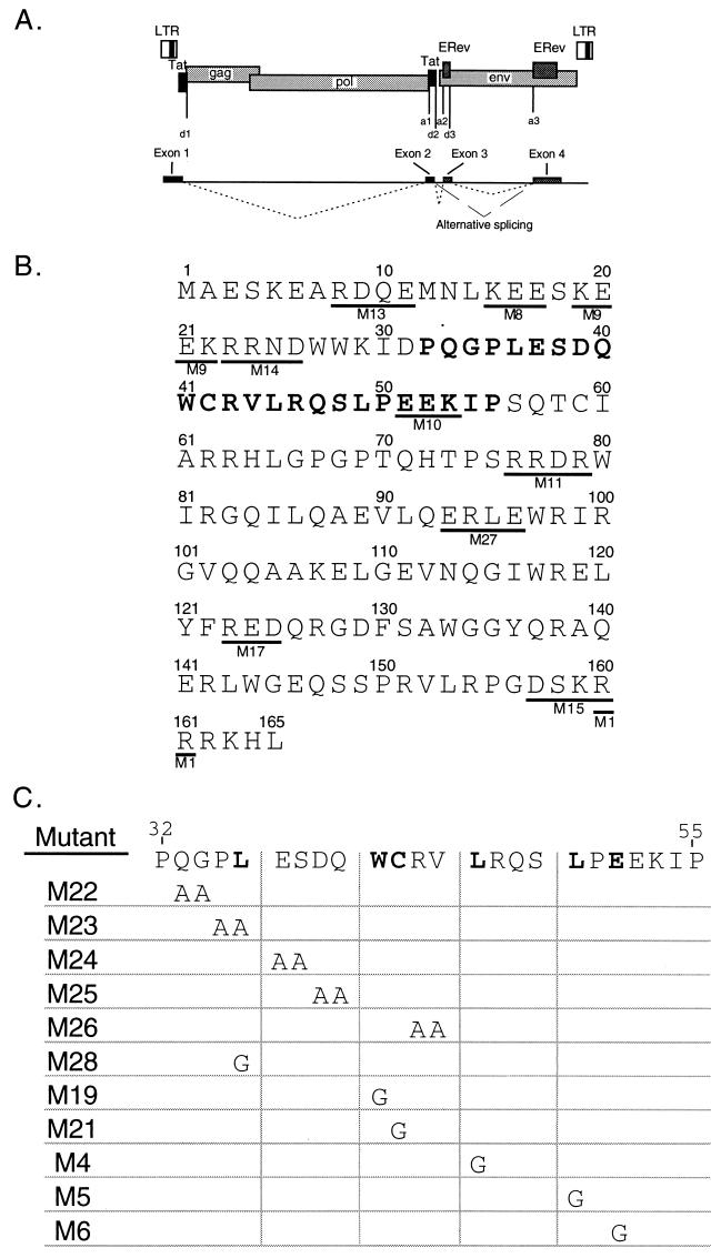
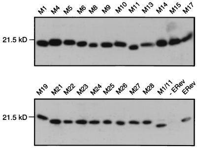
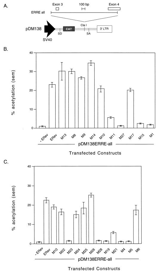
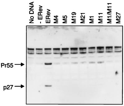
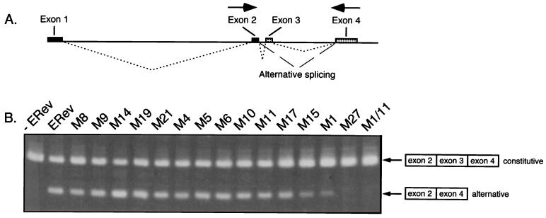
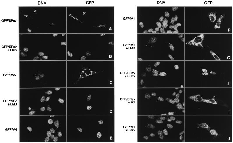
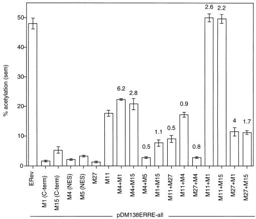
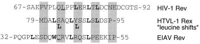
The Rev protein of equine infectious anemia virus (ERev) exports unspliced and partially spliced viral RNAs from the nucleus. Like several cellular proteins, ERev regulates its own mRNA by mediating an alternative splicing event. To determine the requirements for these functions, we have identified ERev mutants that affect RNA export or both export and alternative splicing. Mutants were further characterized for subcellular localization, nuclear-cytoplasmic shuttling, and multimerization. None of the nuclear export signal (NES) mutants are defective for alternative splicing. Furthermore, the NES of ERev is similar in composition but distinct in spacing from other leucine-rich NESs. Basic residues at the C terminus of ERev are involved in nuclear localization, and disruption of the C-terminal residues affects both functions of ERev. ERev forms multimers, and no mutation disrupts this activity. In two mutants with substitutions of charged residues in the middle of ERev, RNA export is affected. One of these mutants is also defective for ERev-mediated alternative splicing but is identical to wild-type ERev in its localization, shuttling, and multimerization. Together, these results demonstrate that the two functions of ERev both require nuclear import and at least one other common activity, but RNA export can be separated from alternative splicing based on its requirement for a functional NES.


Figures

FIG. 1
FIG. 1
(A) Schematic of the EIAV genome showing long terminal repeats (LTR); coding regions for gag, pol, env, and the four exons; and splice donor and splice acceptor sites. Dotted lines connecting exons indicate the ERev-independent splicing pattern used to form the completely spliced ETat-ERev bicistronic transcript (exons 1 to 4). Exons 1 and 2 code for ETat; exons 3 and 4 code for ERev. Dashed lines indicate the ERev-dependent alternative splicing pattern used to form a transcript containing exons 1, 2, and 4. Exon 1 contains nucleotides 211 to 459, exon 2 contains nucleotides 5135 to 5275, exon 3 contains nucleotides 5436 to 5536, and exon 4 contains nucleotides 7234 to 7641. Adapted from Martarano et al. (40). (B) Charged mutations made in EIAV Rev by site-directed mutagenesis. Clusters of 4 amino acids (underlined) in which 3 or 4 residues were charged were mutated to alanine (with the exception of one mutant) by the Kunkel method (29, 30). Mutants are designated by M and a number (shown below the mutated residues). In M1, two arginines (160 and 161) were changed to glycines. Numbers above the sequence show residue positions. The previously defined minimal NES of ERev is indicated by bold characters (17, 39). (C) Site-directed mutations in the NES of EIAV Rev. The wild-type NES (aa 32 to 55) is shown at the top. Specific mutations were generated in the NES in the context of full-length ERev by the Kunkel method. Pairs of residues were mutated to alanines, or single amino acids were changed to glycines.
FIG. 2
FIG. 2
Western blot analysis of 293T lysates to monitor the expression of wild-type or mutated ERev. 293T cells were transfected with 40 μg of expression plasmids encoding wild-type or mutant ERev. Two days after transfection, cell lysates were prepared and normalized to total protein. Proteins were fractionated by SDS-PAGE and then immunoblotted with a rabbit anti-ERev antiserum. Lanes are labeled to show the transfected ERev mutants, wild-type ERev (ERev), or mock transfection (−ERev). The position and size of the relevant molecular mass marker are indicated.
FIG. 3
FIG. 3
(A) Schematic of the ERev-dependent CAT reporter construct pDM138ERRE-all. Shown are the simian virus 40 (SV40) promoter, the HIV-1 second intron splice donor (SD) and splice acceptor (SA) sites, the location of the CAT gene, the unique ClaI cloning site, and the 3′ long terminal repeat (LTR). The sequence from the env region of a Rev-negative EIAV provirus (nucleotides 5278 to 7532) was cloned into the ClaI site and served as the RRE. This fragment, designated ERRE-all, contained EIAV exon 3 and part of exon 4. The EIAV fragment and exons are drawn to scale, but the vector is not. (B) CAT assay of 293 lysates transfected with an ERev reporter construct transactivated by wild-type or mutant ERev. 293 cells were transfected in triplicate with 0.2 μg of the EIAV RRE containing CAT reporter pDM138ERRE-all; 0.2 μg of the β-galactosidase expression vector pCH110; and 1 μg of expression plasmids carrying wild-type, mutant, or no ERev. Two days after transfection, crude lysates were prepared for analysis of CAT activity after normalization for transfection efficiency by a β-galactosidase assay. The activities of the indicated ERev mutants, wild-type ERev (ERev), or reporter alone (−ERev) are reported as mean percent acetylation. The error bars represent the standard error of the mean (sem). (C) CAT assay of 293 lysates transfected with an ERev reporter construct transactivated by wild-type ERev or NES mutants. Transfections and CAT assays were performed as described in panel B. Percent acetylation (mean ± sem) is shown for the indicated NES mutants, wild-type ERev (ERev), or reporter alone (−ERev). (D) Northern analysis of pDM138ERRE-all. 293 cells were transfected with 10 μg of pDM138ERRE-all, 5 μg of either pRS-ERev (lane ERev) or M27 (lane M27), 5 μg of pGL3, and 2 μg of pCH110. Two days after transfection, cytoplasmic RNA was prepared. Cytoplasmic RNA (20 μg) was run on a 1% agarose–formaldehyde gel, transferred, and probed for the CAT gene. The unspliced message is indicated. Differential background signals in the two lanes correlated with CAT activity.
FIG. 3
FIG. 3
(A) Schematic of the ERev-dependent CAT reporter construct pDM138ERRE-all. Shown are the simian virus 40 (SV40) promoter, the HIV-1 second intron splice donor (SD) and splice acceptor (SA) sites, the location of the CAT gene, the unique ClaI cloning site, and the 3′ long terminal repeat (LTR). The sequence from the env region of a Rev-negative EIAV provirus (nucleotides 5278 to 7532) was cloned into the ClaI site and served as the RRE. This fragment, designated ERRE-all, contained EIAV exon 3 and part of exon 4. The EIAV fragment and exons are drawn to scale, but the vector is not. (B) CAT assay of 293 lysates transfected with an ERev reporter construct transactivated by wild-type or mutant ERev. 293 cells were transfected in triplicate with 0.2 μg of the EIAV RRE containing CAT reporter pDM138ERRE-all; 0.2 μg of the β-galactosidase expression vector pCH110; and 1 μg of expression plasmids carrying wild-type, mutant, or no ERev. Two days after transfection, crude lysates were prepared for analysis of CAT activity after normalization for transfection efficiency by a β-galactosidase assay. The activities of the indicated ERev mutants, wild-type ERev (ERev), or reporter alone (−ERev) are reported as mean percent acetylation. The error bars represent the standard error of the mean (sem). (C) CAT assay of 293 lysates transfected with an ERev reporter construct transactivated by wild-type ERev or NES mutants. Transfections and CAT assays were performed as described in panel B. Percent acetylation (mean ± sem) is shown for the indicated NES mutants, wild-type ERev (ERev), or reporter alone (−ERev). (D) Northern analysis of pDM138ERRE-all. 293 cells were transfected with 10 μg of pDM138ERRE-all, 5 μg of either pRS-ERev (lane ERev) or M27 (lane M27), 5 μg of pGL3, and 2 μg of pCH110. Two days after transfection, cytoplasmic RNA was prepared. Cytoplasmic RNA (20 μg) was run on a 1% agarose–formaldehyde gel, transferred, and probed for the CAT gene. The unspliced message is indicated. Differential background signals in the two lanes correlated with CAT activity.
FIG. 4
FIG. 4
Western blot of trans-complementation of a Rev-negative EIAV provirus. D17 cells were mock transfected (no DNA) or transfected with 0.5 μg of Rev-negative EIAV provirus pFL85-11 and 0.5 μg of expression plasmids carrying wild-type ERev (ERev), selected ERev mutants, or no protein (−ERev). Two days after transfection, crude lysates were prepared and analyzed for EIAV p27 by SDS-PAGE and Western blotting. A rabbit anti-EIAV p27gag antibody detected p27 and Pr55, the gag precursor.
FIG. 5
FIG. 5
(A) Schematic of exon splicing in EIAV. The splicing pattern for the constitutively fully spliced message for EIAV (exons 1 to 4) is indicated by dotted lines. Exons 1 and 2 code for ETat; exons 3 and 4 code for ERev. Dashed lines indicate the ERev-dependent skipping of exon 3 to form a transcript containing exons 1, 2, and 4. Arrows represent primers for RT-PCR. (B) RT-PCR analysis of ERev-dependent exon skipping. RNA from cells transfected with 3 μg of pFL85-11 (−ERev) and complemented with 1 μg of expression plasmids carrying wild-type ERev (ERev) and mutant ERev (as indicated) was prepared and converted to cDNA. PCR amplification was done with primers to EIAV exon 2 and exon 4. DNA products were resolved on an agarose gel and visualized by ethidium bromide staining. The skipped exon message (exons 2 and 4, alternative) and the constitutively spliced message (exons 2 to 4, constitutive) are indicated.
FIG. 6
FIG. 6
Localization of GFP-ERev fusion proteins and effects of coexpression or LMB treatment. 3T3 cells were cotransfected with 0.5 μg of GFP-ERev (A, B, H, and I), GFP-M27 (C and D), GFP-M4 (E), or GFP-M1 (F and G) and 2.5 μg of either an empty RSV expression vector (A to G), wild-type ERev (H and J), or M1 (I). On the following day, some cells were treated with 5 nM LMB for 4 h (B, D, and G), and then all cells were fixed with paraformaldehyde and stained with a DNA dye. DNA staining (left panels) and GFP fluorescence (right panels) are shown. Fields are representative of least three independent, blind experiments. Images were acquired on a confocal microscope; a 2× electronic zoom was used on all panels but A. Brightness and contrast were adjusted with Adobe Photoshop.
FIG. 7
FIG. 7
Nonfunctional ERev mutants can be complemented for RNA export. pDM138ERRE-all (0.2 μg), 0.2 μg of pCH110, 0.6 μg of pUC118, and 1 μg of RSV promoter expression vectors were transfected into 293 cells. The RSV vectors expressed ERev, M1, M4, M5, M11, M15, M27, or no protein (to balance the total amount of RSV promoter). CAT assays were performed as previously described. A plus sign indicates that two mutants were coexpressed in trans. The absolute percent acetylation (mean ± standard error of the mean [sem]) is reported for each transfection or cotransfection. The ratios of the cotransfection activity to the sum of the independent activities for two mutants are reported above each cotransfection. C-term, C terminal.
FIG. 8
FIG. 8
Comparison of the NESs from HIV-1 Rev, human T-cell leukemia virus type 1 (HTLV-1) Rex, and ERev. The NESs of HIV-1 Rev (aa 70 to 84), HTLV-1 Rex (aa 80 to 96), and ERev (aa 32 to 55) are shown aligned to best match important residues. Bold residues are required for function in each protein, whereas underlined residues give an intermediate phenotype; the most N-terminal leucine in HIV-1 Rev has been mutated without effect (37). Also shown are the allowable “leucine shifts” in the Rex NES (28).

Similar articles

Cited by

References

    1. Beisel C E, Edwards J F, Dunn L L, Rice N R. Analysis of multiple mRNAs from pathogenic equine infectious anemia virus (EIAV) in an acutely infected horse reveals a novel protein, Ttm, derived from the carboxy terminus of the EIAV transmembrane protein. J Virol. 1993;67:832–842. - PMC - PubMed
    1. Belshan M, Harris M E, Shoemaker A E, Smith T A, Hope T J, Carpenter S. Biological characterization of Rev variation in equine infectious anemia virus. J Virol. 1998;72:4421–4426. - PMC - PubMed
    1. Bogerd H P, Fridell R A, Benson R E, Hua J, Cullen B R. Protein sequence requirements for function of the human T-cell leukemia virus type 1 Rex nuclear export signal delineated by a novel in vivo randomization-selection assay. Mol Cell Biol. 1996;16:4207–4214. - PMC - PubMed
    1. Carroll R, Derse D. Translation of equine infectious anemia virus bicistronic tat-rev mRNA requires leaky ribosome scanning of the tat CTG initiation codon. J Virol. 1993;67:1433–1440. - PMC - PubMed
    1. Chabot B, Blanchette M, Lapierre I, La B H. An intron element modulating 5′ splice site selection in the hnRNP A1 pre-mRNA interacts with hnRNP A1. Mol Cell Biol. 1997;17:1776–1786. - PMC - PubMed

Publication types

Substances

LinkOut - more resources