. 1998 Jul;18(7):3956-65.
doi: 10.1128/MCB.18.7.3956.

A novel phosphorylation-dependent RNase activity of GAP-SH3 binding protein: a potential link between signal transduction and RNA stability

Affiliations

A novel phosphorylation-dependent RNase activity of GAP-SH3 binding protein: a potential link between signal transduction and RNA stability

I E Gallouzi et al. Mol Cell Biol. 1998 Jul.

Abstract

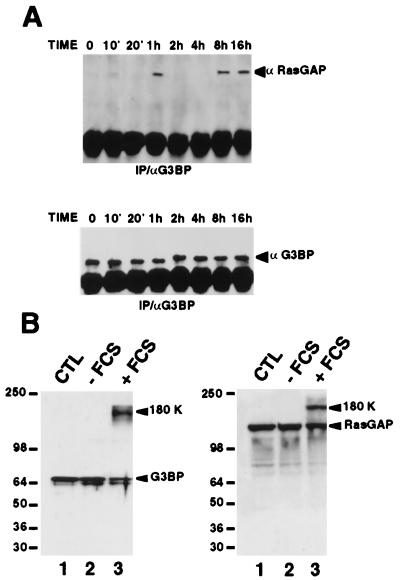
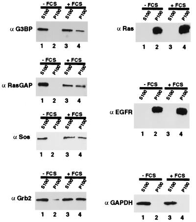
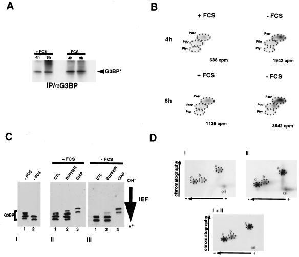
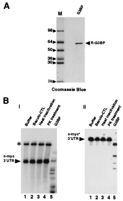
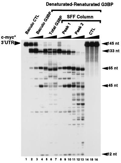
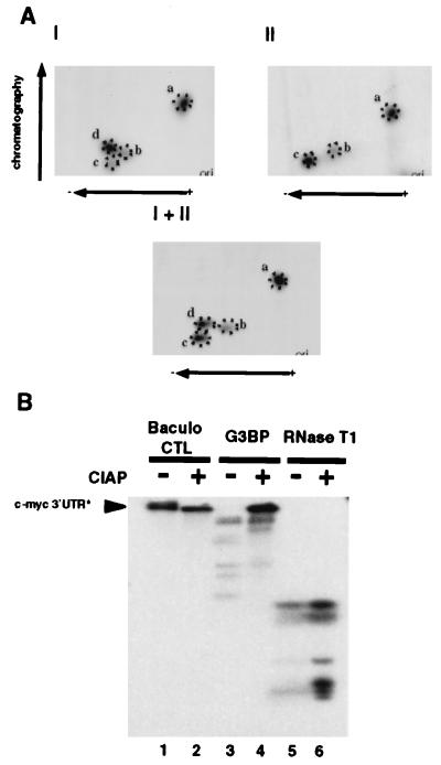
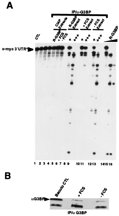
A potential p120 GTPase-activating protein (RasGAP) effector, G3BP (RasGAP Src homology 3 [SH3] binding protein), was previously identified based on its ability to bind the SH3 domain of RasGAP. Here we show that G3BP colocalizes and physically interacts with RasGAP at the plasma membrane of serum-stimulated but not quiescent Chinese hamster lung fibroblasts. In quiescent cells, G3BP was hyperphosphorylated on serine residues, and this modification was essential for its activity. Indeed, G3BP harbors a phosphorylation-dependent RNase activity which specifically cleaves the 3'-untranslated region of human c-myc mRNA. The endoribonuclease activity of G3BP can initiate mRNA degradation and therefore represents a link between a RasGAP-mediated signaling pathway and RNA turnover.


Figures

FIG. 1
FIG. 1
Time course of anti-G3BP antiserum recovery of RasGAP after serum stimulation of quiescent cells showing a physical interaction between RasGAP and G3BP. (A) Anti-RasGAP (upper panel) and anti-G3BP (lower panel) immunoblot analysis of G3BP immunoprecipitates (IP) from cells either unstimulated (lane 0) or stimulated with 10% fetal calf serum for the indicated times. (B) Total extracts obtained from quiescent (lane 2) or dividing (lane 3) cells were treated with EDC, and cross-linked proteins were analyzed by immunoblotting with either anti-G3BP (α G3BP) or anti-RasGAP (α RasGAP) antibody. Lane 1 represents total extracts not treated with EDC. −FCS, without fetal calf serum; +FCS, with fetal calf serum; CTL, control fraction.
FIG. 2
FIG. 2
G3BP associates with RasGAP exclusively in subcellular particulate fractions derived from dividing cells and not quiescent cells. Proteins contained in the S100 and the P100 fractions derived from quiescent cells (lanes 1 and 2, respectively) or dividing cells (lanes 3 and 4, respectively) were immunoblotted with anti-G3BP (α G3BP), anti-RasGAP (α RasGAP), anti-Sos (α Sos), anti-Grb2 (α Grb2), anti-p21ras (α Ras), anti-EGFR (α EGFR), and anti-GAPDH (α GAPDH) antibodies. −FCS, without fetal calf serum; +FCS, with fetal calf serum.
FIG. 3
FIG. 3
G3BP becomes hyperphosphorylated on Ser and Thr residues following serum starvation of CCL39 fibroblasts. (A) Proliferating (+FCS [with fetal calf serum]) and resting (−FCS [without fetal calf serum]) CCL39 fibroblasts were metabolically labeled with 32Pi for 4 or 8 h, and phosphorylated G3BP was affinity purified with an anti-G3BP antibody (α G3BP), separated by gel electrophoresis, transferred to a PVDF membrane, and detected by autoradiography. IP, immunoprecipitates. (B) Phosphoamino acid analysis of labeled G3BP. Equal amounts of G3BP, as estimated by Ponceau red staining, were excised, subjected to Cerenkov counting, and analyzed for phosphoamino acids. Numbers below the panels show total counts per minute incorporated into affinity-purified G3BP. (C) (Panel I) Comparison of G3BP phosphorylation levels between total extracts derived from dividing (lane 1) and quiescent (lane 2) CCL39 fibroblasts. Proteins were subjected to IEF gel analysis as described in Materials and Methods, and G3BP phosphorylation variants were revealed by immunoblotting with an anti-G3BP antibody. (Panels II and III) Total extracts (20 μg) derived from proliferating (panel II) or quiescent (panel III) CCL39 fibroblasts were incubated at 30°C for 1 h in the absence (lane 2) or the presence (lane 3) of 200 U of calf intestinal alkaline phosphatase (IAP) and analyzed as described for panel I. CTL, control fraction. (D) Phosphotryptic peptide mapping of immunopurified 32P-labeled G3BP from dividing (panel I) and quiescent (panel II) cells. Panel I+II represents the analysis of the mixture of the samples in panels I and II. ori, origin.
FIG. 4
FIG. 4
Cleavage of the c-myc 3′-UTR by purified recombinant G3BP. (A) SDS-PAGE analysis of purified recombinant G3BP (R-G3BP) (2 μg) stained with Coomassie blue (lane G3BP). Lane M, molecular size markers (in thousands). (B) The homogeneously 32P-labeled c-myc 3′-UTR was incubated for 10 min at 25°C in 10 μl of 50 mM Tris-HCl (pH 6)–150 mM NaCl–10% glycerol–10 mg of yeast tRNA per ml with 2 μl of buffer (lane 1), 2 μl of baculovirus control fraction (Baculo-CTL) (lane 2), 8 pmol of purified G3BP previously incubated at 100°C for 10 min (lane 3), 8 pmol of purified G3BP digested with proteinase K (PK) (lane 4), or 8 pmol of purified recombinant G3BP (lane 5). (Panel I) Aliquots (6 μl) from each reaction mixture were directly resolved on a nondenaturing polyacrylamide gel as described in Materials and Methods. Labeled RNA was visualized by autoradiography. The asterisk corresponds to abnormal migration of the labeled c-myc 3′-UTR in the native gel. (Panel II) Alternatively, the reaction mixtures were quenched by one phenol-chloroform extraction, and the RNA was electrophoresed on an 8% polyacrylamide–8 M urea gel. The asterisk indicates labeled c-myc.
FIG. 5
FIG. 5
Dose-dependent RNase activity associated with recombinant G3BP after purification under denaturing conditions. Lane 1, 2 μl of baculovirus control fraction (Baculo CTL). The labeled c-myc 3′-UTR was incubated with increasing amounts of purified recombinant G3BP (lane 2, 1.6 pmol; lane 3, 3.2 pmol; and lane 4, 6 pmol of recombinant G3BP). Aliquots of purified recombinant G3BP that were subjected to denaturation in the presence of 6 M urea, renatured, and concentrated over a heparin column were tested for RNase activity (lane 5, 1.6 pmol; lane 6, 3.2 pmol; and lane 7, 6 pmol of G3BP after purification). Denatured purified G3BP was separated over an SFF column into two peaks, and proteins from each peak were renatured, concentrated over a heparin column, and tested for RNase activity (peak 1: lane 8, 1.6 pmol; lane 9, 3.2 pmol; and lane 10, 6 pmol) (peak 2: lane 11, 1.6 pmol; lane 12, 3.2 pmol; and lane 13, 6 pmol). A control fraction (CTL) was obtained by pooling all other fractions not containing G3BP and assessing the RNase activity (lanes 14 to 16). nt, nucleotides; Baculo G3BP, control or recombinant baculovirus G3BP.
FIG. 6
FIG. 6
Dephosphorylation of recombinant G3BP inhibits its ability to cleave the c-myc 3′-UTR RNA. (A) Comparative tryptic phosphopeptide maps of recombinant G3BP and G3BP in quiescent cells. 32P-labeled recombinant G3BP (panel I) and G3BP from quiescent cells (panel II) were (in the gel), and the resulting phosphopeptides were separated in the first dimension by electrophoresis (pH 1.9) and in the second dimension chromatography on thin-layer plates and detected by autoradiography. Panel I+II represents a mixture of the samples in panels I and II. ori, origin. (B) RNase assays performed with 2 μl of baculovirus control fraction (Baculo CTL), 6 pmol of purified recombinant G3BP, or 20 U of sequencing-grade RNase T1 incubated in the absence (lanes 1, 3, and 5, respectively) or the presence (lanes 2, 4, and 6, respectively) of calf intestinal alkaline phosphatase (CIAP) for 1 h at 37°C.
FIG. 7
FIG. 7
RNase activity of G3BP in mammalian cells. (A) RNase assays were performed as described in Materials and Methods with 5 or 25 μl of G3BP immunoprecipitates (IP) of extracts obtained from insect cells infected with baculovirus expressing G3BP (recombinant G3BP [R-G3BP]) (lanes 8 and 9), from dividing cells (with fetal calf serum [+FCS]) (lanes 10 and 11), or from quiescent cells (without fetal calf serum [−FCS]) (lanes 12 and 13) or with 25 μl of G3BP immunoprecipitates of calf intestinal alkaline phosphatase (CIAP)-treated extracts obtained from insect cells infected with baculovirus expressing G3BP (lane 5), from dividing cells (lane 6), or from quiescent cells (lane 7). Lane 1, RNA alone (CTL, control fraction). As controls, RNase assays were performed with 25 μl of protein G-Sepharose (without antibody) incubated with extracts obtained from insect cells infected with baculovirus expressing G3BP (lane 2), from dividing cells (lane 3), or from quiescent cells (lane 4). α-G3BP, antibody to G3BP. (B) Immunoblot analysis of G3BP in immunoprecipitates (IP) of extracts obtained from insect cells infected with baculovirus expressing G3BP (Baculo CTL), from dividing cells (+FCS), or from quiescent cells (−FCS). The asterisk indicates labeled RNA. Lanes 14 to 16, RNase assays with recombinant G3BP.

References

    1. Blanchard J M, Piechaczyk M, Dani C, Chambard J C, Franchl A, Pouyssegur J, Jeanteur P. c-myc gene is transcribed at high rate in G0-arrested fibroblasts and is post-transcriptionally regulated in response to growth factors. Nature. 1985;317:443–445. - PubMed
    1. Boyle W J, van der Geer P, Hunter T. Phosphopeptide mapping and phosphoamino acid analysis by two-dimensional separation on thin-layer cellulose plates. Methods Enzymol. 1991;201:110–149. - PubMed
    1. Brannan C I, Perkins A S, Vogel K S, Ratner N, Nordlund M L, Reid S W, Buchberg A M, Jenkins N A, Parada L F, Copeland N G. Targeted disruption of the neurofibromatosis type-1 gene leads to developmental abnormalities in heart and various neural crest-derived tissues. Genes Dev. 1994;8:1019–1029. . (Erratum, 8:2792.) - PubMed
    1. Bryant S S, Mitchell A L, Collins F, Miao W, Marshall M, Jove R. N-terminal sequences contained in the Src homology 2 and 3 domains of p120 GTPase-activating protein are required for full catalytic activity toward Ras. J Biol Chem. 1996;271:5195–5199. - PubMed
    1. Buday L, Downward J. Epidermal growth factor regulates p21ras through the formation of a complex of receptor, Grb2 adapter protein, and Sos nucleotide exchange factor. Cell. 1993;73:611–620. - PubMed

Publication types

LinkOut - more resources