. 1998 Jul;18(7):3966-73.
doi: 10.1128/MCB.18.7.3966.

Binding of Shp2 tyrosine phosphatase to FRS2 is essential for fibroblast growth factor-induced PC12 cell differentiation

Affiliations

Binding of Shp2 tyrosine phosphatase to FRS2 is essential for fibroblast growth factor-induced PC12 cell differentiation

Y R Hadari et al. Mol Cell Biol. 1998 Jul.

Abstract

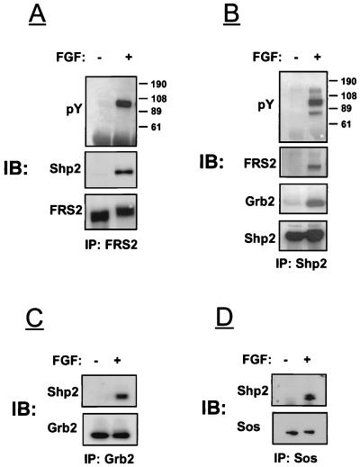
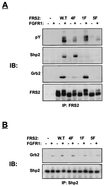
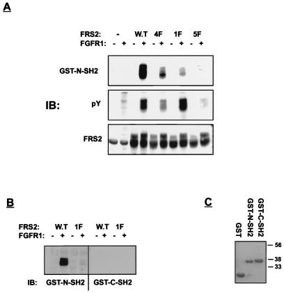
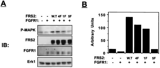
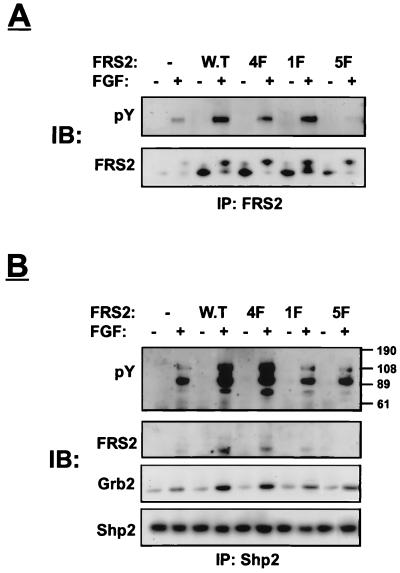
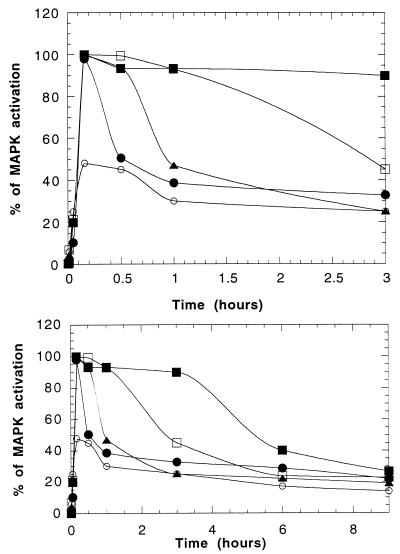
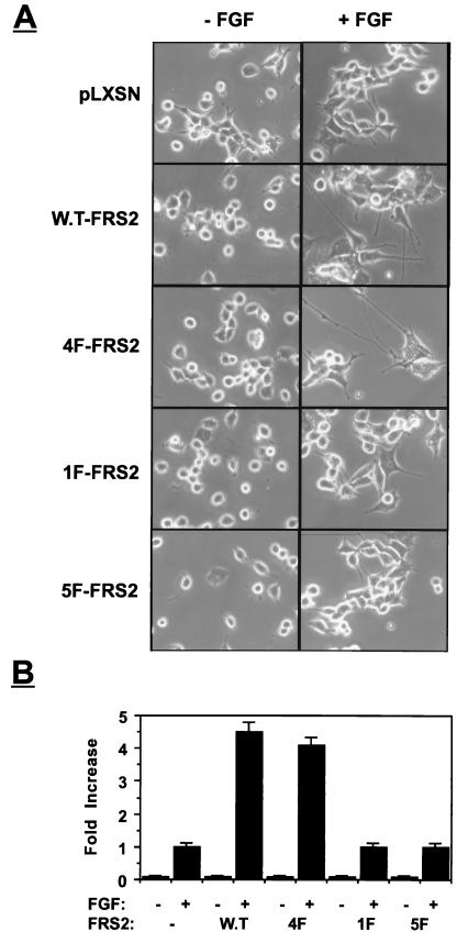
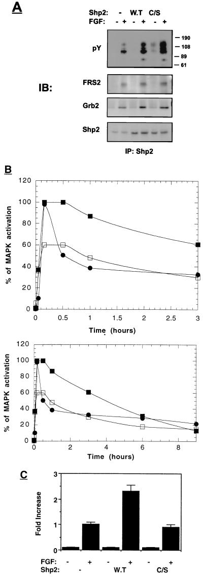
FRS2 is a lipid-anchored docking protein that plays an important role in linking fibroblast growth factor (FGF) and nerve growth factor receptors with the Ras/mitogen-activated protein (MAP) kinase signaling pathway. In this report, we demonstrate that FRS2 forms a complex with the N-terminal SH2 domain of the protein tyrosine phosphatase Shp2 in response to FGF stimulation. FGF stimulation induces tyrosine phosphorylation of Shp2, leading to the formation of a complex containing Grb2 and Sos1 molecules. In addition, a mutant FRS2 deficient in both Grb2 and Shp2 binding induces a weak and transient MAP kinase response and fails to induce PC12 cell differentiation in response to FGF stimulation. Furthermore, FGF is unable to induce differentiation of PC12 cells expressing an FRS2 point mutant deficient in Shp2 binding. Finally, we demonstrate that the catalytic activity of Shp2 is essential for sustained activation of MAP kinase and for potentiation of FGF-induced PC12 cell differentiation. These experiments demonstrate that FRS2 recruits Grb2 molecules both directly and indirectly via complex formation with Shp2 and that Shp2 plays an important role in FGF-induced PC12 cell differentiation.


Figures

FIG. 1
FIG. 1
FGF induces association between FRS2 and Shp2 in NIH 3T3 cells. NIH 3T3 cells were starved overnight, stimulated with aFGF, and extracted with lysis buffer. Lysates from stimulated or unstimulated cells were immunoprecipitated with anti-FRS2 (A), anti-Shp2 (B), anti-Grb2 (C), or anti-Sos1 (D) antibodies. Samples were analyzed by immunoblotting (IB) with the indicated antibodies.
FIG. 2
FIG. 2
Phosphorylated tyrosine 436 on FRS2 binds to Shp2. Human 293 cells were transiently transfected with expression vectors for wild-type (W.T) FRS2 or FRS2 mutants (1 μg) together with FGFR1 expression vector (0.1 μg). After 36 h, the cells were lysed and subjected to immunoprecipitation with anti-FRS2 (A) or anti-Shp2 (B) antibodies. Samples were resolved by SDS-PAGE (10% gel), transferred to a nitrocellulose filter, and immunoblotted (IB) with the indicated antibodies.
FIG. 3
FIG. 3
Shp2 binds to tyrosine-phosphorylated FRS2 via its amino-terminal SH2 domain. Human 293 cells were transiently transfected with expression vectors for wild-type (W.T) FRS2 and FRS2 mutants as described in legend to Fig. 2. (A) Total cell lysates were resolved by SDS-PAGE (10% gel), transferred to a nitrocellulose filter, and immunoblotted (IB) with the indicated antibodies. The nitrocellulose filter was incubated with GST–N-SH2 (3 μg/ml) together with monoclonal anti-GST antibodies as described in Materials and Methods. After washing, the nitrocellulose filter was blotted with anti-mouse horseradish peroxidase-conjugated antibodies. (B) Total cell lysates of cells transfected with wild-type or 1F FRS2 (same samples as in panel A) were immunoblotted with either GST–N-SH2 or GST–C-SH2 fusion protein. (C) GST alone (3 μg/ml) or GST–N-SH2 and GST–C-SH2 fusion proteins were purified over a glutathione agarose column, resolved by SDS-PAGE (10% gel), and stained with Coomassie blue.
FIG. 4
FIG. 4
Activation of MAP kinase in cells overexpressing FRS2 mutants. Human 293 cells were transiently transfected with expression vectors for wild-type (W.T) FRS2 and FRS2 mutants (0.2 μg) together with FGFR1 (0.01 μg) and Erk1 (2 μg) expression vectors. After 36 h, the cells were lysed, and cellular proteins were resolved by SDS-PAGE (10% gel), transferred to a nitrocellulose filter, and immunoblotted (IB) with the indicated antibodies (A). Quantitation of MAP kinase (MAPK) activity was carried out with a phosphorimager (B). Similar results were obtained in three different experiments.
FIG. 5
FIG. 5
Analysis of PC12 cells overexpressing FRS2 or FRS2 mutants. Constructs encoding for wild-type (W.T) FRS2 or FRS2 mutants were cloned into the pLXSN expression vector, and a high-titer stock of viruses was produced. Parental PC12 cells were infected with the different viruses. Cells were selected for 2 weeks in medium supplemented with Geneticin (0.5 mg/ml). Pools of selected cultures were used in this experiment. The cells were starved overnight and treated with aFGF (100 ng/ml for 5 min). Cell lysates derived from stimulated or unstimulated cells were subjected to immunoprecipitation with anti-FRS2 antibodies (A) or anti-Shp2 antibodies (B). The samples were resolved by SDS-PAGE (10%), transferred to a nitrocellulose filter, and immunoblotted (IB) with the indicated antibodies.
FIG. 6
FIG. 6
Kinetics of MAP kinase activation in PC12 cells transfected with vector alone (•), wild-type FRS2 (▪), 4F mutant (□), 1F mutant (▴), and 5F mutant (○). The upper and lower panels show the early and late time points of MAP kinase (MAPK) activation, respectively.
FIG. 7
FIG. 7
Neurite outgrowth in PC12 cells induced by overexpression of FRS2 and FRS2 mutants. PC12 cells expressing pLXSN vector alone or cells overexpressing wild-type FRS2 (W.T-FRS2) or FRS2 mutants were grown in the presence or absence of aFGF (100 ng/ml) and heparin (5 μg/ml). Neurite outgrowth was detected (A) and quantitated (B) following induction for 72 h. Neurite outgrowth was quantitated by scoring the number of cells with neurites longer then the size of two cell bodies. The length of the neurites was measured, and the average neurite length was calculated for each cell line. Similar results were obtained in four different experiments.
FIG. 8
FIG. 8
MAP kinase activation and neurite outgrowth in cells overexpressing wild-type or catalytically inactive Shp2. (A) PC12 cells were stably transfected with an expression vector that directs the synthesis of wild-type (W.T) or Cys/Ser inactive mutant (C/S) Shp2. The cells were starved overnight, stimulated with aFGF (100 ng/ml for 5 min), and extracted with lysis buffer. Lysates from stimulated or unstimulated cells were immunoprecipitated with anti-Shp2 antibodies and immunoblotted (IB) with the indicated antibodies. (B) PC12 cells expressing vector alone (•) or wild-type (▪) or Cys/Ser inactive mutant (□) Shp2 were stimulated with aFGF (100 ng/ml) for different time periods. The upper and lower panels show the early and late time points of MAP kinase (MAPK) activation, respectively. Quantitation of MAP kinase activation was determined as described in Materials and Methods. (C) PC12 cells expressing the pLXSN vector alone (control) and cells overexpressing wild-type or Cys/Ser inactive mutant Shp2 were grown in the absence or presence of aFGF (100 ng/ml) and heparin (5 μg/ml). Neurite outgrowth was quantitated following induction for 72 h. Quantification of neurite outgrowth was calculated as described in Materials and Methods.

References

    1. Bar-Sagi D, Feramisco J R. Microinjection of the ras oncogene protein into PC12 cells induces morphological differentiation. Cell. 1985;42:841–848. - PubMed
    1. Basilico C, Moscatelli D. The FGF family of growth factors and oncogenes. Adv Cancer Res. 1992;59:115–165. - PubMed
    1. Batzer A G, Rotin D, Urena J M, Skolnik E Y, Schlessinger J. Hierarchy of binding sites for Grb2 and Shc on the epidermal growth factor receptor. Mol Cell Biol. 1994;14:5192–5201. - PMC - PubMed
    1. Bennett A M, Hausdorff S F, O’Reilly A M, Freeman R M, Neel B G. Multiple requirements for SHPTP2 in epidermal growth factor-mediated cell cycle progression. Mol Cell Biol. 1996;16:1189–1202. - PMC - PubMed
    1. Chen Y, Grall D, Salcini A E, Pelicci P G, Pouyssegur J, Van-Obberghen-Schilling E. Shc adaptor proteins are key transducers of mitogenic signaling mediated by the G protein-coupled thrombin receptor. EMBO J. 1996;15:1037–1044. - PMC - PubMed

Publication types

MeSH terms

Substances