. 1998 Jul;18(7):4043-52.
doi: 10.1128/MCB.18.7.4043.

Prohibitin family members interact genetically with mitochondrial inheritance components in Saccharomyces cerevisiae

Affiliations

Prohibitin family members interact genetically with mitochondrial inheritance components in Saccharomyces cerevisiae

K H Berger et al. Mol Cell Biol. 1998 Jul.

Abstract

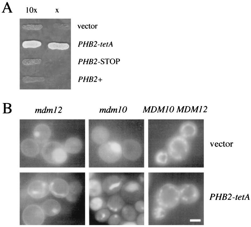
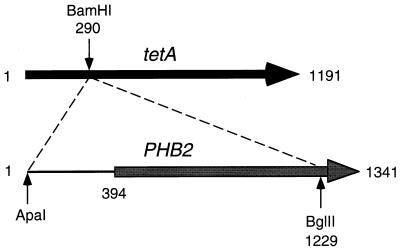
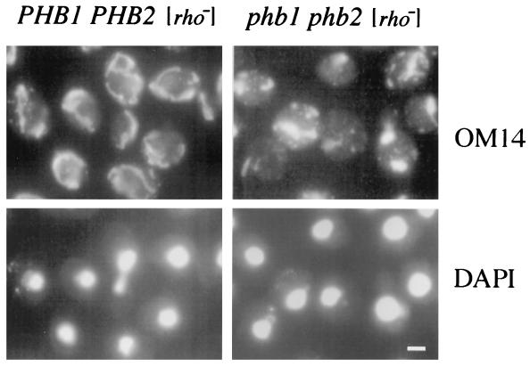
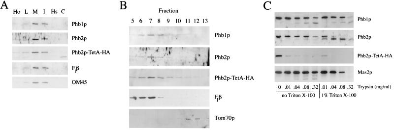
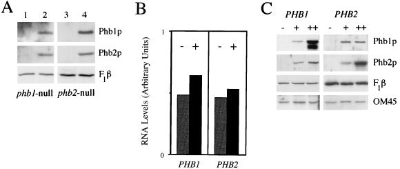
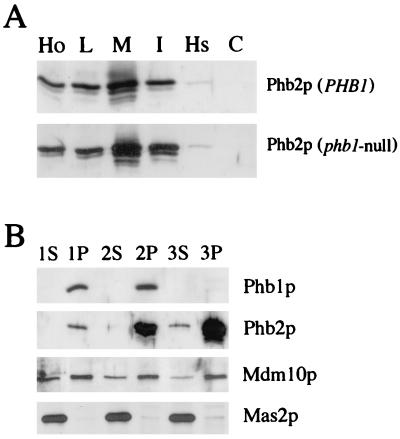
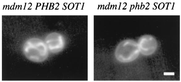
Phb2p, a homolog of the tumor suppressor protein prohibitin, was identified in a genetic screen for suppressors of the loss of Mdm12p, a mitochondrial outer membrane protein required for normal mitochondrial morphology and inheritance in Saccharomyces cerevisiae. Phb2p and its homolog, prohibitin (Phb1p), were localized to the mitochondrial inner membrane and characterized as integral membrane proteins which depend on each other for their stability. In otherwise wild-type genetic backgrounds, null mutations in PHB1 and PHB2 did not confer any obvious phenotypes. However, loss of function of either PHB1 or PHB2 in cells with mitochondrial DNA deleted led to altered mitochondrial morphology, and phb1 or phb2 mutations were synthetically lethal when combined with a mutation in any of three mitochondrial inheritance components of the mitochondrial outer membrane, Mdm12p, Mdm10p, and Mmm1p. These results provide the first evidence of a role for prohibitin in mitochondrial inheritance and in the regulation of mitochondrial morphology.


Figures

FIG. 1
FIG. 1
Plasmid-mediated suppression of growth and mitochondrial morphology defects. (A) Suppression of the mutant growth defect. Equal numbers of mdm12-null cells harboring the high-copy-number plasmids indicated were plated on selective medium lacking leucine and incubated at 30°C (semipermissive temperature). Lane x shows cells plated at a 10-fold dilution relative to cells shown in lane 10x. Plasmids were vector, YEp13; PHB2-tetA, the smallest suppressing subclone; PHB2-STOP, the subclone with an in-frame stop codon between PHB2 and tetA; and PHB2+, a vector with the wild-type PHB2 gene. (B) Partial suppression of mitochondrial morphology defects. Mitochondria were visualized by fluorescence microscopy following DASPMI uptake. Cells harbored either vector (YE13) or the PHB2-tetA plasmid. Representative wild-type MDM10 MDM12 (strain MYY290), mdm12-null (MYY623), and mdm10-null (MYY503) cells are shown. Bar = 2 μm.
FIG. 2
FIG. 2
Schematic diagram of the suppressing PHB2-tetA clone. PHB2 sequences were cloned as an in-frame gene fusion with the 3′ portion of tetA in the 2μm-based vector YEp13, as described in Materials and Methods. Illustrated is the smallest suppressing subclone (plasmid pKB38), generated by deletion of the insert DNA up to the unique ApaI site 5′ of PHB2. Numbering refers to nucleotides.
FIG. 3
FIG. 3
Altered mitochondrial morphology in phb1 phb2 [rho] cells. Cells with deficiencies of mitochondrial DNA which possessed either wild-type (left panels) or null (right panels) alleles of PHB1 and PHB2 were grown in YPD liquid medium at 30°C, fixed, and processed for indirect immunofluorescence microscopy. Mitochondria were visualized with antibodies to mitochondrial outer membrane protein OM14, and nuclear DNA was stained with DAPI. Bar = 2 μm.
FIG. 4
FIG. 4
Localization of Phb1p, Phb2p, and Phb2p-TetA. (A) Immunoblot analysis of subcellular fractions. Subcellular fractions were obtained from mdm12-null cells harboring the PHB2-tetA suppressor. F1β and OM45 were used as markers for the mitochondrial inner and outer membranes, respectively. Samples in the Phb2p-TetA-HA section were immunoblotted with antibodies that recognize the HA epitope of the ∼68-kDa fusion protein. These antibodies also recognize an unrelated, endogenous yeast protein of lower molecular mass that fractionates with the cytosol. Ho, total cell homogenate; L, low-speed pellet; M, mitochondrial pellet; I, intermediate-speed pellet; Hs, high-speed pellet; C, cytosolic fraction. (B) Phb1p, Phb2p, and Phb2p-TetA are localized to the mitochondrial inner membrane. Inner and outer mitochondrial membranes were resolved by sucrose density gradient centrifugation. Fractions were numbered in the order collected, from 1 (bottom) to 17 (top). Shown are fractions from the middle of the gradient which contained the peak concentrations of mitochondrial inner and outer membrane proteins. Control proteins were F1β, a mitochondrial inner membrane protein, and Tom70p, a mitochondrial outer membrane protein. Equal amounts of protein were loaded for each fraction. (C) Trypsin treatment of mitoplasts defines the topology of Phb1p, Phb2p, and Phb2p-TetA-HA in the mitochondrial inner membrane. Mitoplasts were digested with various amounts of trypsin either with or without the addition of 1% Triton X-100. Proteins were resolved by SDS-PAGE and analyzed by immunoblotting. Mas2p is a mitochondrial matrix protein protected from trypsin in the absence of detergent.
FIG. 5
FIG. 5
Phb1p and Phb2p are interdependent. (A) Immunoblot analysis of total cell protein extracts from phb1-null (lanes 1 and 2) and phb2-null (lanes 3 and 4) cells harboring a centromere-based vector with no insert (lanes 1 and 3), with wild-type PHB1 (lane 2), or with wild-type PHB2 (lane 4). Protein extracts were blotted with antibodies against Phb1p, Phb2p, or F1β, as indicated. (B) Quantitation of Northern analysis of PHB1 and PHB2. Levels of PHB1 RNA in phb2-null cells harboring either centromere-based vector alone (−) or CEN-PHB2 (+) were compared (left side), as were PHB2 RNA levels in phb1-null cells harboring either vector (−) or CEN-PHB1 (+) (right side). PHB1 and PHB2 RNA levels were standardized by using MDM1 RNA as a control for recovery; levels of this message did not vary with mutations in PHB1 and/or PHB2. (C) Overexpression of Phb1p and Phb2p. Levels of Phb1p, Phb2p, F1β, and OM45 in protein extracts were analyzed by immunoblotting. Protein extracts in the left section (PHB1) were from phb1-null cells harboring either vector alone (−), centromere-based vector containing wild-type PHB1 (+), or 2μm-based vector containing PHB1 (++). The right section (PHB2) shows protein extracts from phb2-null cells harboring either vector alone (−), centromere-based vector containing wild-type PHB2 (+), or 2μm-based vector containing PHB2 (++).
FIG. 6
FIG. 6
Localization and membrane association of overexpressed Phb2p. (A) Subcellular distribution of overexpressed Phb2p in PHB1 and phb1-null cells. Subcellular fractions are the same as those described in the legend for Fig. 4. (B) Membrane association of Phb1p and Phb2p. Mitochondria were extracted with 0.1 M Na2CO3 and supernatant (S) and pellet (P) fractions were isolated by centrifugation. 1, PHB1 PHB2 cells harboring vector only (MYY291-YEp13); 2, PHB1 PHB2 cells harboring high-copy-number PHB2 (MYY291-YEp13::PHB2); 3, phb1-null cells harboring high-copy-number PHB2 (MYY632-YEp13::PHB2). As previously documented (39), antiserum against Mdm10p recognizes two species: authentic Mdm10p, which is associated with the pellet, and an unrelated polypeptide of slightly higher molecular weight which fractionates with the supernatant.
FIG. 7
FIG. 7
Double mutant phb2 mdm12 cells harboring the SOT1 suppressor mutation are viable and exhibit the suppressed mitochondrial phenotype. SOT1-mutation-suppressed mdm12-null cells which carried either PHB2 (strain MYY629) or null mutation phb2 (strain MYY648) were grown in YPD at 23°C, stained with DASPMI, and examined by fluorescence microscopy. Bar = 2 μm.

References

    1. Allard J D, Bertrand K P. Membrane topology of the pBR322 tetracycline resistance protein. J Biol Chem. 1992;267:17809–17819. - PubMed
    1. Atencio D P, Yaffe M P. MAS5, a yeast homolog of DnaJ involved in mitochondrial protein import. Mol Cell Biol. 1992;12:283–291. - PMC - PubMed
    1. Austriaco N R., Jr Review: to bud until death: the genetics of aging in the yeast, Saccharomyces. Yeast. 1996;12:623–630. - PubMed
    1. Baudin A, Ozier-Kalogeropoulos O, Denouel A, Lacroute F, Cullin C. A simple and efficient method for direct gene deletion in Saccharomyces cerevisiae. Nucleic Acids Res. 1993;21:3329–3330. - PMC - PubMed
    1. Becherer K A, Rieder S D, Emr S D, Jones E W. Novel syntaxin homologue, Pep12p, required for the sorting of lumenal hydrolases to the lysosome-like vacuole in yeast. Mol Biol Cell. 1996;12:283–291. - PMC - PubMed

Publication types

MeSH terms