. 1998 Jul;18(7):4053-69.
doi: 10.1128/MCB.18.7.4053.

Spa2p interacts with cell polarity proteins and signaling components involved in yeast cell morphogenesis

Affiliations

Spa2p interacts with cell polarity proteins and signaling components involved in yeast cell morphogenesis

Y J Sheu et al. Mol Cell Biol. 1998 Jul.

Abstract

The yeast protein Spa2p localizes to growth sites and is important for polarized morphogenesis during budding, mating, and pseudohyphal growth. To better understand the role of Spa2p in polarized growth, we analyzed regions of the protein important for its function and proteins that interact with Spa2p. Spa2p interacts with Pea2p and Bud6p (Aip3p) as determined by the two-hybrid system; all of these proteins exhibit similar localization patterns, and spa2Delta, pea2Delta, and bud6Delta mutants display similar phenotypes, suggesting that these three proteins are involved in the same biological processes. Coimmunoprecipitation experiments demonstrate that Spa2p and Pea2p are tightly associated with each other in vivo. Velocity sedimentation experiments suggest that a significant portion of Spa2p, Pea2p, and Bud6p cosediment, raising the possibility that these proteins form a large, 12S multiprotein complex. Bud6p has been shown previously to interact with actin, suggesting that the 12S complex functions to regulate the actin cytoskeleton. Deletion analysis revealed that multiple regions of Spa2p are involved in its localization to growth sites. One of the regions involved in Spa2p stability and localization interacts with Pea2p; this region contains a conserved domain, SHD-II. Although a portion of Spa2p is sufficient for localization of itself and Pea2p to growth sites, only the full-length protein is capable of complementing spa2 mutant defects, suggesting that other regions are required for Spa2p function. By using the two-hybrid system, Spa2p and Bud6p were also found to interact with components of two mitogen-activated protein kinase (MAPK) pathways important for polarized cell growth. Spa2p interacts with Ste11p (MAPK kinase [MEK] kinase) and Ste7p (MEK) of the mating signaling pathway as well as with the MEKs Mkk1p and Mkk2p of the Slt2p (Mpk1p) MAPK pathway; for both Mkk1p and Ste7p, the Spa2p-interacting region was mapped to the N-terminal putative regulatory domain. Bud6p interacts with Ste11p. The MEK-interacting region of Spa2p corresponds to the highly conserved SHD-I domain, which is shown to be important for mating and MAPK signaling. spa2 mutants exhibit reduced levels of pheromone signaling and an elevated level of Slt2p kinase activity. We thus propose that Spa2p, Pea2p, and Bud6p function together, perhaps as a complex, to promote polarized morphogenesis through regulation of the actin cytoskeleton and signaling pathways.


Figures
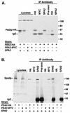
FIG. 1
FIG. 1
Spa2p and Pea2p coimmunoprecipitate. Proteins were prepared from SPA2 PEA2::HA, SPA2 PEA2::myc, spa2Δ PEA2::HA, and spa2Δ PEA2::myc strains (Y2003, Y2004, Y2005, and Y2006, respectively) and immunoprecipitated with the indicated antibodies. The total yeast lysates and immunoprecipitates were analyzed on immunoblots. (A) Immunoblot probed with anti-HA MAb 16B12. The doublet at approximately 60 kDa corresponds to Pea2p::HA; it is observed in lysates from the SPA2 PEA2::HA strain but not the SPA2 PEA2::myc strain. The doublet is also observed in IP with anti-HA antibody or anti-Spa2p antiserum from the SPA2 PEA2::HA lysate (fifth and seventh lanes from the left). Anti-Spa2p antiserum failed to precipitate Pea2p::HA from the spa2Δ PEA2::HA lysate (last lane). +, presence of an allele (for PEA2::HA and PEA2::myc) or wild-type copy of the gene (for SPA2). (B) Immunoblot probed with affinity-purified anti-Spa2p antiserum. Spa2p migrates as a 190-kDa band that is not present in the spa2Δ lysates. The band was detected in an IP with anti-HA antibody or anti-Spa2p antiserum from the SPA2 PEA2::HA lysate or in IP with anti-c-myc antibody or anti-Spa2p antiserum from the SPA2 PEA2::myc lysate. IgG marks the position of immunoglobulin heavy chain. Pre-imm, preimmune serum.
FIG. 2
FIG. 2
Spa2p interacts with Pea2p through a region containing SHD-II. (A and B) Filters showing the two-hybrid interactions. The dark patches indicate protein-protein interactions that result in expression of β-Gal. (A) LexA::Spa2p and AD::Pea2p interact with each other but not AD::Kar3p or LexA::Cik1p, two coiled-coil proteins. (B) Examples of interactions between AD::Pea2p and LexA fusions containing different regions of Spa2p. (C) Summary of mapping of the Pea2p-interacting region of Spa2p. Each horizontal bar represent a segment of Spa2p-coding sequence fused to the LexA plasmid (pSH2-1); the end residues for each Spa2p segment are labeled. These fusions were tested for interaction with the AD::PEA2 construct, and the strength of interaction were assigned as ++++ for the strongest interaction and − for interaction not detectable above background as judged from replicas of whole transformation plates. The summarized Pea2p-interacting region is shaded. The structure of Spa2p is presented above the fusions as described by Roemer et al. (71). CC, coiled coil A from residue 281 to 428; SHD-II, residue 429 to 535; a.a., amino acid. The first subregion of SHD-II is predicted to be coiled coil as well (coiled coil B).
FIG. 3
FIG. 3
HA::SPA2 deletion constructs and their relative cellular protein levels. (A) Diagram of HA::Spa2p deletions. Each horizontal bar represents a segment of HA::Spa2p. The end residues for each segment are labeled according to the position in the wild-type protein. A summary of deletion analysis results is presented to the right of the constructs. For more detailed phenotypic analysis, see Table 3. The structure of Spa2p is presented above the deletions as described by Gehrung and Snyder (30) and Roemer et al. (71). The shaded regions below SHD-I and SHD-II correspond to the MEK-interacting region and Pea2p-interacting region, respectively (see also Fig. 2 and 6). (B) Immunoblot analysis of proteins from HA::Spa2p deletion strains. Cell lysates were prepared from a spa2Δ strain (Y2007) containing the indicated HA::SPA2 deletion constructs. Equal amounts of protein were loaded in each well and fractionated by SDS–8% PAGE. The immunoblot was prepared and probed with anti-HA MAb 16B12. Two separate samples from two different experiments are shown for the 1-430 and 1-530 constructs. The 110-kDa band is a protein that cross-reacts with 16B12 ascites; this band is also present in Fig. 1A. NT, not tested.
FIG. 4
FIG. 4
Immunolocalization of HA::Spa2p and Pea2p::myc in strains containing Spa2p deletion constructs. spa2Δ PEA2::myc strains containing the different HA::SPA2 deletion constructs were treated with α-factor and stained with anti-HA antibodies (top) or anti-c-myc antibodies (bottom). Examples of the full-length protein (a) and five different constructs, i.e., pHA::spa2(1-736) (b), pHA::spa2(1-530) (c), pHA::spa2(1-430) (d), pHA::spa2(1-13, 265-552) (e), and pHA::spa2(1-13, 265-1466) (f), are shown. Although each of the different mutants exhibits polarization defects, fields that contain polarized cells are shown.
FIG. 5
FIG. 5
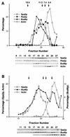
Velocity sedimentation analysis of Spa2p, Pea2p, and Bud6p. Cell lysates of a PEA2::myc BUD6::HA strain were prepared in the absence (A) or presence (B) of detergent and subjected to centrifugation in a 5 to 20% sucrose gradients. Fractions were collected and probed with anti-Spa2p antibodies, anti-c-myc antibodies (to detect Pea2p::myc), anti-HA antibodies (to detect Bud6p::HA), or an anti-actin MAb (C4). The S values of markers included in the same gradients are indicated at the top; these are thyroglobulin (19.4S), catalase (11.3S), aldolase (7.4S), and bovine serum albumin (4.4S). The amount of each immunoreactive protein was quantified for each fraction and is shown above the immunoblots. Only the top two-thirds of the gradient is shown, as no immunoreactive material is detected in the bottom third of the gradient. Note that in panel A the Pea2p peak contains material with a lower S value than Spa2p and Bud6p; this is likely to reflect its association with a degradation product of Spa2p that is not shown.
FIG. 6
FIG. 6
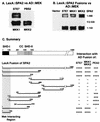
Spa2p interacts with MEKs through its N-terminal region containing the conserved domain SHD-I. (A and B) Filters of two-hybrid assays. (A) Interaction of LexA::Spa2p with AD constructs of Ste7p, Pbs2p, Mkk1p, and Mkk2p. (B) Different Spa2p fusions were tested for interaction with each of the AD::MEK constructs in panel A and with AD vector. (C) Summary of the interaction analysis for the different fusions. For each construct, the interactions were tested on colonies of hundreds of transformants and on patches from at least six transformants. Not shown is the interaction of AD::Spa2p(1-150) with LexA::Ste7p; these fusions interact strongly.
FIG. 7
FIG. 7
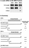
The N-terminal nonkinase domains of MEKs interact with Spa2p. (A) LexA::Spa2p fusions were tested for interactions with full-length, N-terminal (N-Term), or C-terminal (C-Term) AD constructs of Ste7p, Mkk1p, and Mkk2p. (B) Specific constructs from panel A and all fusions tested. The kinase domains of Ste7p and Mkk1p are indicated. NT, not tested.
FIG. 8
FIG. 8
Slt2p kinase activities in wild-type cells (WT) and spa2 mutants. (A) In vitro phosphorylation of MBP by Slt2p immunoprecipitated from spa2Δ and congenic wild-type cells. (B) MBP phosphorylation by Slt2p immunoprecipitated from spa2(1-2, 116-1466) strain (shown as spa2ΔSHD-I) and an isogenic wild-type strain. The relative kinase activities were quantified and are shown below each autoradiograph.
FIG. 9
FIG. 9
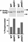
Levels of hyperphosphorylated Swi6p in wild-type (WT), spa2Δ, and pea2Δ strains. Equal amounts of proteins from cell lysates of wild-type (Y604 and Y762), spa2Δ (Y602), and pea2Δ (Y2002) strains grown to early log phase (OD600 = 0.3) were fractionated by SDS–8% PAGE. The immunoblot was prepared and probed with affinity-purified anti-Swi6p antiserum. The percentages of hyperphosphorylated Swi6p are shown below the blots. The relative amounts of proteins in the upper and lower bands were quantified by transmittence-reflectance scanning densitometry.
FIG. 10
FIG. 10
Summary of the different interactions between constituents of the 12S complex and signaling components. Proteins that contact one another have been shown to be physically associated by co-IP or sedimentation analysis. Proteins that interact as determined by the two-hybrid system are indicated by lines with arrowheads at each end. Components that were analyzed in this study are shaded. For a further description of the different components, see reference .

Similar articles

Cited by

References

    1. Adams A, Pringle J. Relationship of actin and tubulin distribution to bud growth in wild-type and morphogenetic-mutant Saccharomyces cerevisiae. J Cell Biol. 1984;98:934–945. - PMC - PubMed
    1. Amberg D C, Zahner J E, Mulholland J W, Pringle J R, Botstein D. Aip3p/Bud6p, a yeast actin-interacting protein that is involved in morphogenesis and the selection of bipolar budding sites. Mol Biol Cell. 1997;8:729–753. - PMC - PubMed
    1. Arkowitz R A, Lowe N. A small conserved domain in the yeast Spa2p is necessary and sufficient for its polarized localization. J Cell Biol. 1997;138:17–36. - PMC - PubMed
    1. Ayscough K R, Stryker J, Pokala N, Sanders M, Crews P, Drubin D G. High rate of actin filament turnover in budding yeast and roles for actin in establishment and maintenance of cell polarity revealed using the actin inhibitor latrunculin-A. J Cell Biol. 1997;137:399–416. - PMC - PubMed
    1. Baudin A, Ozier-Kalogeropoulos O, Denouel A, Lacroute F, Cullin C. A simple and efficient method for direct gene deletion in Saccharomyces cerevisiae. Nucleic Acids Res. 1993;21:3329–3330. - PMC - PubMed

Publication types

MeSH terms

Substances