. 1998 Jul;18(7):4141-8.
doi: 10.1128/MCB.18.7.4141.

Functional conservation of the transportin nuclear import pathway in divergent organisms

Affiliations

Functional conservation of the transportin nuclear import pathway in divergent organisms

M C Siomi et al. Mol Cell Biol. 1998 Jul.

Abstract

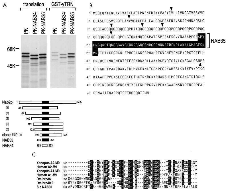
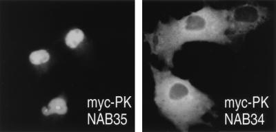
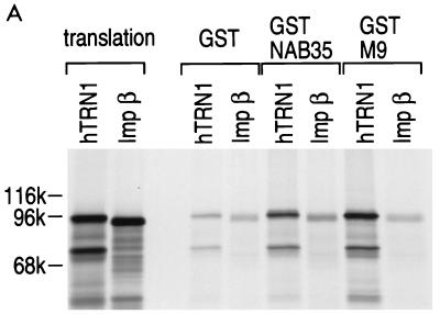
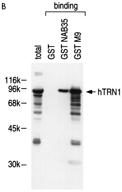
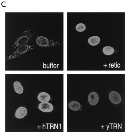
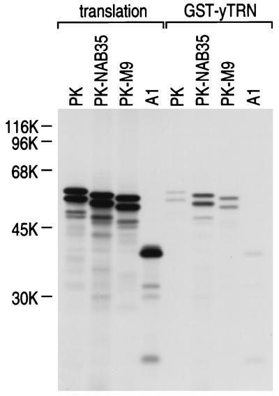
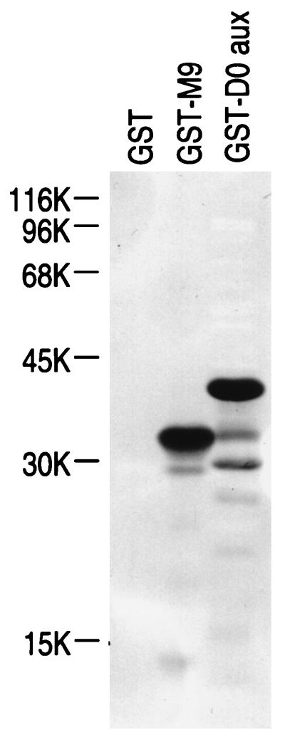
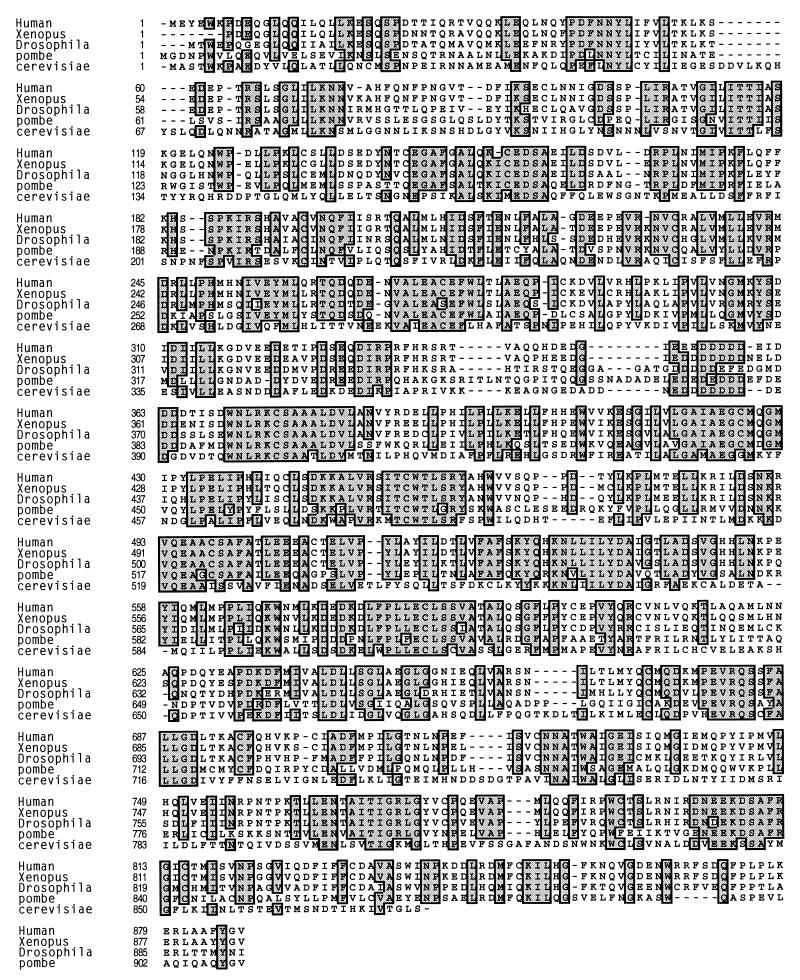
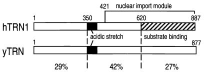
Human transportin1 (hTRN1) is the nuclear import receptor for a group of pre-mRNA/mRNA-binding proteins (heterogeneous nuclear ribonucleoproteins [hnRNP]) represented by hnRNP A1, which shuttle continuously between the nucleus and the cytoplasm. hTRN1 interacts with the M9 region of hnRNP A1, a 38-amino-acid domain rich in Gly, Ser, and Asn, and mediates the nuclear import of M9-bearing proteins in vitro. Saccharomyces cerevisiae transportin (yTRN; also known as YBR017c or Kap104p) has been identified and cloned. To understanding the nuclear import mediated by yTRN, we searched with a yeast two-hybrid system for proteins that interact with it. In an exhaustive screen of the S. cerevisiae genome, the most frequently selected open reading frame was the nuclear mRNA-binding protein, Nab2p. We delineated a ca.-50-amino-acid region in Nab2p, termed NAB35, which specifically binds yTRN and is similar to the M9 motif. NAB35 also interacts with hTRN1 and functions as a nuclear localization signal in mammalian cells. Interestingly, yTRN can also mediate the import of NAB35-bearing proteins into mammalian nuclei in vitro. We also report on additional substrates for TRN as well as sequences of Drosophila melanogaster, Xenopus laevis, and Schizosaccharomyces pombe TRNs. Together, these findings demonstrate that both the M9 signal and the nuclear import machinery utilized by the transportin pathway are conserved in evolution.


Figures

FIG. 1
FIG. 1
NAB35 is the target sequence of S. cerevisiae mRNA-binding protein, Nab2p, for yTRN interaction. (A) GST-yTRN on glutathione-Sepharose was incubated with myc-PK, myc-PK-NAB34 (PK-NAB34), or myc-PK-NAB35 (PK-NAB35) translated in vitro in the presence of [35S]methionine. The bound fraction was analyzed by SDS-PAGE and visualized by fluorography (GST-yTRN). Products of the translation reaction are shown as translation. Numbers at the left are molecular size standards (in kilodaltons). Schematic drawings of full-length Nab2p protein (3), Nab2p clones selected in an exhaustive genomic two-hybrid screen, NAB35 (shown by a black box), and NAB34 are shown below. The numbers in parentheses are the numbers of identical clones obtained by the screen. The numbers at the beginning and the end of the fragments indicate the first and the last amino acid numbers, respectively, for each fragment according to reference . (B) The NAB35 region in the Nab2p amino acid sequence (3) is shown by a black box. The starting points of clones selected by the screen and the ending point of clone 49 are indicated by arrowheads. (C) Alignment of M9 and M9-like sequences from a variety of organisms. Identical residues found in five or more sequences are boxed in black. The point mutation of Gly-274 to Ala in human A1-M9, indicated by a white circle, abolishes both NLS and NES activities of A1 (24).
FIG. 2
FIG. 2
NAB35-bearing protein is localized in the nucleus of mammalian cells. Transfection of COS7 cells was carried out with either myc-PK-NAB35 or myc-PK-NAB34, and immunofluorescence microscopy was carried out with an anti-myc antibody. Note that myc-PK-NAB35 is clearly localized in the nucleus in mammalian cells in contrast to myc-PK-NAB34.
FIG. 3
FIG. 3
hTRN1 mediates the nuclear import of NAB35-bearing proteins. (A) GST-NAB35 binds hTRN1 in vitro. GST, GST-NAB35, or GST-M9 (24) on glutathione-Sepharose was incubated with either hTRN1 or importin β (Imp β) translated in vitro in the presence of [35S]methionine. The bound fraction was analyzed by SDS-PAGE and visualized by fluorography. Products of in vitro translation are shown as translation. The positions of molecular mass markers are indicated on the left (in kilodaltons). (B) GST, GST-NAB35, or GST-M9 on glutathione-Sepharose was incubated with the cytoplasmic fraction of HeLa cells in the presence of 400 mM NaCl. After extensive washing, the bound fraction was analyzed by SDS-PAGE and visualized by immunoblotting with D45 (binding) (35). The cytoplasmic fraction used in this experiment is indicated in the lane labeled total. The position of hTRN1 is indicated with an arrow on the right, and the positions of molecular mass markers are indicated (in kilodaltons) on the left. (C) hTRN1 and yTRN mediate the nuclear import of GST-NAB35. Digitonin-permeabilized HeLa cells were incubated with GST-NAB35 (100 μg/ml) in buffer alone (buffer) or in the presence of either rabbit reticulocyte lysate (retic), hTRN1 (50 μg/ml), or yTRN (50 μg/ml). Import was detected with an anti-GST monoclonal antibody, followed by indirect immunofluorescence with fluorescein isothiocyanate-conjugated goat anti-mouse immunoglobulin G.
FIG. 3
FIG. 3
hTRN1 mediates the nuclear import of NAB35-bearing proteins. (A) GST-NAB35 binds hTRN1 in vitro. GST, GST-NAB35, or GST-M9 (24) on glutathione-Sepharose was incubated with either hTRN1 or importin β (Imp β) translated in vitro in the presence of [35S]methionine. The bound fraction was analyzed by SDS-PAGE and visualized by fluorography. Products of in vitro translation are shown as translation. The positions of molecular mass markers are indicated on the left (in kilodaltons). (B) GST, GST-NAB35, or GST-M9 on glutathione-Sepharose was incubated with the cytoplasmic fraction of HeLa cells in the presence of 400 mM NaCl. After extensive washing, the bound fraction was analyzed by SDS-PAGE and visualized by immunoblotting with D45 (binding) (35). The cytoplasmic fraction used in this experiment is indicated in the lane labeled total. The position of hTRN1 is indicated with an arrow on the right, and the positions of molecular mass markers are indicated (in kilodaltons) on the left. (C) hTRN1 and yTRN mediate the nuclear import of GST-NAB35. Digitonin-permeabilized HeLa cells were incubated with GST-NAB35 (100 μg/ml) in buffer alone (buffer) or in the presence of either rabbit reticulocyte lysate (retic), hTRN1 (50 μg/ml), or yTRN (50 μg/ml). Import was detected with an anti-GST monoclonal antibody, followed by indirect immunofluorescence with fluorescein isothiocyanate-conjugated goat anti-mouse immunoglobulin G.
FIG. 4
FIG. 4
hnRNP A1 does not bind yTRN. GST-yTRN on glutathione-Sepharose was incubated with either myc-PK (PK), myc-PK-NAB35 (PK-NAB35), myc-PK-M9 (PK-M9), or myc-A1 (A1) translated in vitro in the presence of [35S]methionine. The bound fraction was analyzed by SDS-PAGE and visualized by fluorography (GST-yTRN). Products of in vitro translation are shown as translation. The positions of molecular mass markers are indicated (in kilodaltons) on the left.
FIG. 5
FIG. 5
hTRN1 interaction with hnRNP D0 protein. GST, GST-M9, and GST-D0 aux (2 μg in each lane) bound to nitrocellulose blots were probed with 35S-labeled hTRN1. Note that GST-D0 aux can interact with hTRN1 under conditions in which GST-M9 is capable of binding hTRN1. The positions of molecular mass markers are indicated (in kilodaltons) on the left.
FIG. 6
FIG. 6
Sequence alignment of TRNs from divergent organisms. hTRN1 (Human) (5, 11, 33) was aligned with TRNs from X. laevis (Xenopus), D. melanogaster (Drosophila), Schizosaccharomyces pombe (pombe; accession no. g2656009), and Saccharomyces cerevisiae (cerevisiae) (2, 33). Identical amino acids found in four or more sequences are indicated with gray boxes. The numbers to the left of the amino acid sequences indicate amino acid positions.
FIG. 7
FIG. 7
Schematic representation of hTRN1 and yTRN. The structures of hTRN1 and yTRN are schematically shown, the solid boxes representing the acidic stretch in both proteins, and the cross-hatched box representing the substrate (M9) binding domain (11, 33) in hTRN1. The nuclear import module (28) in hTRN1 is also indicated. The percent identities of three regions of hTRN1 and yTRN are shown.

Similar articles

Cited by

References

    1. Adam S A, Marr R S, Gerace L. Nuclear protein import in permeabilized mammalian cells requires soluble cytoplasmic factors. J Cell Biol. 1990;111:807–816. - PMC - PubMed
    1. Aitchison J D, Blobel G, Rout M P. Kap104p: a karyopherin involved in the nuclear transport of messenger RNA binding proteins. Science. 1996;274:624–627. - PubMed
    1. Anderson J T, Wilson S M, Datar K V, Swanson M S. NAB2: a yeast nuclear polyadenylated RNA-binding protein essential for cell viability. Mol Cell Biol. 1993;13:2730–2741. - PMC - PubMed
    1. Bischoff F R, Görlich D. RanBP1 is crucial for the release of RanGTP from importin β-related nuclear transport factors. FEBS Lett. 1997;419:249–254. - PubMed
    1. Bischoff, F. R., S. Nakielny, and G. Dreyfuss. Unpublished results.

Publication types

MeSH terms

Substances

Associated data

LinkOut - more resources