. 1998 Jul;18(7):4157-64.
doi: 10.1128/MCB.18.7.4157.

The C-terminal domain of Sin1 interacts with the SWI-SNF complex in yeast

Affiliations

The C-terminal domain of Sin1 interacts with the SWI-SNF complex in yeast

J Pérez-Martín et al. Mol Cell Biol. 1998 Jul.

Abstract

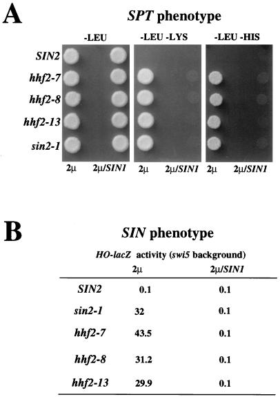
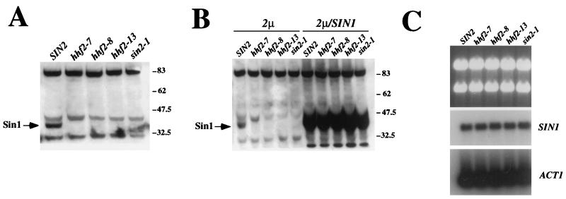
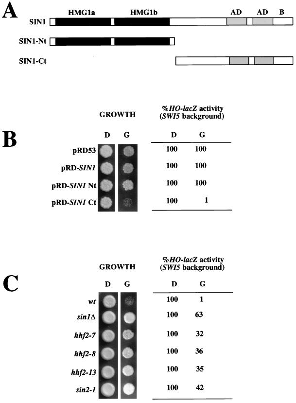
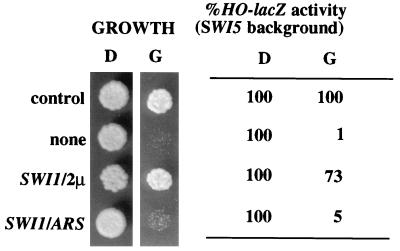
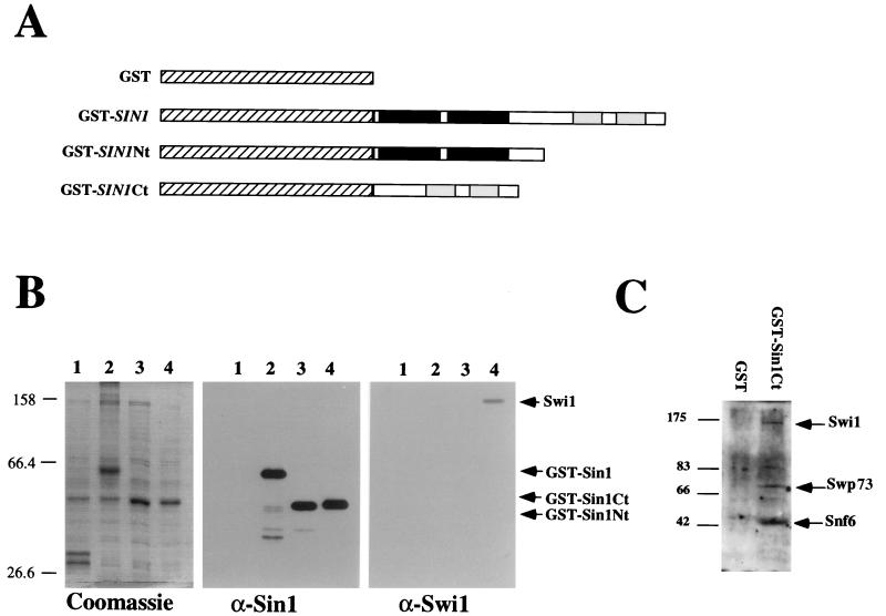
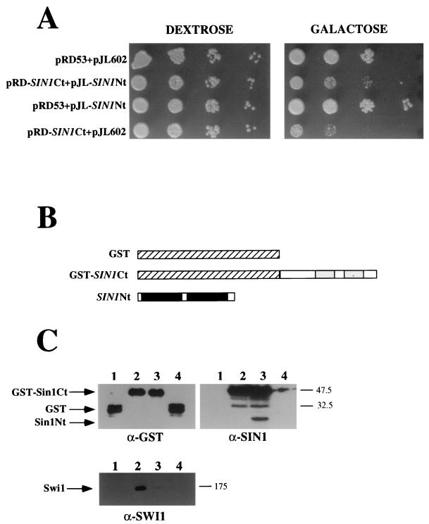
In the yeast Saccharomyces cerevisiae, the SWI-SNF complex has been proposed to antagonize the repressive effects of chromatin by disrupting nucleosomes. The SIN genes were identified as suppressors of defects in the SWI-SNF complex, and the SIN1 gene encodes an HMG1-like protein that has been proposed to be a component of chromatin. Specific mutations (sin mutations) in both histone H3 and H4 genes produce the same phenotypic effects as do mutations in the SIN1 gene. In this study, we demonstrate that Sin1 and the H3 and H4 histones interact genetically and that the C terminus of Sin1 physically associates with components of the SWI-SNF complex. In addition, we demonstrate that this interaction is blocked in the full-length Sin1 protein by the N-terminal half of the protein. Based on these and additional results, we propose that Sin1 acts as a regulatable bridge between the SWI-SNF complex and the nucleosome.


Figures

FIG. 1
FIG. 1
SIN1 is a high-copy suppressor of the sin mutations in histone H3 and H4 genes. Wild-type (SIN2; JJY14) or H4 histone mutant (hhf2-7, JJY19; hhf2-8, JJY20; hhf2-13, JJY21) and H3 histone mutant (sin2-1; JJY22) cells were transformed with YEp13 (2μm) or YEp13-SIN1 (2μm/SIN1). (A) Spt phenotype. Two microliters of a cell suspension (approximately 5 × 10⁶ cells/ml) from each culture was spotted onto minimal-medium plates (lacking leucine, lacking leucine and lysine, or lacking leucine and histidine) and incubated at 30°C for 3 days. Cells with a histone sin mutation show an Spt phenotype (i.e., the ability to suppress a δ element insertion in the HIS4 and LYS2 promoters) and are able to grow without lysine or histidine. A high dose of the SIN1 gene suppresses this phenotype. (B) Sin phenotype. Exponentially growing cultures were assayed for β-galactosidase activity, expressed as Miller units. Cells with a histone sin mutation show a Sin phenotype; that is, they are able to express the HO gene in the absence of the Swi5 protein, one of the activators of this promoter (note that all strains used in the experiment shown in this figure carry the swi5::hisG mutation). This ability is suppressed by a high-copy vector carrying the SIN1 gene.
FIG. 2
FIG. 2
Sin1 levels are lower in histone sin mutants. (A) Western blot with an anti-Sin1 polyclonal antibody from wild-type (SIN2; JJY14), H4 histone mutant (hhf2-7, JJY19; hhf2-8, JJY20; hhf2-13, JJY21), and H3 histone mutant (sin2-1; JJY22) cells. The arrow indicates the Sin1 protein. (B) Overexpression of Sin1 protein in histone sin mutant cells. The strains used are the same as those shown in panel A but were transformed with a control high-copy-number plasmid (2 μm) or the same plasmid carrying the wild-type SIN1 locus (2μm/SIN1). (C) Northern analysis of strains used in panel A. The upper panel shows the agarose gel stained with ethidium bromide, while the middle and bottom panels show blots of the same gel after hybridization with a SIN1 (middle) or an ACT1 (bottom) probe. Numbers to the right of panels A and B indicate molecular weight standards (in thousands).
FIG. 3
FIG. 3
SIN1 Ct half overexpression causes cell growth defects and low HO expression. (A) Schematic representation of the Sin1 protein with its most relevant characteristics. The Nt half includes two regions with similarities to mammalian HMG1 (HMG1a and HMG1b) protein. The Ct includes two regions rich in acidic domains (similar to those found in several other HMG-like proteins [AD]) and a region rich in positively and negatively charged residues (B). The panel also shows the Nt and Ct derivatives. (B) Overexpression of the Sin1 Ct causes slow growth and low HO expression. To score growth, 2 μl of a suspension (approximately 5 × 10⁶ cells/ml) of JJY10 cells transformed with the indicated plasmids was spotted onto minimal medium lacking uracil and containing either dextrose (D) or galactose (G). Plates were incubated at 30°C for 3 days. HO-lacZ activity was determined by assaying for β-galactosidase activity in exponentially growing liquid cultures in either dextrose or galactose. The expression of the HO-lacZ reporter was normalized to that of the control (JJY10 transformed with pRD53 in dextrose), which ranged between 110 and 90 Miller units. β-Galactosidase activity of cells grown in galactose was similar to that of cells grown in dextrose. (C) The defects produced by Sin1 Ct overexpression are alleviated by the same kinds of mutations that suppress swi defects. The plasmid pRD-SIN1Ct was introduced in JJY10 (wild type [wt]), JJY23 (sin1Δ::TRP1), JJY24 (hhf2-7), JJY25 (hhf2-8), JJY26 (hhf2-13), and JJY27 (sin2-1) cells, and cultures of these strains were scored for both growth and HO-lacZ expression as described above. The Sin1 Ct protein was expressed at similar levels in all strains, as assessed by Western blotting (data not shown).
FIG. 4
FIG. 4
High dose of SWI1 suppresses the Sin1 Ct-associated defects. JJY10 cells were transformed with pJL602 and YEp24 (control), pJL-SIN1Ct and YEp24 (none), pJL-SIN1Ct and pBD1 (SWI1/2μm), and pJL-SIN1Ct and pBD12 (SWI1/ARS). Cell growth and HO-lacZ activity were scored as in Fig. 3.
FIG. 5
FIG. 5
Interactions between the Ct half of Sin1 and the Swi1 protein. (A) Schematic representation of fusion proteins used. GST portions are indicated as hatched boxes. The proposed HMG1 boxes in Sin1 are highlighted in black, and the two acidic tracts are shaded. (B) The Swi1 protein copurifies with the Ct half of Sin1. Proteins obtained by the GST-affinity purification procedure (see Materials and Methods) were separated on SDS–10% polyacrylamide gels and either stained with Coomassie (left panel) or transferred to Immobilon membrane (Millipore), probed with the antiserum indicated, and detected with a secondary antibody and the Amersham enhanced chemiluminescence detection kit (center and right panels). The samples were obtained from cells overexpressing GST alone (lane 1), GST-SIN1 fusion (lane 2), GST-Sin1 Nt fusion (lane 3), and GST-Sin1 Ct fusion (lane 4). In spite of its lower estimated molecular weight, the GST-Sin1 Ct fusion migrates more slowly than does the GST-Sin1 Nt fusion, perhaps due to its high content of charged residues. (C) Western blot of GST-affinity eluates from cells overexpressing GST or the GST-Sin1 Ct fusion probed with anti-Swi1, anti-Swp73, and anti-Snf6. The numbers to the left of the blots are molecular weight standards (in thousands).
FIG. 6
FIG. 6
The Sin1 Nt half interacts with the Sin1 Ct half. (A) Overexpression of the Sin1 Nt half alleviates the defects associated with the overexpression of Sin1 Ct. Cells (JJY10) carrying the following plasmids—pRD53 and pJL602 (controls), pRD-SIN1Ct and pJL-SIN1Nt (Sin1 Ct and Sin1 Nt as independent polypeptides), pRD53 and pJL-SIN1Nt (Sin1 Nt alone), pRD-SIN1Ct and pJL602 (Sin1 Ct alone)—were spotted in 10-fold serial dilutions into media lacking uracil and leucine with either dextrose or galactose and incubated for 3 days at 30°C. (B) Scheme showing the protein fusions used in panel C. Note that Sin1 Nt half was not fused to GST protein in the following experiments. (C) Sin1 Nt interacts with GST-Sin1 Ct. Proteins obtained by the GST-affinity purification procedure were loaded into an SDS–12% polyacrylamide gel (GST and Sin1 blots) or an SDS–8.5% polyacrylamide gel (Swi1 blot) and treated as described in the legend for Fig. 4B. The samples were obtained from cells expressing GST (lane 1), GST-Sin1 Ct (lane 2), GST-Sin1 Ct and SIN1 Nt (lane 3), or GST and Sin1 Nt (lane 4). The numbers to the right of the blots are molecular weight standards (in thousands).

References

    1. Bernues J, Querol E, Martinez P, Barrios A, Espel E, Llobevas J. Detection by chemical cross-linking of the interaction between high mobility group protein 1 and histone oligomers in free solution. J Biol Chem. 1983;258:11020–11024. - PubMed
    1. Bianchi M E, Falciola L, Ferrari S, Lilley D M J. The DNA binding site of HMG1 protein is composed of two similar segments (HMG boxes), both of which have counterparts in other eukaryotic regulatory proteins. EMBO J. 1992;3:1055–1063. - PMC - PubMed
    1. Bonne-Andrea C, Harper F, Sobczac J, de Recondo A M. Rat liver HMG1: a physiological nucleosome assembly factor. EMBO J. 1984;3:1193–1199. - PMC - PubMed
    1. Bustin M, Lehn D A, Landsman D. Structural features of the HMG chromosomal proteins and their genes. Biochim Biophys Acta. 1990;1049:231–243. - PubMed
    1. Cairns B R, Henry N L, Kornberg R D. TFG3/TF30/ANC1, a component of the yeast SWI/SNF complex that is similar to the leukemogenic proteins ENL and AF-9. Mol Cell Biol. 1996;16:3308–3316. - PMC - PubMed

Publication types

MeSH terms

LinkOut - more resources